| [1] |
Ohtsubo K., Marth J.D., Cell, 2006, 126(5), 855—867
|
| [2] |
Akama T. O., Nakagawa H., Sugihara K., Narisawa S., Ohyama C., Nishimura S., O’Brien D., Moremen K., Millan J., Fukuda M., Science, 2002, 295(5552), 124—127
|
| [3] |
Wang J.H., Tong Y., Zhu Y., Tian H., Gao X. D., Pharm. Biotechnol., 2011, 18(1), 77—80
|
|
(王家红, 童玥, 朱玥, 田浤, 高向东. 药物生物技术, 2011, 18(1), 77—80)
|
| [4] |
Vavasseur F., Yang J. M., Dole K., Paulsen H., Brockhausen I., Glycobiology, 1995, 5(3), 351—357
|
| [5] |
Isailovic D., Kurulugama R. T., Plasencia M. D., Stokes S., Kurulugama R., Pungpapong V., Zhang M., Kyselova Z., Goldman R., Mechref Y., Novotny M., Clemmer D., Journal of Proteome Research, 2008, 7(3), 1109—1117
|
| [6] |
Xia L., Ju T., Westmuckett A., An G., Ivanciu L., McDaniel J., Lupu F., Cummings R., McEver R., Journal of Cell Biology, 2004, 164(3), 451—459
|
| [7] |
Fukuda M.,Biochimica et Biophysica Acta(BBA)-General Subjects, 2002, 1573(3), 394—405
|
| [8] |
Iwai T., Kudo T., Kawamoto R., Kubota T., Togayachi A., Hiruma T., Okada T., Kawamoto T., Morozum K., Narimatsu H., Procee-dings of the National Academy of Sciences of the United States of America, 2005, 102(12), 4572—4577
|
| [9] |
Okamoto T., Yoneyama M. S., Hatakeyama S., Mori K., Yamamoto H., Koie T., Saitoh H., Yamaya K., Funyu T., Fukuda M., Ohyama C., Tsuboi S., Molecular Medicine Reports, 2013, 7(2), 359—364
|
| [10] |
Lin Y. R., Reddy B. V., Irvine K. D., Developmental Dynamics: an Official Publication of the American Association of Anatomists, 2008, 237(12), 3703—3714
|
| [11] |
Hollingsworth M. A., Swanson B. J., Nature Reviews Cancer, 2004, 4(1), 45—60
|
| [12] |
Ju T., Cummings R. D., Proceedings of the National Academy of Sciences, 2002, 99(26), 16613—16618
|
| [13] |
Kawar Z. S., Johnson T. K., Natunen S., Lowe J., Cummings R., Glycobiology, 2008, 18(6), 441—446
|
| [14] |
Cummings R. D., Pierce J. M., Chemistry & Biology, 2014, 21(1), 1—15
|
| [15] |
Royle L., Mattu T. S., Hart E., Langridge J., Merry A., Murphy N., Harvey D., Dwek R., Rudd P., Anal. Biochem., 2002, 304(1), 70—90
|
| [16] |
Harvey D. J., Expert Review of Proteomics, 2005, 2(1), 87—101
|
| [17] |
Karlsson N. G., Thomsson K. A., Glycobiology, 2008, 19(3), 288—300
|
| [18] |
Miura Y., Kato K., Takegawa Y., Kurogochi M., Furukawa J., Shinohara Y., Nagahori N., Amano M., Hinou H., Nishimura S., Anal. Chem., 2010, 82(24), 10021—11029
|
| [19] |
Yamada K., Hyodo S., Matsuno Y., Kinoshita M., Maruyama S., Osaka Y., Casal E., Lee Y., Kakehi K., Anal. Biochem., 2007, 371(1), 52—61
|
| [20] |
Langkilde N. C., Wolf H., Clausen H., Ørntoft T. F., Cancer Research, 1992, 52(18), 5030—5036
|
| [21] |
Maniatis S., Zhou H., Reinhold V., Anal. Chem., 2010, 82(6), 2421—2425
|
| [22] |
Wang C., Fan W., Zhang P., Wang Z., Huang L., Proteomics, 2011, 11(21), 4229—4242
|
| [23] |
Kudelka M. R., Antonopoulos A., Wang Y., Duong D., Song X., Seyfried N., Dell A., Haslam S., Cummings R., Ju T., Nature Methods, 2016, 13(1), 81—86
|
| [24] |
Alvarez-Manilla G., Warren N. L., Abney T., Atwood J., Azadi P., York W., Pierce M., Orlando R., Glycobiology, 2007, 17(7), 677—687
|
| [25] |
Jang-Lee J., North S. J., Sutton-Smith M., Goldberg D., Panico M., Morris H., Haslam S., Dell A., Methods in Enzymology, 2006, 415, 59—86
|
| [26] |
Huang Y., Konse T., Mechref Y., Novotny M., Rapid Communications in Mass Spectrometry, 2002, 16(12), 1199—1204
|
| [27] |
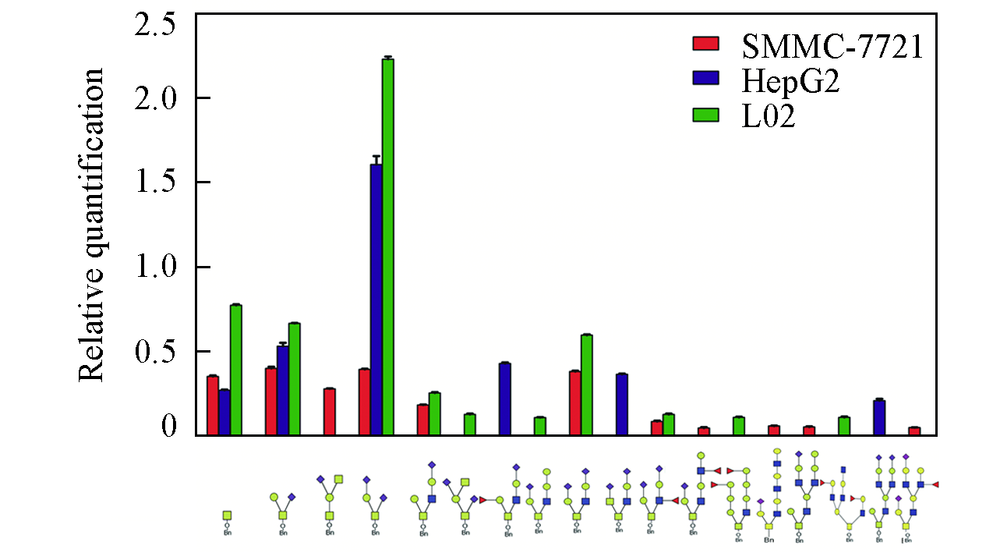
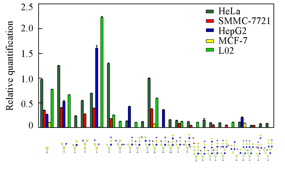
Gu X., Quanlitation and Quantitation Comparative Analysis of N- and O-glycomics from SMMC-7721 and L02 cells, Northwest University, Xi’an, 2016
|
|
(顾笑. SMMC-7721与L02细胞系N-和O-糖组定性定量比较分析, 西安: 西北大学, 2016)
|
| [28] |
Domon B., Costello C. E., Glycoconjugate Journal, 1988, 5(4), 397—409
|
| [29] |
Dube D. H., Bertozzi C. R., Nature Reviews Drug Discovery, 2005, 4(6), 477—488
|
| [30] |
Wang Y., Jobe S. M., Ding X., Choo H., Archer D., Mi R., Ju T., Cummings R., Proceedings of the National Academy of Sciences, 2012, 109(40), 16143—16148
|
| [31] |
Brockhausen I., EMBO Reports, 2006, 7(6), 599—604
|
| [32] |
Brockhausen I.,Biochimica et Biophysica Acta(BBA)-General Subjects, 1999, 1473(1), 67—95
|
| [33] |
Tian E., Ten Hagen K G., Glycoconjugate Journal, 2009, 26(3), 325—334
|
 ), 王仲孚(