

高等学校化学学报 ›› 2021, Vol. 42 ›› Issue (1): 133.doi: 10.7503/cjcu20200548
所属专题: 分子筛功能材料 2021年,42卷,第1期
陈晓, 申博渊, 熊昊, 魏飞( )
)
收稿日期:2020-08-10
出版日期:2021-01-10
发布日期:2021-01-12
通讯作者:
魏飞
E-mail:wf-dce@tsinghua.edu.cn
基金资助:
CHEN Xiao, SHEN Boyuan, XIONG Hao, WEI Fei()
Received:2020-08-10
Online:2021-01-10
Published:2021-01-12
Contact:
WEI Fei
E-mail:wf-dce@tsinghua.edu.cn
Supported by:摘要:
分子筛和金属有机骨架(MOF)材料以其独特的孔道和骨架结构在催化、 储能、 干燥及净化和吸附分离等领域有着广泛应用, 对其结构的原子尺度表征对于深入理解其构效关系具有重要意义. 但其大孔道结构和有机骨架使得它们对电子束辐照极为敏感, 在常规透射电镜成像模式下结构会很快被破坏变为非晶, 从而无法获得孔道和骨架的原子排列信息. 最近发展起来的基于积分差分相位衬度扫描透射电子显微(iDPC-STEM)技术在电子敏感材料和轻元素组分成像方面展现出明显优势, 使得对多孔骨架材料及烃池物种的表征成为了可能. 本文综述了本课题组近期利用该技术对分子筛和MOF材料原子尺度结构方面的研究. 将iDPC-STEM技术应用到ZSM-5分子筛催化剂中, 实现了对该分子筛的原子级骨架结构的成像分析. 在MOF体系中, 利用该技术观察到MIL-101骨架内部有机连接体与金属节点的配位方式, 在此基础上解析了MIL-101结构中有机配体的连接和金属节点的苯环结构, 并观察了MOF的原子级表面、 界面和缺陷等局域结构特征. 最后对iDPC-STEM技术在原子尺度成像方面的应用潜力进行了总结与展望.
中图分类号:
TrendMD:
陈晓, 申博渊, 熊昊, 魏飞. 电子束敏感材料的原子尺度结构研究. 高等学校化学学报, 2021, 42(1): 133.
CHEN Xiao, SHEN Boyuan, XIONG Hao, WEI Fei. Atomic Scale Structure of Beam Sensitive Materials. Chem. J. Chinese Universities, 2021, 42(1): 133.
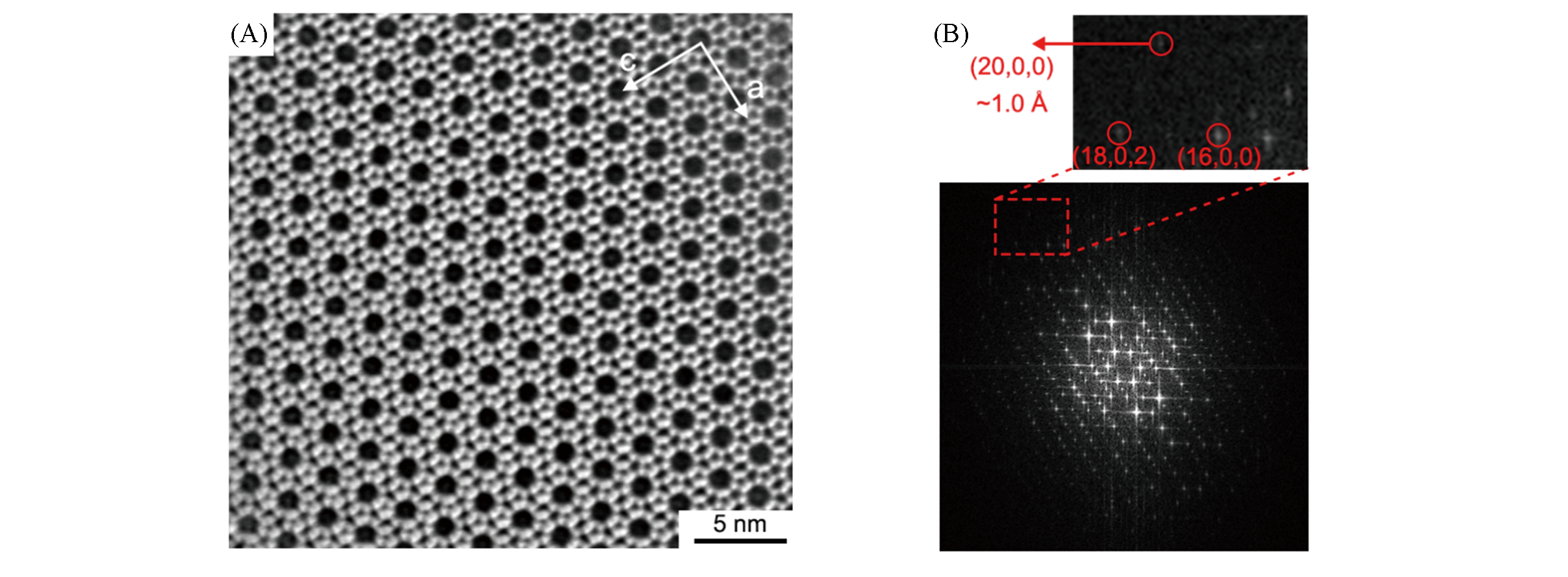
Fig.1 iDPC?STEM image(A) and corresponding FFT pattern(B) of ZSM?5 skeleton [010] crystal orientation[40](20,0,0) plane indicates an information transfer of about 1 ?.Copyright 2020, Wiley?VCH.
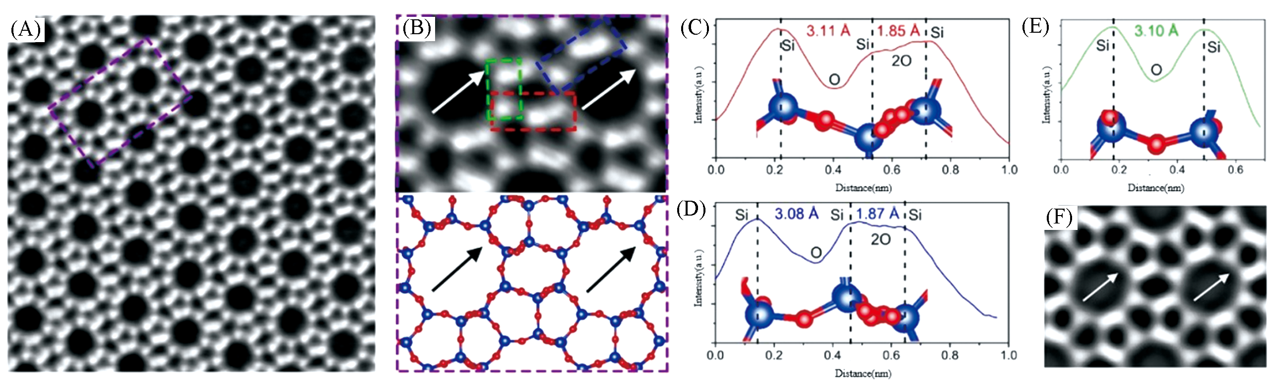
Fig.2 Atomic resolution iDPC‐STEM imaging of the [010] projection of ZSM‐5[40](A, B) Magnified iDPC‐STEM images of ZSM‐5 showing the atomic‐resolution straight channels as the elliptical Si10‐rings; (C, D) profile analysis of three structural units on the framework marked as three colored frames in (B); (E) intensity profiles exhibit the contrasts and projected distances between different atoms; (F) integration mapping of the projected electrostatic potential in ZSM‐5 framework from the [010] projection.Copyright 2020, Wiley?VCH.
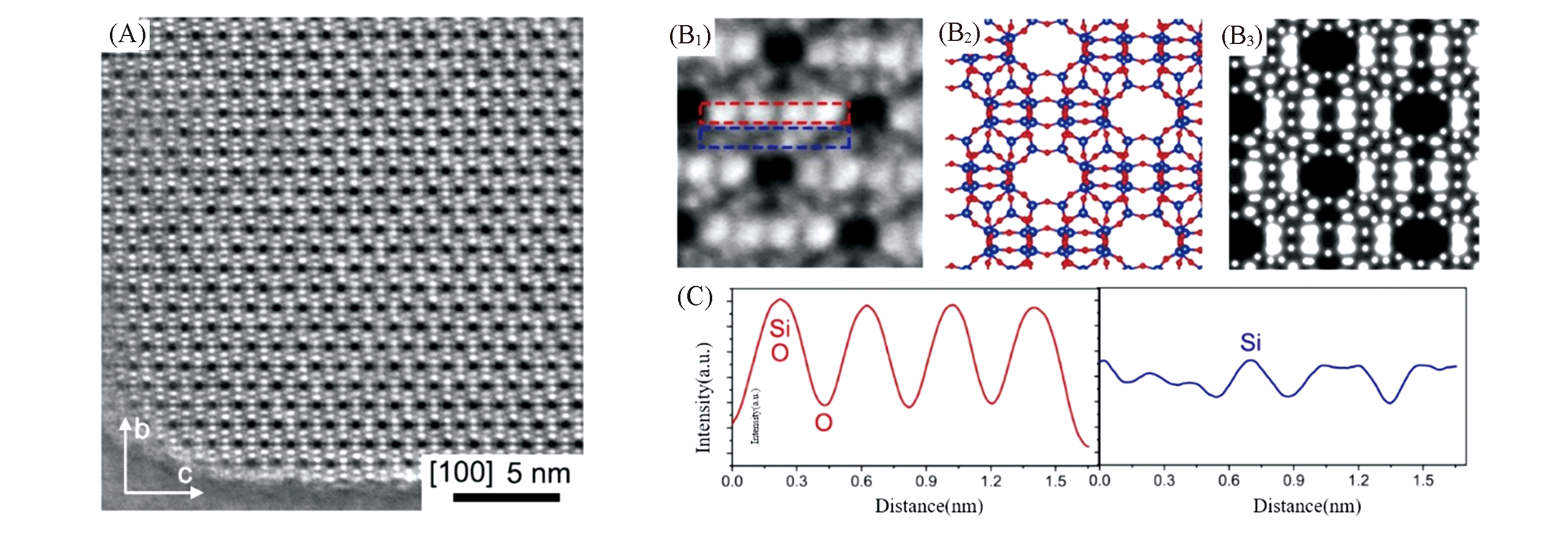
Fig.3 iDPC‐STEM image (A) and detailed analysis(B1―B3, C) from the [100] direction[40]The imaged structure is consistent with the structural model and calculated electrostatic potential(B1—B3). The intensity profiles (C) show the detailed atomic position of each element.Copyright 2020, Wiley?VCH.
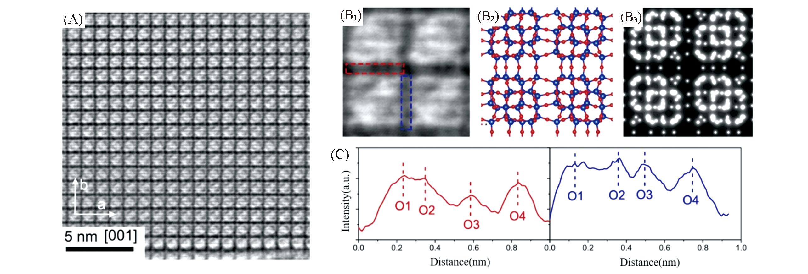
Fig.4 iDPC‐STEM image from [001] projection of ZSM?5[40](A—C) iDPC‐STEM image and detailed analysis (B1—B3, C) from the [001] direction. Especially, the intensity profiles (C) show the capacity of iDPC‐STEM for imaging light O bridges, where the positions of four O atoms are clearly marked.Copyright 2020, Wiley?VCH .
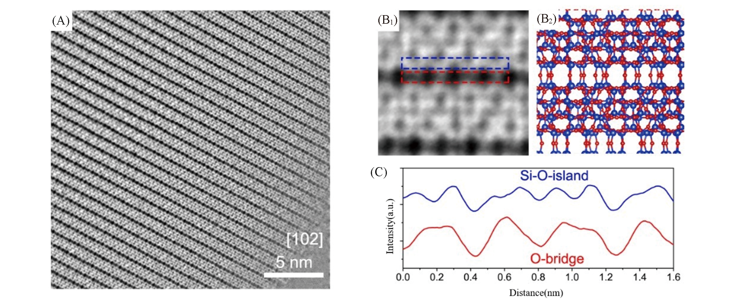
Fig.5 iDPC?STEM image from [102] projection of ZSM?5(A―C)[40]The higher‐index surface was found out to give a more obvious imaging of O bridges as shown in the profile analysis.Copyright 2020, Wiley?VCH.
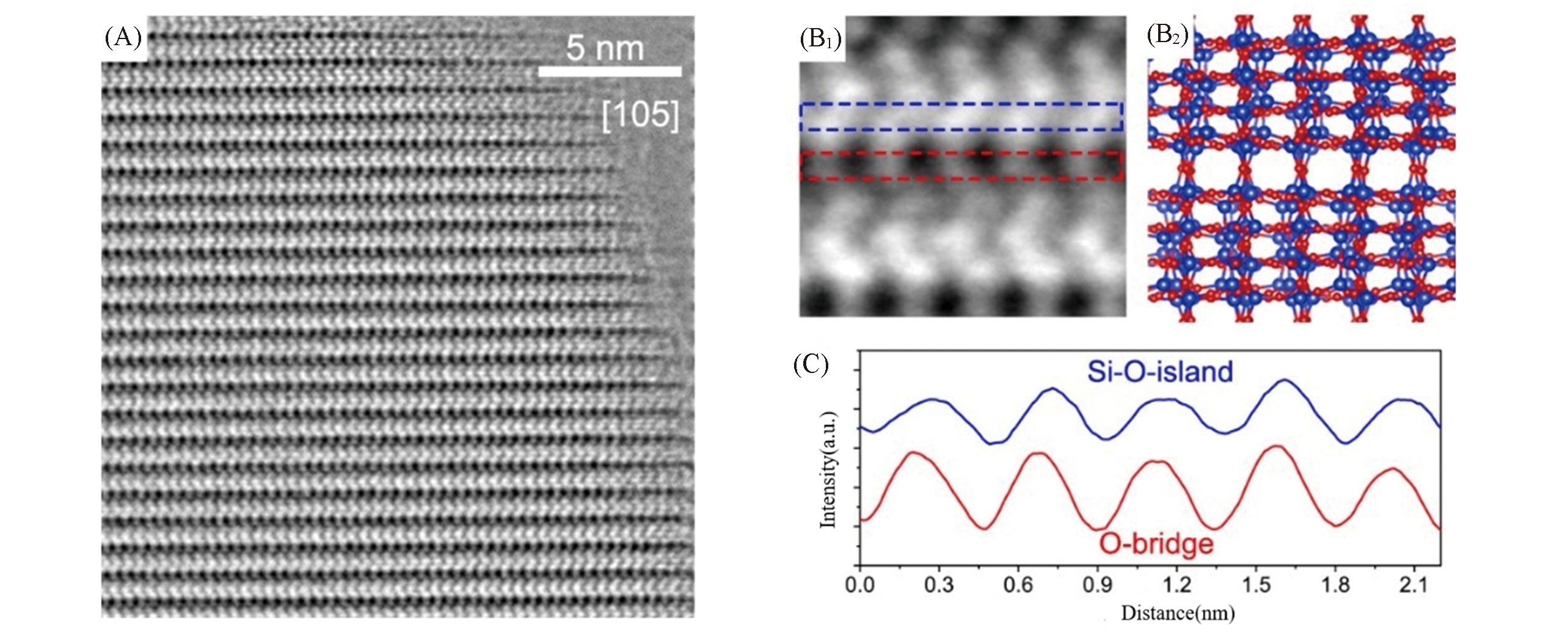
Fig.6 iDPC?STEM image from [105] projection of ZSM?5[40]iDPC‐STEM image(A) and detailed analysis (B1—B2, C) from the [105] direction.Copyright 2020, Wiley?VCH.
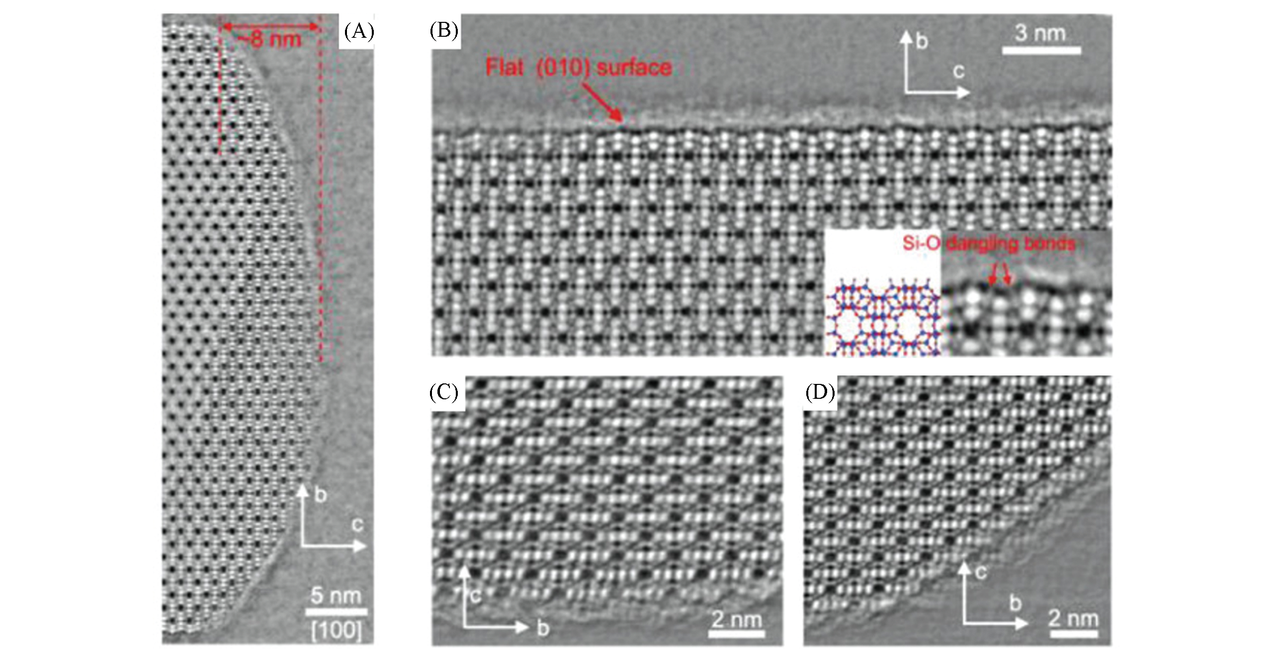
Fig.7 iDPC‐STEM images of the surface terminations from the [100] projection of ZSM?5[40]It exhibits the atomically flat (010) surface with clear Si—O dangling bonds.Copyright 2020, Wiley?VCH.
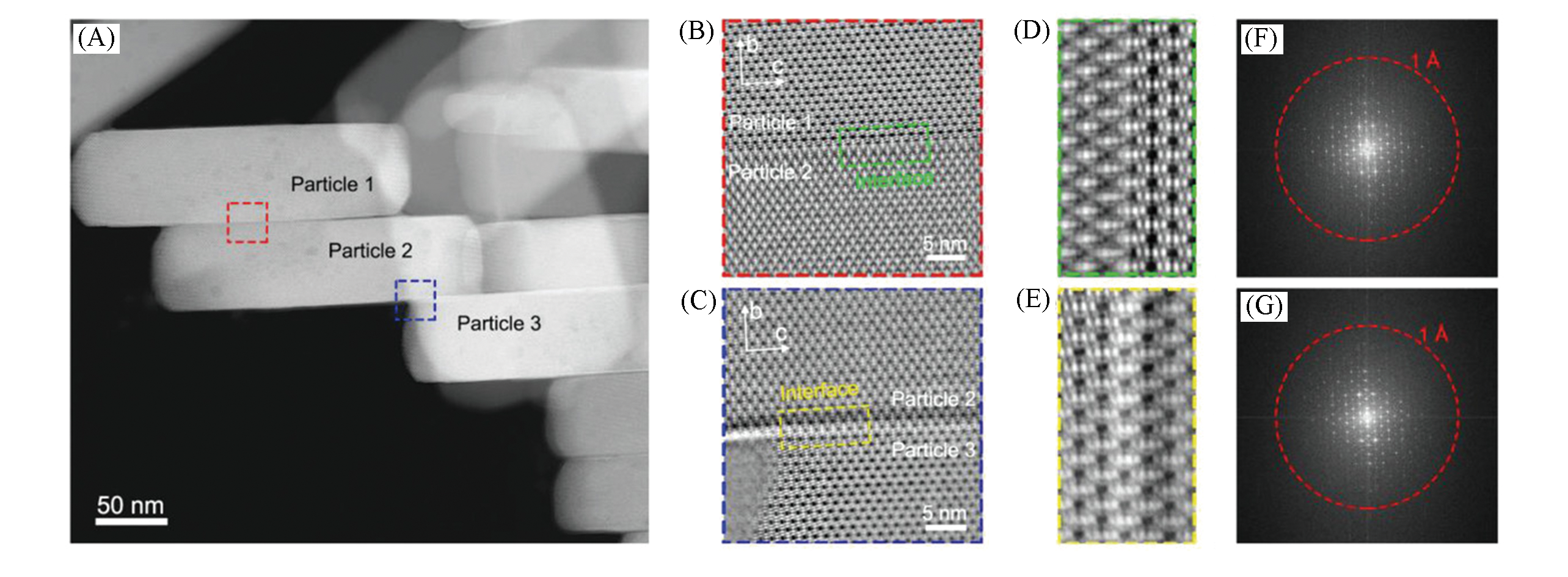
Fig.8 iDPC‐STEM imaging of the (010) interfaces between ZSM‐5 particles[40](A) HAADF‐STEM image of assembled ZSM‐5 particles. These particles were stacked from the [010] direction, and their flat (010) surfaces were in close contact with each other; (B, C) iDPC‐STEM images of the (010) interfaces in the areas marked in (A); (D, E) Zoom‐in interface areas of iDPC‐STEM images marked in (B,C); (F,G) FFT patterns corresponding to (B), (F) and (C), (G), respectively. FFT pattern of adjacent particles shows single set of diffraction spots indicating the coherent alignment of ZSM‐5 lattices by the interactions between these (010) surfaces.Copyright 2020, Wiley‐VCH.
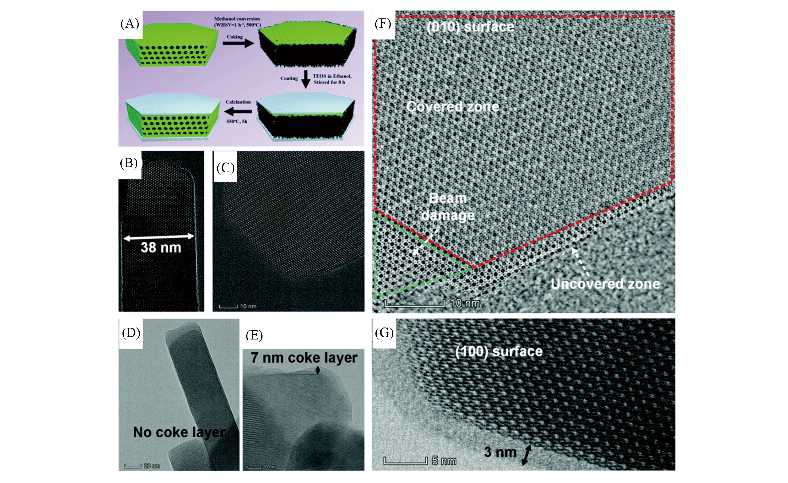
Fig.9 Designed scheme of the silica deposition process and the results of TEM images[11](A) Designed scheme of the silica deposition process. Aberration-corrected HRTEM images of pristine ZSM-5(ZSM-5-P): (100) surface(B) and (010) surface(C). Aberration-corrected HRTEM images of coked ZSM-5-P (ZSM-CP): (100) surface(D) and (010) surface(E). Image of surface-selective coated ZSM-5 after the reaction (coked ZSM-5-OC): aberration-corrected STEM image of the (010) surface(F). (G) Aberration-corrected HRTEM image of the (100) surface.Copyright 2019, Royal Society of Chemistry.
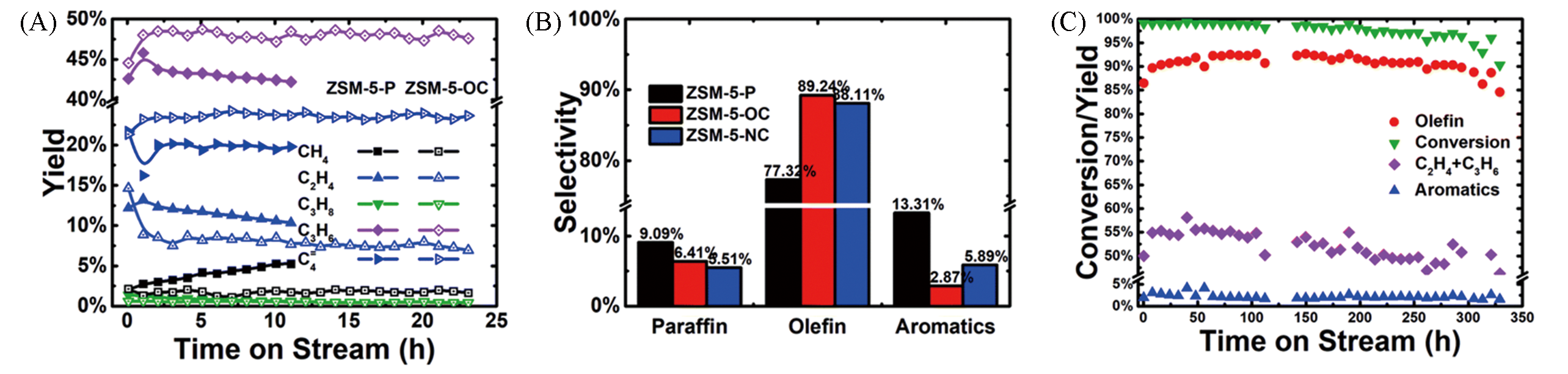
Fig.10 Comparison of the catalytic performance of various ZSM?5 catalysts in the MTO reaction[11](A) The carbon?based yield of different species (methanol conversion remains at 100%); (B) olefins, aromatics and paraffin selectivity, (TOS=9 h); (C) lifetime test of ZSM?5?OC(WHSV=1 h-1, reaction temperature: 500 °C, the N2 flow rate is 30 SCCM). Mass balance (outlet carbon/inlet carbon) was kept at about 98%.Copyright 2019, Royal Society of Chemistry.
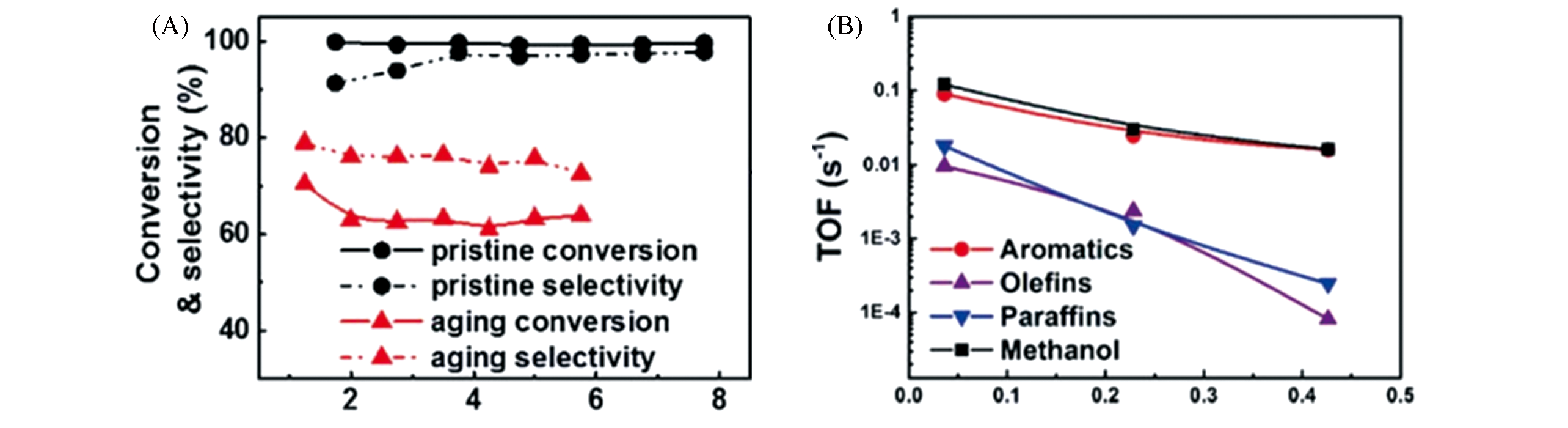
Fig.11 Methanol conversion and aromatic selectivity of UZ?5 and UZ5 samples as a function of time?on?stream[9,10](A) Methanol conversion and aromatic selectivity of pristine and aging samples as a function of time?on?stream (reaction temperature: 475 ℃, reaction pressure: atmospheric pressure, WHSV: 0.8 h-1 ); (B) turnover frequencies (TOFs) to different products vs. acid density.Copyright 2016, Royal Society of Chemistry.
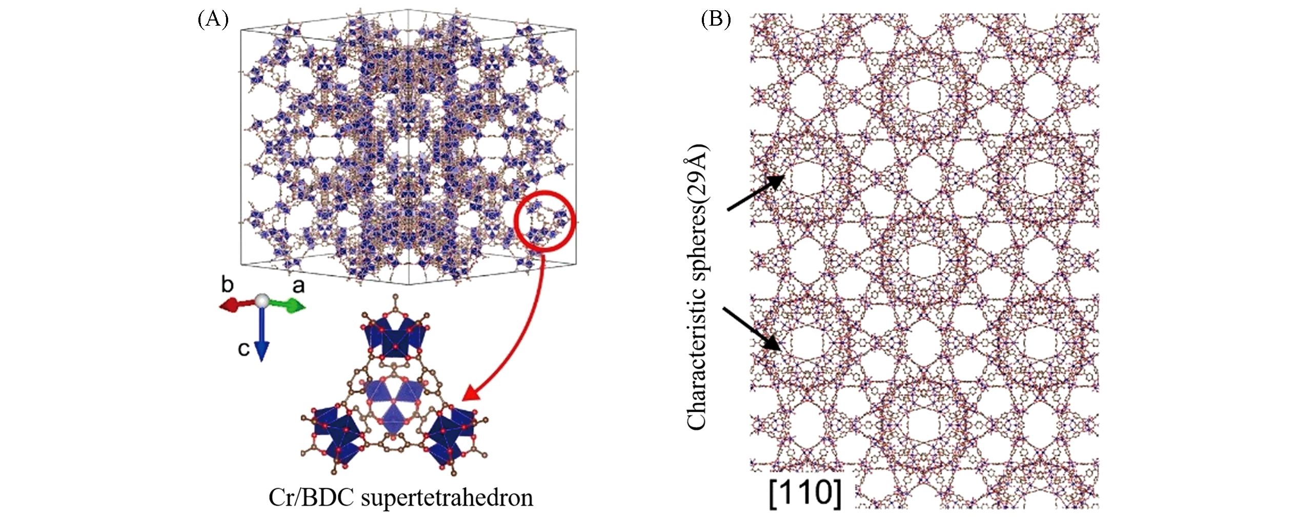
Fig.12 Structure model of MIL?101 framework[43](A) Framework model of MIL?101; (B) structural model of MIL?101 viewed from the <110> projection.Copyright 2020, Nature Publishing Group.
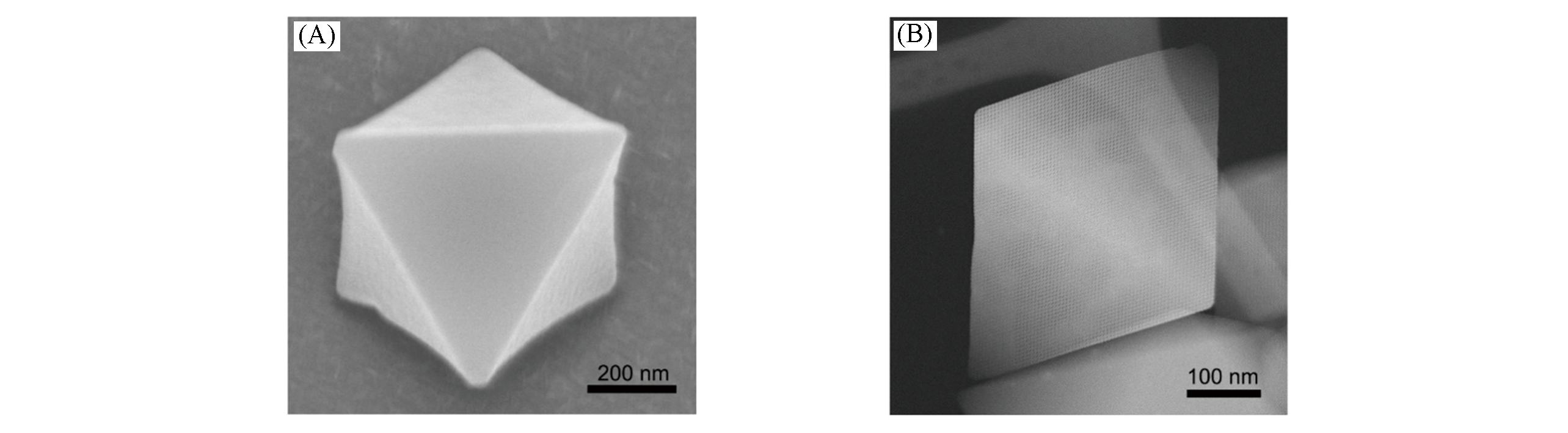
Fig.13 SEM(A) and ADF?STEM(B) images of MIL?101[43](A) SEM image showing the octahedral shape of a MIL?101 crystal. Scale bar: 200 nm; (B) ADF?STEM image of MIL?101 from the <110> projection.Copyright 2020, Nature Publishing Group.
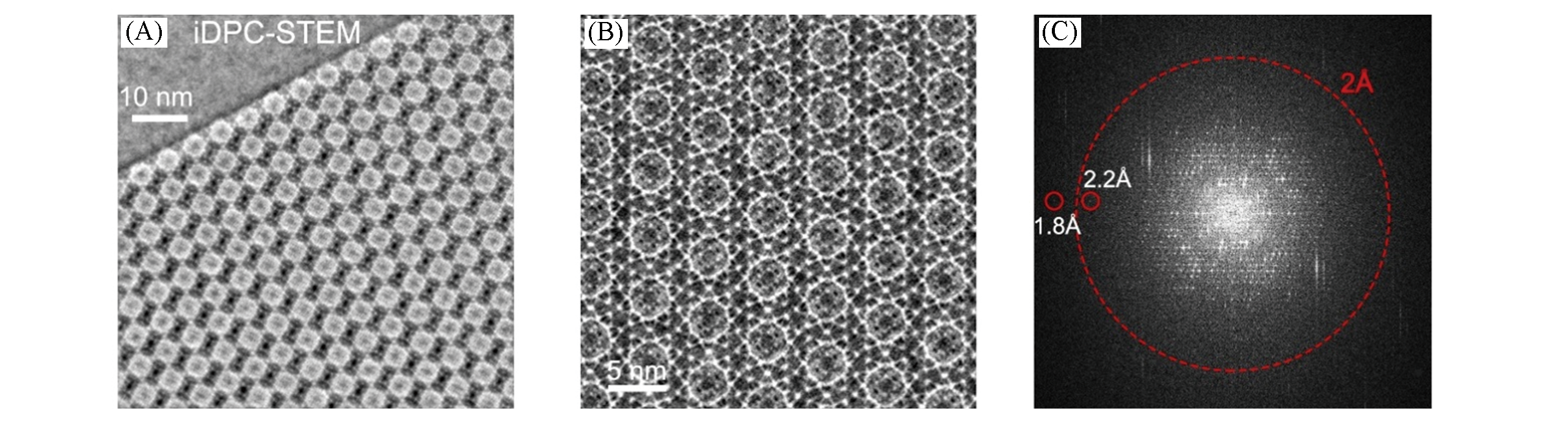
Fig.14 iDPC?STEM imaging of MIL?101 [110] projection with an ultrahigh resolution(A, B) and the corresponding FFT pattern of image(C)[43]The corresponding FFT showing an information transfer up to 1.8 ?.Copyright 2020, Nature Publishing Group.
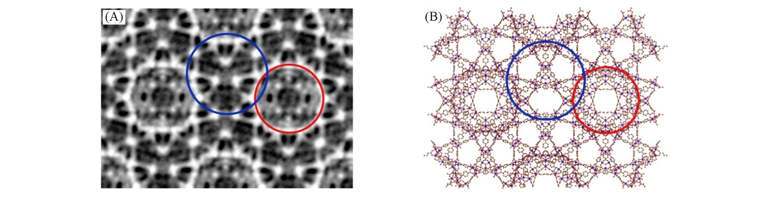
Fig.15 Imaging the atomic MIL?101 lattice in the averaged iDPC?STEM image(A) and the corresponding model structure(B)[43](A, B) The red and blue circles indicate two types of cages with 29 and 34 ? sizes, respectively.Copyright 2020, Nature Publishing Group.
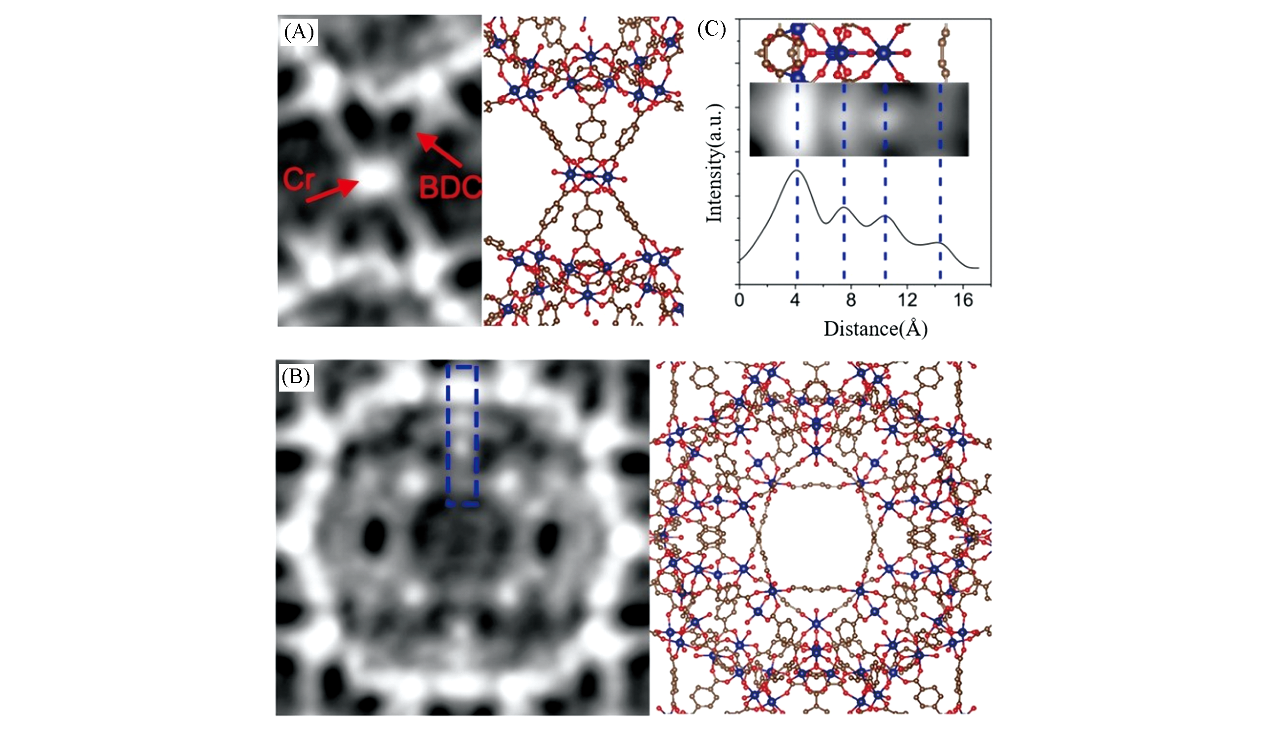
Fig.16 Detailed structure observation and strength profile analysis of MIL?101[43](A) Imaging the super tetrahedrons formed by Cr nodes and BDC linkers; (B) imaging the structure in 29 ? cage in the MIL?101; (C) profile analysis of image(exp) and simulation(sim) of the detailed structures in the blue frame in (B), which helps to resolve the Cr columns in the frameworks.Copyright 2020, Nature Publishing Group.
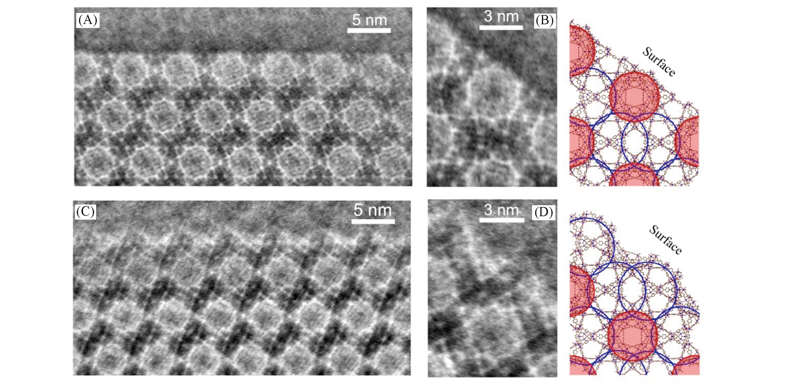
Fig.17 Two different {111} surface analysis of MIL?101 crystal[43](A, C) Two types of surface terminations with two types of cages exposed on the {111} surfaces. (B, D) Structures of single unit cells at two types of surface terminations and the corresponding structural models, respectively.Copyright 2020, Nature Publishing Group.
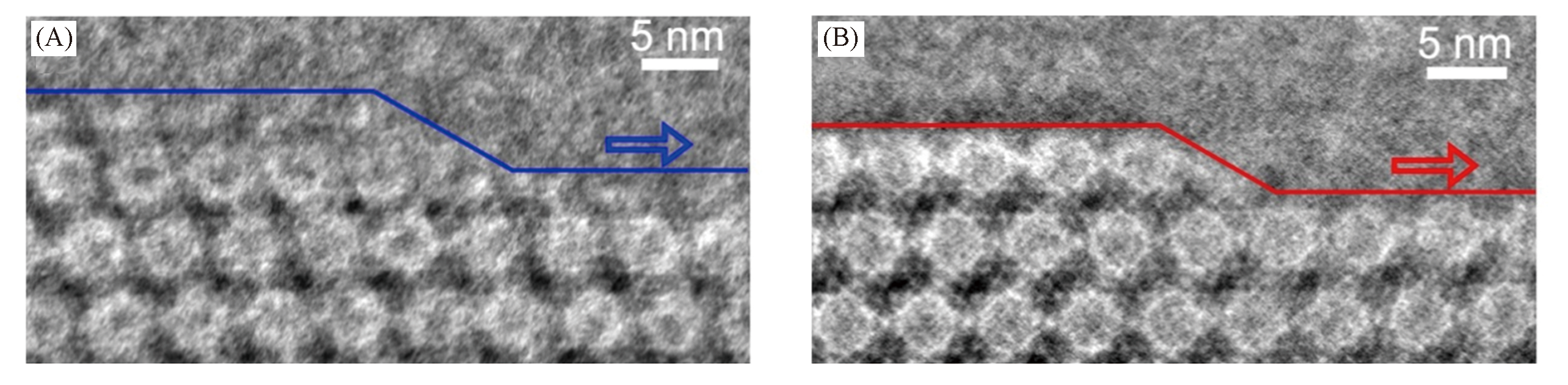
Fig.18 Surface steps and step?edge sites of the MIL?101 crystals with two types of surface terminations, respectively[43]Copyright 2020, Nature Publishing Group.
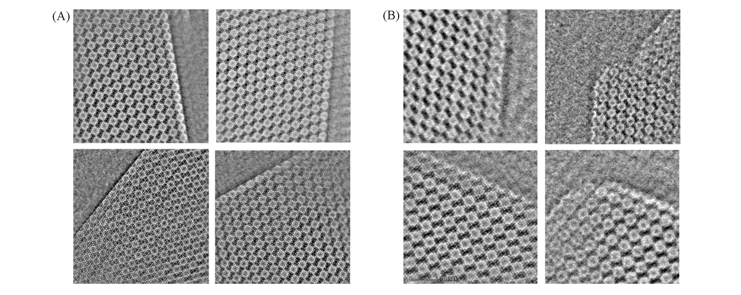
Fig.19 More images of the two {111} surfaces of MIL?101[43]Copyright 2020, Nature Publishing Group.
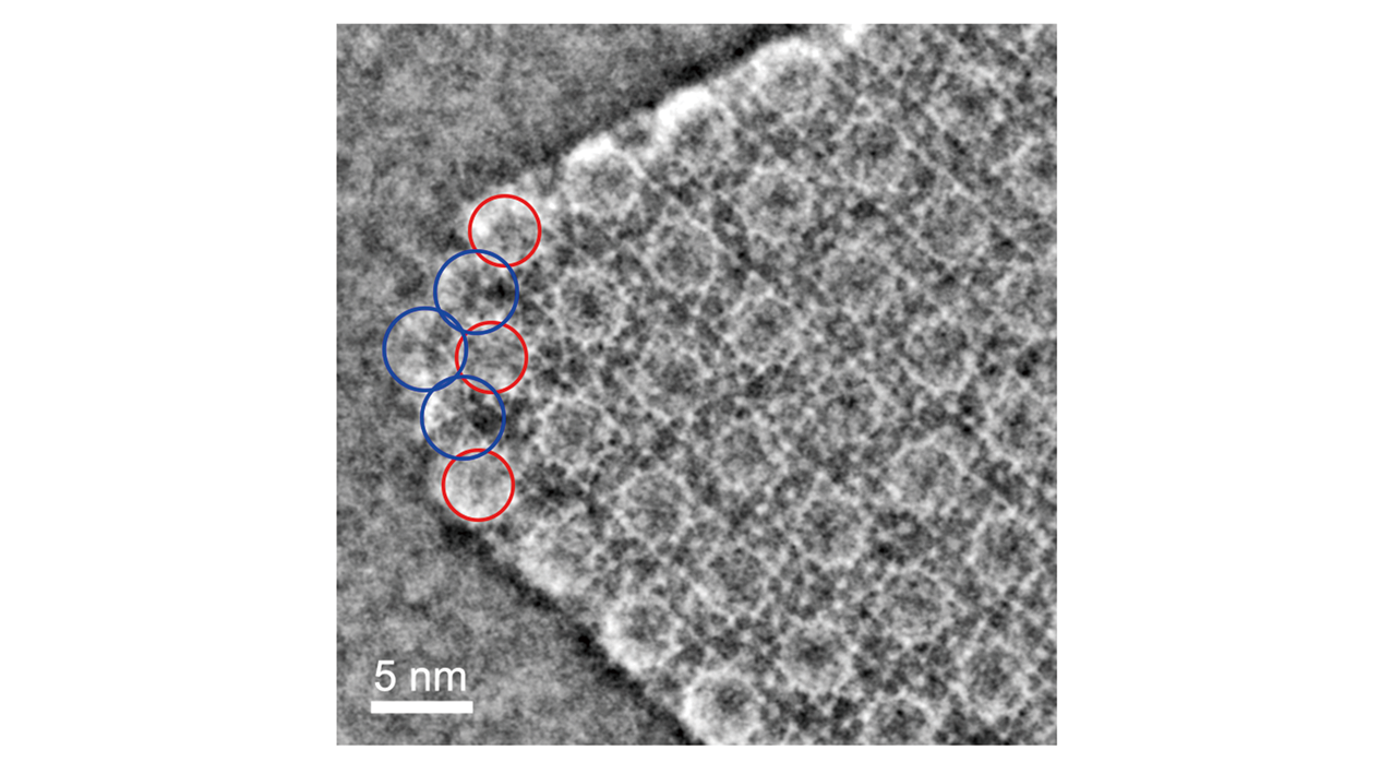
Fig.20 iDPC?STEM image showing the terminations of a corner in the octahedral MIL?101 crystal[43]The 34 ? cages (marked by the blue circles) were exposed at this corner.Copyright 2020, Nature Publishing Group.
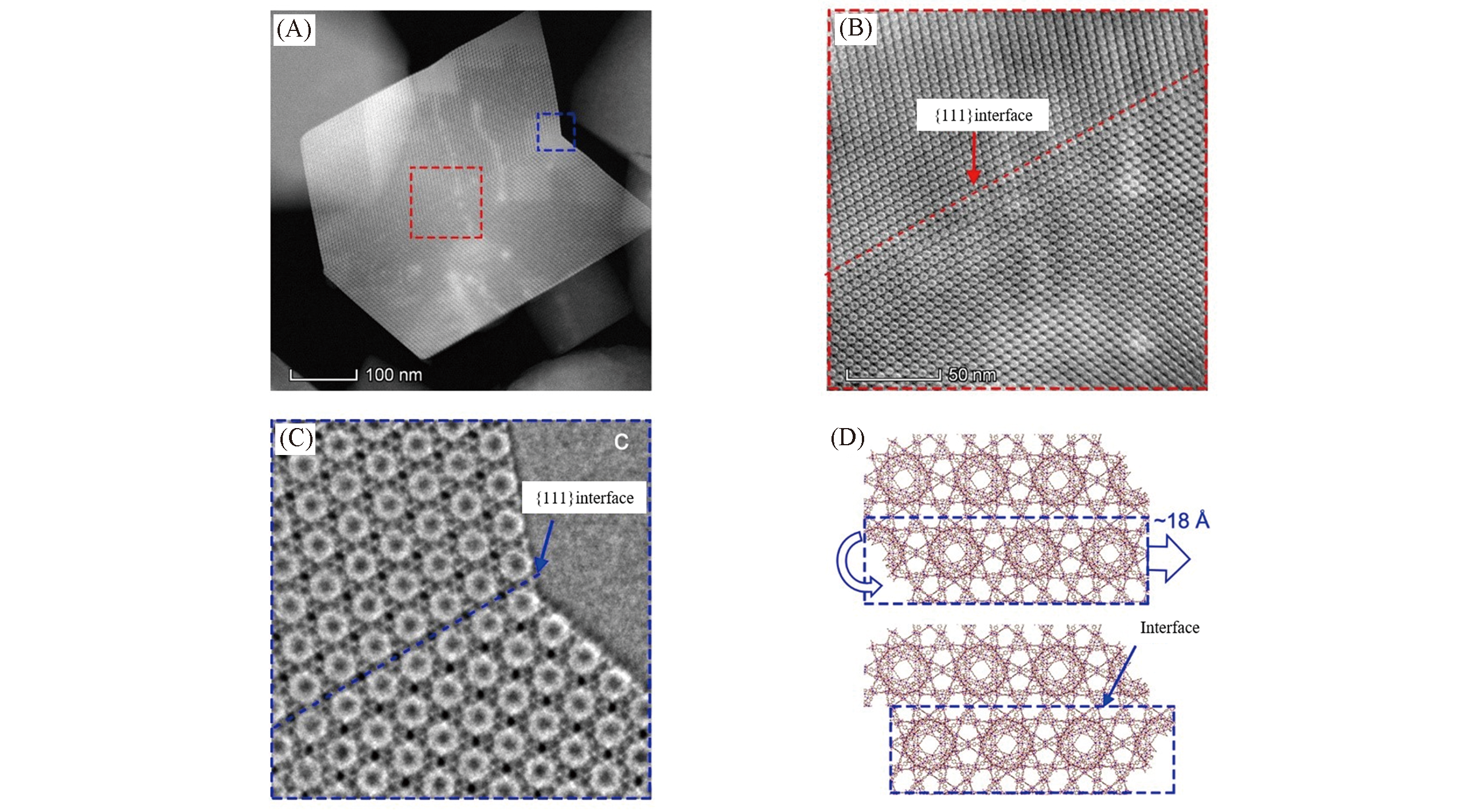
Fig.21 Observation and analysis of MIL?101 twin interface structure[43](A) ADF?STEM image of a twin?crystal of MIL?101; (B) ADF?STEM image of the {111} interface marked as the red frame in image (A); (C) iDPC?STEM image of the {111} interface marked as the blue frame in image (A). The structural model of the twin?plane interface imaged in image (C).Copyright 2020, Nature Publishing Group.
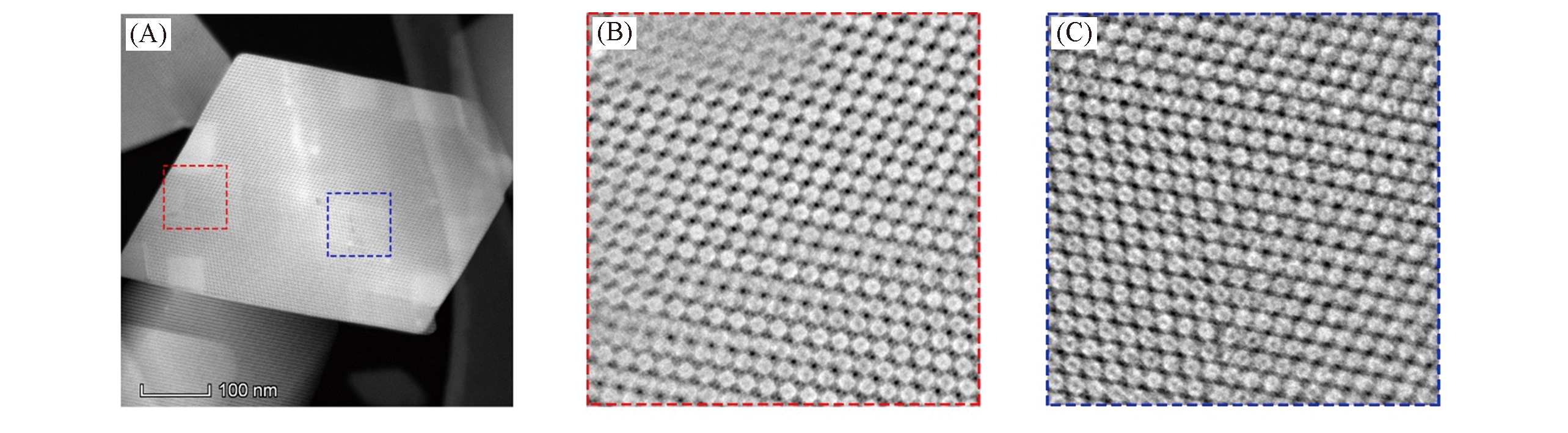
Fig.22 Observation and analysis of mismatch defect structure at MIL?101 twin interface[43](A) ADF?STEM image of a MIL?101 crystal; (B, C) iDPC?STEM images showing the defects in this MIL?101 crystal. Copyright 2020, Nature Publishing Group.
| 1 | National Bureau of Statistics of the People’s Republic of China. The 2017 Statistical Bulletin of the People’s Republic of China on the National Economic and Social Development, 2018(中华人民共和国国家统计局. 中华人民共和国2017年国民经济和社会发展统计公报, 2018) |
| 2 | Olson D. H., Kokotailo G. T., Lawton S. L., J. Phys. Chem., 1981, 85, 2238—2243 |
| 3 | Yoshio O., Catal. Rev., 1992, 34, 179—226 |
| 4 | Conte M., Lopez-Sanchez J. A., He Q., David J. M., Graham J. H., Catal. Sci. Technol., 2011, 2, 105—112 |
| 5 | Ni Y., Sun A., Wu X., Hai G., Hu J., Li T., Li G., Micropor. Mesopor. Mater., 2011, 143, 435—442 |
| 6 | Shen K., Qian W., Wang N., Su C., Wei F., J. Am. Chem. Soc., 2013, 135, 15322—15325 |
| 7 | Shen K., Qian W., Wang N., Zhang J. G., Wei F., J. Mater. Chem. A, 2013, 1, 3272—3275 |
| 8 | Liu Y., Zhou X., Pang X., Jin Y., Meng X., Zheng X., Gao X., Xiao F., ChemCatChem, 2013, 5, 1517—1523 |
| 9 | Ma Y., Wang N., Qian W. Z., Wang Y., Zhang J., Wei F., RSC Adv., 2016, 6, 81198—81202 |
| 10 | Ma Y., Cai D., Li Y., Wang N., Muhammad U., Carlsson A., Tang D., Qian W. Z., Wang Y., Su D. S., Wei F., RSC Adv., 2016, 6, 74797—74801 |
| 11 | Cai D., Wang N., Chen X., Ma Y., Hou Y., Li X., Zhang C., Chen Z., Song W., Arslan M. T., Li Y., Wang Y., Qian W., Wei F., Nanoscale, 2019, 11, 8096—8101 |
| 12 | Li H. L., Eddaoudi M., O'Keeffe M., Yaghi O. M., Nature, 1999, 402, 276—279 |
| 13 | Yaghi O. M., O'Keeffe M., Ockwig N. W. , Chae H. K., Eddaoudi M., Kim J., Nature, 2003, 423, 705—714 |
| 14 | Furukawa H., Cordova K. E., O'Keeffe M., Yaghi O. M., Science, 2013, 341, 974—986 |
| 15 | Li P., Vermeulen N. A., Malliakas C. D., Yaghi O. M., Science, 2017, 356, 624—627 |
| 16 | Eddaoudi M., Kim J., Rosi N. L., Vodak D. T., O'Keeffe M., Yaghi O. M., Science, 2002, 295, 469—472 |
| 17 | Rosi N. L., Eckert J., Eddaoudi M., Vodak D. T., Kim J., O'Keeffe M., Yaghi O. M., Science, 2003, 300, 1127—1129 |
| 18 | Li J. R., Kuppler R. J., Zhou H. C., Chem. Soc. Rev., 2009, 38, 1477—1504 |
| 19 | Lee J. Y., Farha O. K., Roberts J., Scheidt K. A., Nguyen SonBinh T., Hupp Joseph T., Chem. Soc. Rev., 2009, 38(5), 1450—1459 |
| 20 | Sumida K., Rogow D. L., Mason J. A., McDonald T. M., Bloch E. D., Herm Z. R., Bae T. H., Long J. R., Chem. Rev., 2012, 112, 724—781 |
| 21 | Ikuno T., Zheng J., Vjunov A., Sanchez-Sanchez M., Ortuñ O. M. A., Pahls D. R., Fulton J. L., Camaioni D. M., Li Z., Ray D., Mehdi B. L., Browning N. D., Farha O. K., Hupp J. T., Cramer C. J., Gagliardi L., Lercher J. A., J. Am. Chem. Soc., 2017, 139, 10294—10301 |
| 22 | Lebedev O. I., Millange F., Serre C., Tendeloo G. V., Fe´rey G., Chem. Mater., 2005, 17, 6525—6527 |
| 23 | Cravillon J., Münzer S., Lohmeier S. J., Feldhoff A., Huber K., Wiebcke M., Chem. Mater., 2009, 21, 1410—1412 |
| 24 | Zhu L., Zhang D., Xue M., Li H., Qiu S., Cryst. Eng. Comm., 2013, 15, 9356—9359 |
| 25 | Meledina M., Turner S., Maria F., Leus K., Lobato I., Ramachandran R. K., Detavernier J., Christophe D. C., Van D. V. P., Tendeloo G. V., Part Part Syst Char., 2016, 33, 382—387 |
| 26 | Mayoral A., Mahugo R., Sanchez-Sanchez M., Díaz I., ChemCatChem, 2017, 9, 3497—3502 |
| 27 | Wiktor C., Meledina M., Turner S., Lebedev O. I., Fischer Roland A., J. Mater. Chem. A, 2017, 5, 14969—14989 |
| 28 | Egerton R. F., Li P., Malac M., Micron., 2004, 35, 399—409 |
| 29 | Ugurlu O., Haus J., Gunawan A. A., Thomas M. G., Maheshwari S., Tsapatsis M., Mkhoyan K. A., Phys. Rev., 2011, 83, 113408 |
| 30 | Egerton R. F., Microsc. Res. Tech., 2012, 75, 1550—1556 |
| 31 | Garcia A., Raya A. M., Mariscal M. M., Rodrigo E., Miriam H., Sergio I. M., Giovanni S., Pedro L. G., Miguel J. Y., Arturo P., Ultramicroscopy, 2014, 146, 33—38 |
| 32 | Chen Q., Dwyer C., Sheng G., Zhu C., Li X., Zheng C., Zhu Y., Adv. Mater., 2020, 32, 1907619 |
| 33 | Chapman, N. J., J. Phys. D: Appl. Phys., 1984, 17, 623—647 |
| 34 | Chapman J. N., McFadyen I. R., McVitie S., IEEE T Magn, 1990, 26, 1506—1511 |
| 35 | Shibata N., Findlay S. D., Kohno Y., Sawada H., Ikuhara Y., Nat. Phys., 2012, 8, 611—615 |
| 36 | Lazić I., Bosch E. G. T., Lazar S., Ultramicroscopy, 2016, 160, 265—280 |
| 37 | Lazić I., Bosch E. G. T., Adv. Imag. Elect. Phys., 2017, 199, 75—184 |
| 38 | Yücelen E., Lazić I., Bosch E. G. T., Sci. Rep., 2018, 8,1—10 |
| 39 | Rose H., Ultramicroscopy, 1977, 2, 251—267 |
| 40 | Shen B., Chen X., Cai D., Xiong H., Liu X., Meng C., Han Y., Wei F., Adv. Mater., 2020, 32, 1906103 |
| 41 | Ferey G., Mellot⁃Draznieks C., Serre C., Millange F., Dutour J., Surble S., Margiolaki I., Science, 2005, 309, 2040—2042 |
| 42 | Li X., Wang J., Liu X., Liu L., Han Y., J. Am. Chem. Soc., 2019, 141,12021—12028 |
| 43 | Shen B., Chen X., Shen K., Xiong H., Wei F., Nature Commun., 2020,11, 2692 |
| [1] | 姚伊婷, 吕佳敏, 余申, 刘湛, 李昱, 李小云, 苏宝连, 陈丽华. 等级孔微孔-介孔Fe2O3/ZSM-5中空分子筛催化材料的制备及催化苄基化性能[J]. 高等学校化学学报, 2022, 43(8): 20220090. |
| [2] | 李玉龙, 谢发婷, 管燕, 刘嘉丽, 张贵群, 姚超, 杨通, 杨云慧, 胡蓉. 基于银离子与DNA相互作用的比率型电化学传感器用于银离子的检测[J]. 高等学校化学学报, 2022, 43(8): 20220202. |
| [3] | 丁杨, 王万辉, 包明. 多孔骨架固定分子催化剂催化CO2加氢制备甲酸研究进展[J]. 高等学校化学学报, 2022, 43(7): 20220309. |
| [4] | 李志光, 齐国栋, 徐君, 邓风. Sn-Al-β分子筛酸性在葡萄糖转化反应中作用的固体NMR研究[J]. 高等学校化学学报, 2022, 43(6): 20220138. |
| [5] | 陈玮琴, 吕佳敏, 余申, 刘湛, 李小云, 陈丽华, 苏宝连. 有机杂化介孔Beta分子筛的合成及在苯甲醇烷基化反应中的应用[J]. 高等学校化学学报, 2022, 43(6): 20220086. |
| [6] | 鲁聪, 李振华, 刘金露, 华佳, 李光华, 施展, 冯守华. 一种新的镧系金属有机骨架材料的合成、 结构及荧光检测性质[J]. 高等学校化学学报, 2022, 43(6): 20220037. |
| [7] | 李加富, 张凯, 王宁, 孙启明. 分子筛限域单原子金属催化剂的研究进展[J]. 高等学校化学学报, 2022, 43(5): 20220032. |
| [8] | 孟祥龙, 杨歌, 郭海玲, 刘晨光, 柴永明, 王纯正, 郭永梅. 纳米分子筛的合成及硫化氢吸附性能[J]. 高等学校化学学报, 2022, 43(3): 20210687. |
| [9] | 邢珮琪, 陆通, 李光华, 王力彦. 两个镉(II)金属有机骨架的可控合成与结构相关性[J]. 高等学校化学学报, 2022, 43(10): 20220218. |
| [10] | 魏李娜, 彭莉, 朱锋, 顾鹏飞, 顾学红. 中空纤维Au-CeZr/FAU催化膜的制备及在富氢气氛CO选择性氧化反应中的应用[J]. 高等学校化学学报, 2022, 43(10): 20220175. |
| [11] | 李文, 乔珺一, 刘鑫垚, 刘云凌. 含萘基团的锆金属有机骨架材料对水中硝基芳烃爆炸物的荧光检测性能[J]. 高等学校化学学报, 2022, 43(1): 20210654. |
| [12] | 罗强强, 金少青, 孙洪敏, 杨为民. 液相酸溶液后补钛合成Ti-MWW分子筛[J]. 高等学校化学学报, 2021, 42(9): 2742. |
| [13] | 李海勃, 肖长发, 江龙, 黄云, 淡宜. MCM-41分子筛负载氯化铝催化丙烯酸甲酯与1-辛烯共聚[J]. 高等学校化学学报, 2021, 42(9): 2974. |
| [14] | 李奕川, 朱国富, 王宇, 柴永明, 刘晨光, 何盛宝. 基底表面性质与前驱液化学环境对原位定向构筑钛硅分子筛膜的影响[J]. 高等学校化学学报, 2021, 42(9): 2934. |
| [15] | 张旭, 阙家乾, 侯月新, 吕佳敏, 刘湛, 雷坤皓, 余申, 李小云, 陈丽华, 苏宝连. 等级孔介孔-微孔TS-1分子筛单晶的合成及催化氯丙烯环氧化性能[J]. 高等学校化学学报, 2021, 42(8): 2529. |
| 阅读次数 | ||||||
|
全文 |
|
|||||
|
摘要 |
|
|||||