高等学校化学学报 ›› 2025, Vol. 46 ›› Issue (11): 20250166.doi: 10.7503/cjcu20250166

• 分析化学 • 上一篇    下一篇

新型荧光碳点的一步法制备及细胞成像性能

范红婷1, 徐佳彤1, 齐春轩2(), 马恒昌1()   

  1. 1.西北师范大学化学化工学院, 兰州 730070
    2.宝鸡文理学院化学化工学院, 宝鸡 721013
  • 收稿日期:2025-06-13 出版日期:2025-11-10 发布日期:2025-08-05
  • 通讯作者: 齐春轩,马恒昌 E-mail:qichunxuan@163.com;mahczju@hotmail.com
  • 基金资助:
    国家自然科学基金(22165026);2025年甘肃省高校研究生“创新之星”项目(2025CXZX-275)

One-step Preparation of Novel Fluorescent Carbon Dots for Cell Imaging

FAN Hongting1, XU Jiatong1, QI Chunxuan2(), MA Hengchang1()   

  1. 1.College of Chemistry and Chemical Engineering,Northwest Normal University,Lanzhou 730070,China
    2.College of Chemistry and Chemical Engineering,Baoji University of Arts and Sciences,Baoji 721013,China
  • Received:2025-06-13 Online:2025-11-10 Published:2025-08-05
  • Contact: QI Chunxuan, MA Hengchang E-mail:qichunxuan@163.com;mahczju@hotmail.com
  • Supported by:
    the National Natural Science Foundation of China(22165026);the Innovative Star Project for Graduate Students in Gansu Province Universities 2025, China(2025CXZX-275)

摘要:

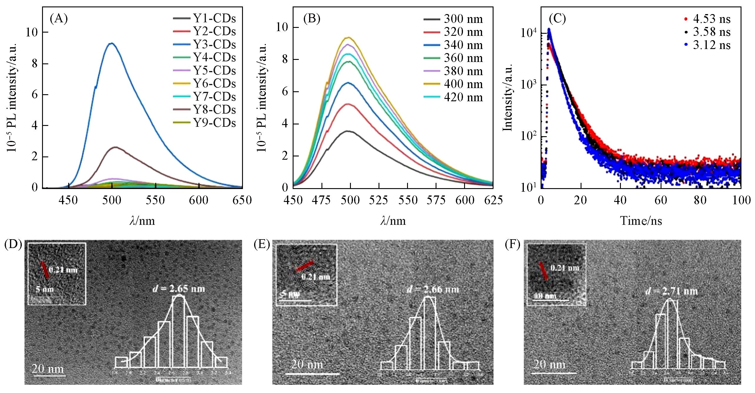
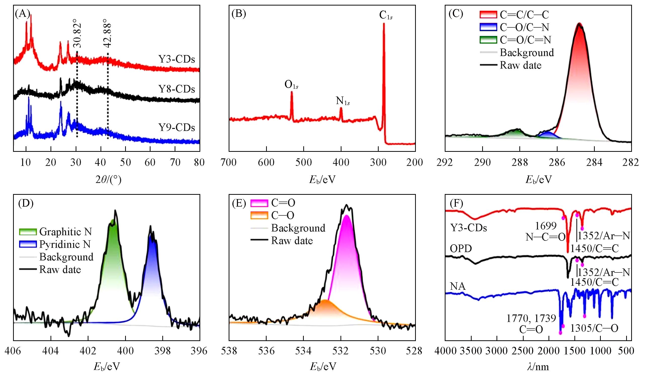
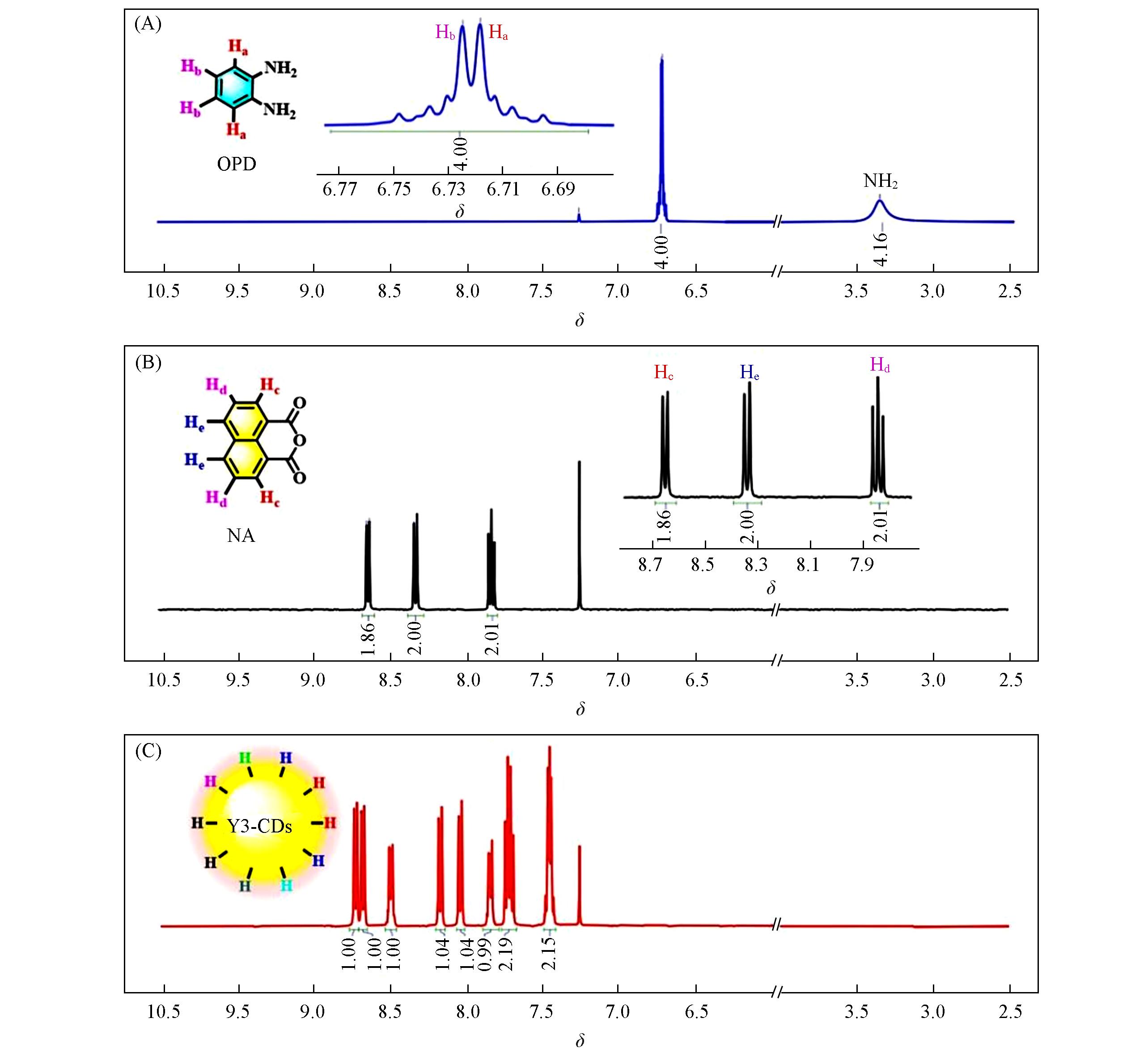
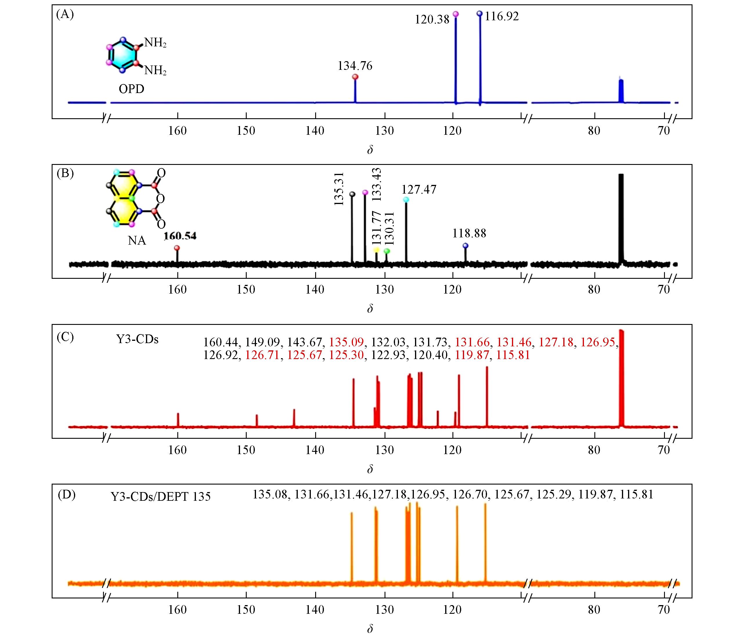
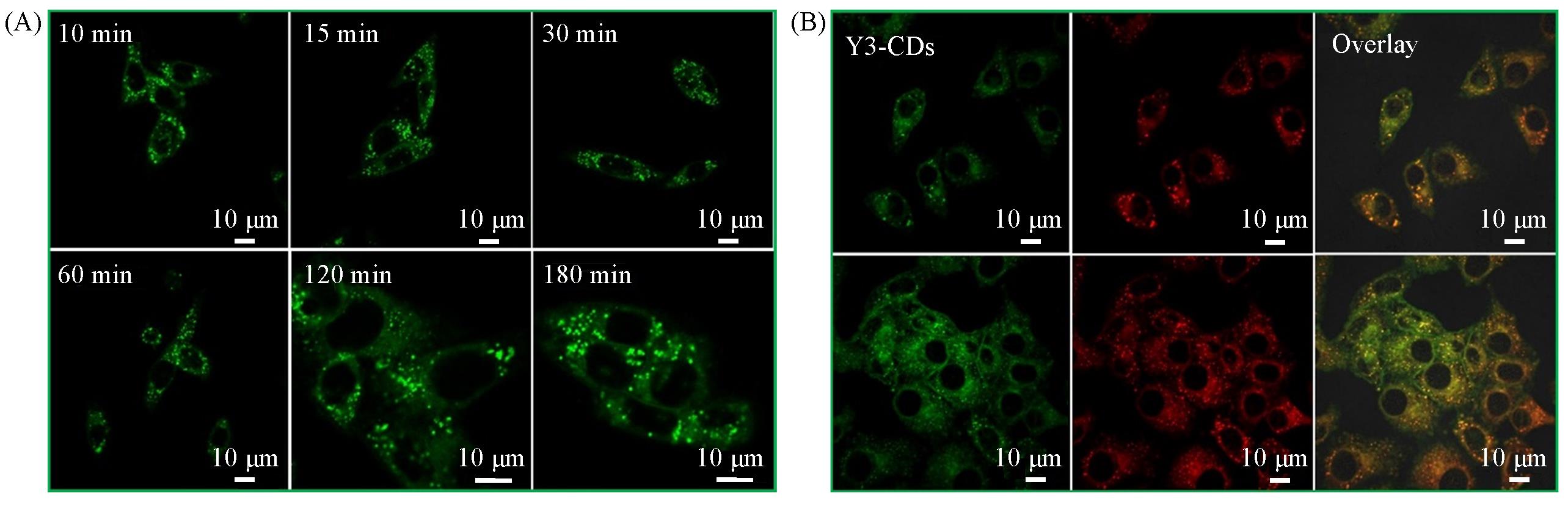
以1,8-萘二酸酐(NA)和1,2-邻苯二胺(OPD)为前驱体, 采用一步水热法快速制备了一种新型荧光碳点. 通过调控反应条件, 合成了具有优异发光性能的黄色荧光碳点Y3-CDs. 高分辨透射电镜(HRTEM)表征结果显示, Y3-CDs的平均粒径为2.6 nm, 晶格间距为0.21 nm, 对应的是X射线粉末衍射(XRD)谱图中 42.88o处石墨烯衍射峰的(100)晶面. 光电子能谱元素(XPS)分析结果表明, 碳点中存在sp2 /sp3 杂化碳(C=C/C—C, 284.8 eV)、 碳氮键(C—N, 286.5 eV)和羰基碳(C=O, 288.4 eV). 进一步利用荧光光谱(FL)、 紫外-可见吸收光谱(UV-Vis)和荧光寿命对Y3-CDs的发光性能进行了表征, 其表现出不依赖于激发的发射行为, 表明Y3-CDs为单一发射中心. 红外光谱(FTIR)和核磁共振波谱(NMR)揭示了更详细的碳原子和氢原子存在状态, 1H NMR表明所有氢原子存在于碳核表面, 表现出邻位取代模式和偶联效应. 13C NMR和DEPT 135谱图表明存在10种不同的叔碳原子. 将Y3-CDs应用于细胞示踪及细胞成像, 结果表明, Y3-CDs具有优异的细胞成像能力和抗光漂白性能, 即使在连续照射180 min后, Y3-CDs仍保持强烈的荧光, 可实现长时间HeLa细胞成像. 同时, 细胞共染实验表明, Y3-CDs具有优异的脂滴选择性成像能力, 皮尔森相关系数为0.846.

关键词: 碳点, 核磁共振分析, 抗光漂白, 细胞示踪, 脂滴成像

Abstract:

In this study, novel fluorescent carbon dots(CDs) were rapidly synthesized via a one-step hydrothermal method using 1,8-naphthalic anhydride(NA) and 1,2-phenylenediamine(OPD) as precursors. By optimizing reaction conditions, yellow fluorescent carbon dots(Y3-CDs) with superior luminescence performance were prepared. The particle sizes and morphology of the Y3-CDs were characterized by high-resolution transmission electron microscopy(HRTEM), revealing an average diameter of approximately 2.6 nm and an interlayer spacing of 0.21 nm. This spacing corresponds to the d-spacing of graphene(100) planes and is consistent with the diffraction peak observed at 42.88o in X-ray powder diffraction(XRD) analysis. X-ray photoelectron spectroscopy(XPS) confirmed the presence of sp2 /sp3 hybridized carbons(C=C/C—C, 284.8 eV), carbon-nitrogen bonds(C—N, 286.5 eV), and carbonyl carbons(C=O, 288.4 eV). The optical properties of the Y3-CDs were further investigated using fluorescence spectrometry(FL), ultraviolet-visible absorption spectroscopy(UV-Vis), and lifetime measurements. The fluorescence spectra exhibited non-excitation-dependent emission behavior, indicating a single emission center. Fourier-transform infrared spectroscopy(FTIR) and nuclear magnetic resonance(NMR) provided detailed structural insights. Due to their high purity, the characteristic hydrogen(H) and carbon(C) atoms could be clearly resolved and analyzed. 1H NMR results demonstrated that all H atoms were located on the surface of the carbon core, exhibiting ortho-substitution patterns and coupling effects. 13C NMR and DEPT 135 spectra confirmed the presence of ten distinct tertiary carbon atoms. The cell-tracking and imaging performance of Y3-CDs were systematically evaluated. Results demonstrated that Y3-CDs possess excellent cell imaging capabilities and anti-photobleaching properties. Even after 180 minutes of continuous irradiation, Y3-CDs retained strong fluorescence, enabling long-term tracking of HeLa cells. Co-localization experiments further highlighted their selective lipid droplet imaging ability, with a Pearson’s correlation coefficient of 0.846.

Key words: Carbon dots, NMR analysis, Anti-photobleaching, Cell tracking, Lipid droplet imaging

中图分类号: 

TrendMD: