

高等学校化学学报 ›› 2022, Vol. 43 ›› Issue (12): 20220575.doi: 10.7503/cjcu20220575
董明杰, 王璇, 董海峰( ), 张学记()
收稿日期:2022-08-30
出版日期:2022-12-10
发布日期:2022-10-21
通讯作者:
董海峰,张学记
E-mail:hfdong@szu.edu.cn;zhangxueji@szu.edu.cn
基金资助:
DONG Mingjie, WANG Xuan, DONG Haifeng(), ZHANG Xueji()
Received:2022-08-30
Online:2022-12-10
Published:2022-10-21
Contact:
DONG Haifeng, ZHANG Xueji
E-mail:hfdong@szu.edu.cn;zhangxueji@szu.edu.cn
Supported by:摘要:
成像技术的迅速发展使科学家和临床医生能够准确地了解癌症的发病机制和病理过程, 并根据患者的情况制定个性化的治疗策略. 将各种成像与治疗试剂整合为一体的癌症诊疗平台, 可以同时用于癌症的诊断和治疗, 受到了广泛的关注. 金属-有机框架材料(MOFs)是由有机配体和金属离子/离子簇自组装而成的一种有趣而独特的多孔有机-无机杂化材料. 由于其易于后修饰、 孔隙和结构可设计、 功能可调等特点, 已被证明具有成为癌症诊疗药物负载平台的巨大潜力. 本文介绍了将诊疗药物负载到MOFs中的策略, 并综合评述了在磁共振成像、 计算机断层扫描成像、 正电子发射断层扫描成像、 光学成像和光声成像等多种成像技术指导下, MOFs作为癌症诊断和治疗平台的发展概况. 此外, 还讨论了MOFs在癌症诊疗和临床转化方面当前面临的挑战和发展前景.
中图分类号:
TrendMD:
董明杰, 王璇, 董海峰, 张学记. 金属-有机框架材料在癌症诊疗中的应用. 高等学校化学学报, 2022, 43(12): 20220575.
DONG Mingjie, WANG Xuan, DONG Haifeng, ZHANG Xueji. Applications of Metal-organic Frameworks in Cancer Theranostics. Chem. J. Chinese Universities, 2022, 43(12): 20220575.
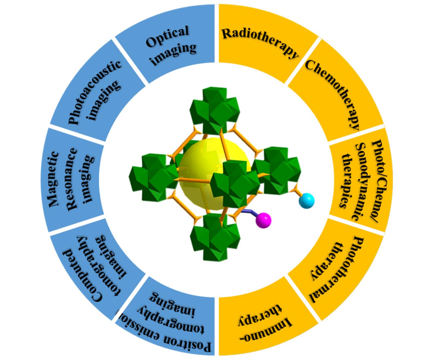
Fig.1 Schematic illustration of MOFs as cancer theranostic nanoplatforms
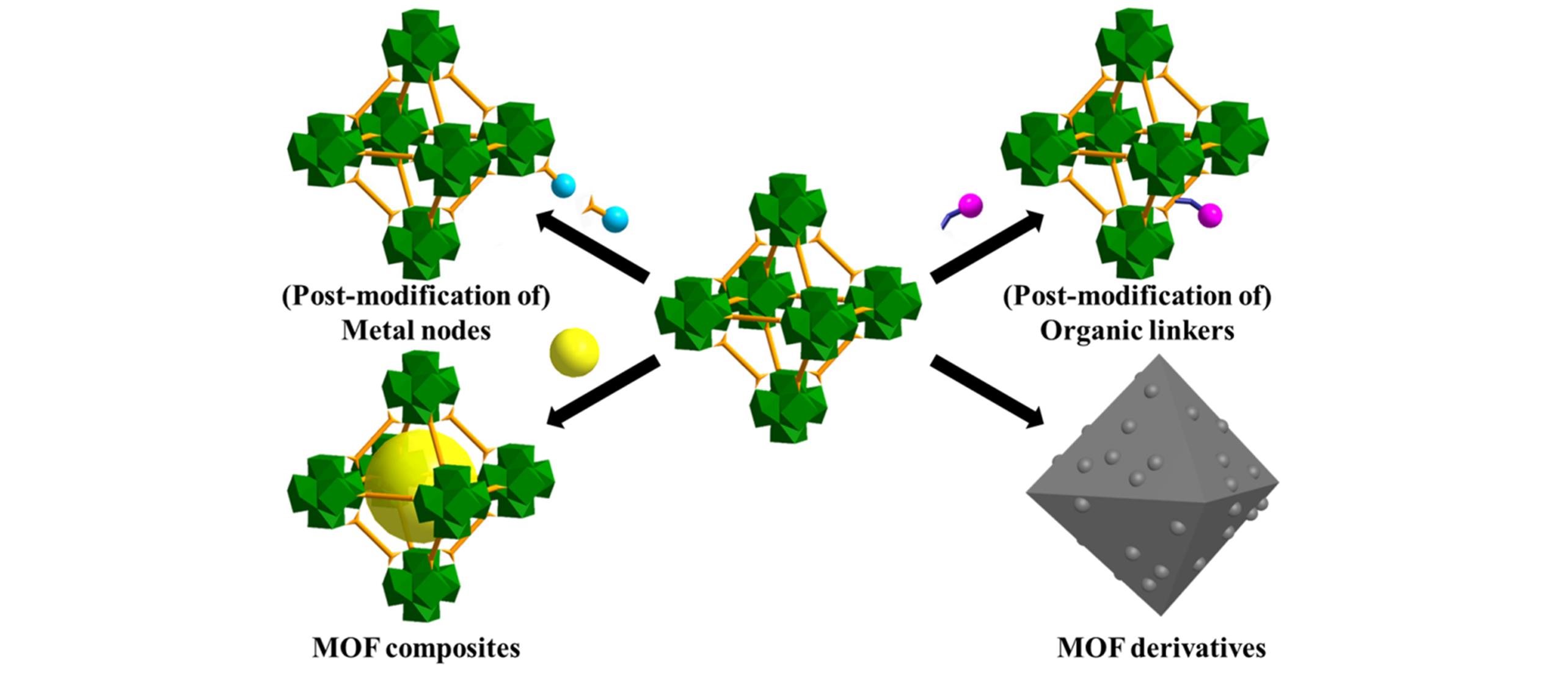
Fig.2 Four common strategies for loading theranostic agents into MOFs
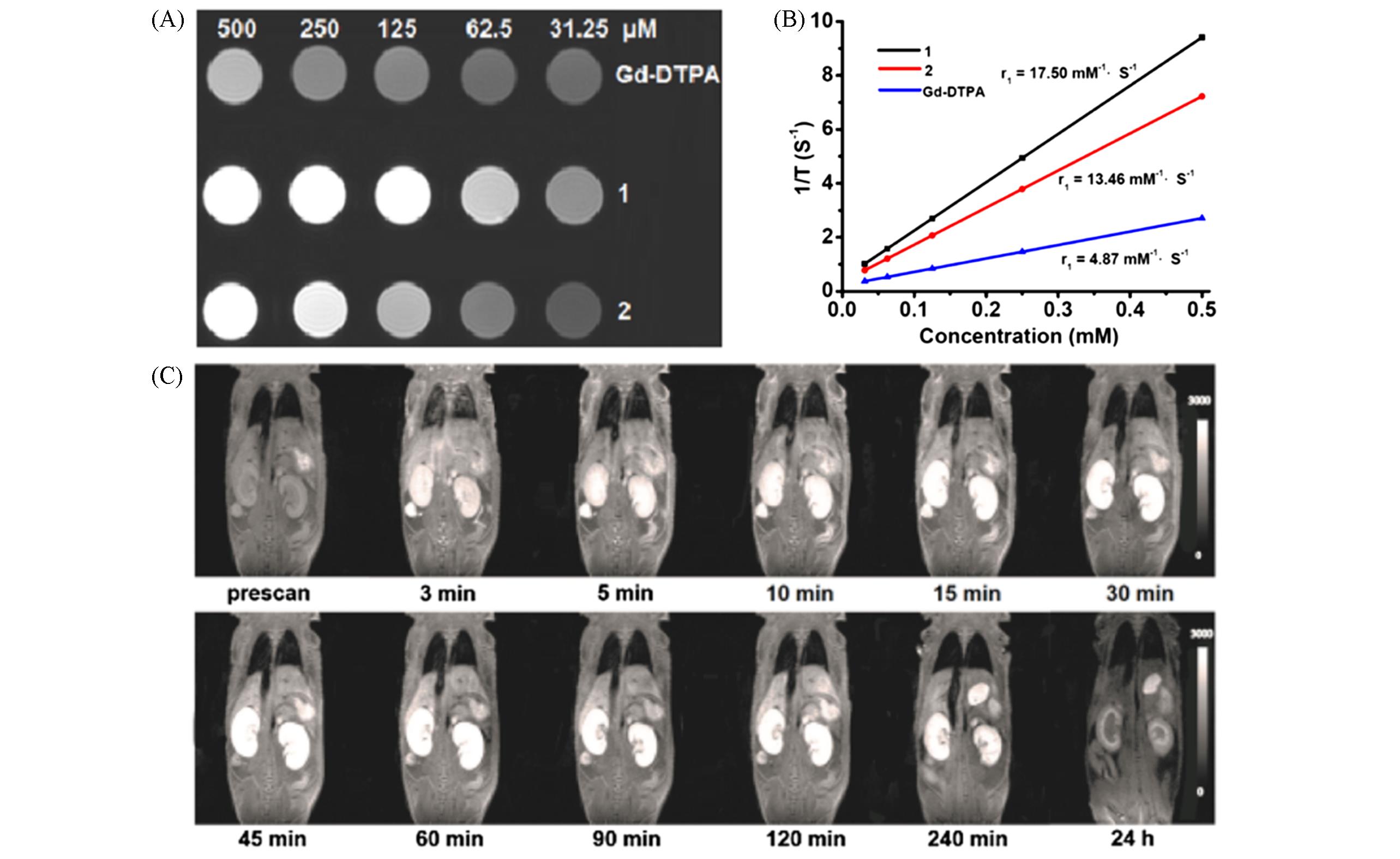
Fig.3 MRI imaging properties of ([Mn2(Cmdcp)2(H2O)2]·H2O) n (1) and [Gd(Cmdcp)(H2O)3](NO3)·3H2O) n[43](A) T1-weighted MRI images of {[Mn2(Cmdcp)2(H2O)2]·H2O} n (1), {[Gd(Cmdcp)(H2O)3](NO3)·3H2O} n (2) and Gd-DTPA of varying concentrations in water; (B) 1/T1-concentration plots of {[Mn2(Cmdcp)2(H2O)2]·H2O} n (1), {[Gd(Cmdcp)(H2O)3](NO3)·3H2O} n (2) and Gd-DTPA; (C) MR signal intensity from a dynamic study of normal kidneys after intravenous administration of {[Mn2(Cmdcp)2(H2O)2]·H2O} n (1).Copyright 2017, American Chemical Society.

Fig.4 CT imaging properties of UiO⁃PDT[55](A) Schematic diagram of the fabrication of UiO-PDT and its application for CT imaging in rat orthotopic hepatoma model; (B) relations between CT values of UiO-PDT and its different concentrations.Copyright 2017, the Royal Society of Chemistry.
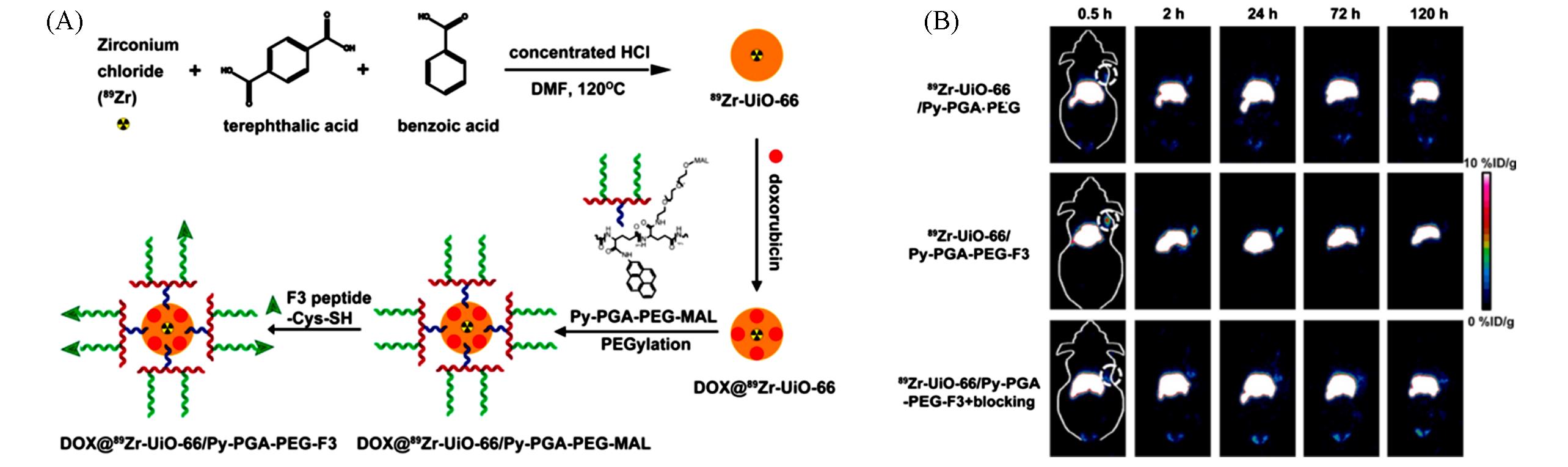
Fig.5 Synthesis and PET imaging properties of 89Zr⁃UiO⁃66/Py⁃PGA⁃PEG⁃F3[58](A) Scheme for the synthesis of 89Zr-UiO-66/Py-PGA-PEG-F3 conjugates; (B) coronal PET images of MDA-MB-231 tumour bearing mice at different time points after being injected with 89Zr-UiO-66/Py-PGA-PEG-F3, 89Zr-UiO-66/Py-PGA-PEG, and 89Zr-UiO-66/Py-PGA-PEG-F3 with excess F3 peptide for blocking.Copyright 2017, American Chemistry Society.
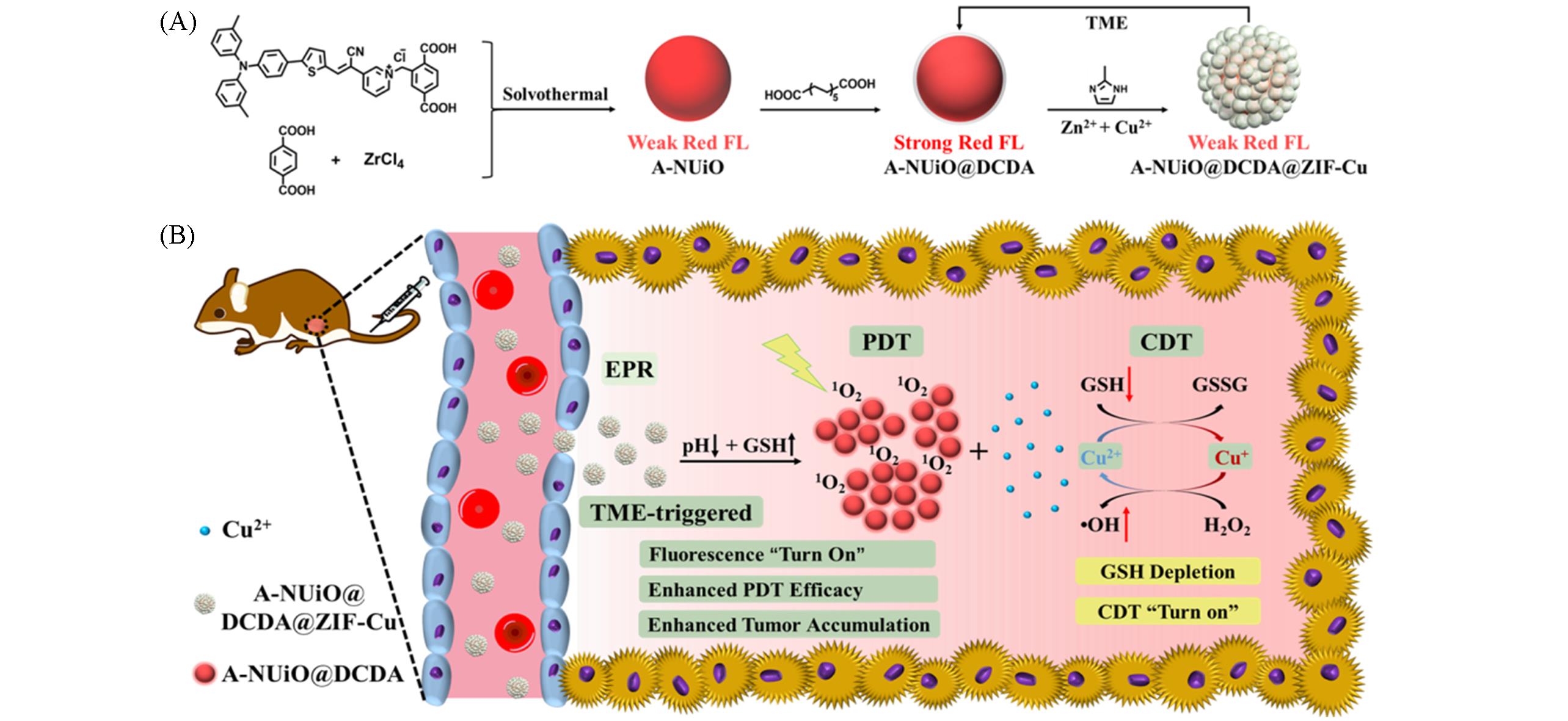
Fig.6 Schematic illustration of the synthesis process and theranostic features of A⁃NUiO@DCDA@ZIF⁃Cu[66](A) Synthesis process of A-NUiO@DCDA@ZIF-Cu; (B) A-NUiO@DCDA@ZIF-Cu features relating to TME stimuli-responses, enhancing tumor accumulation, and combination therapy.Copyright 2022, American Chemical Society.
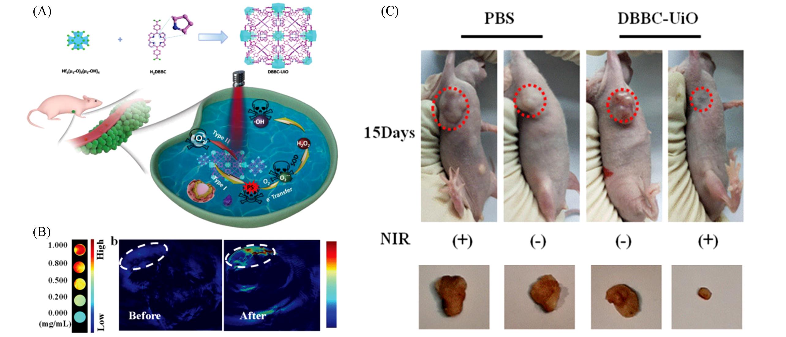
Fig.7 Synthesis, PA imaging and tumor therapy properties of DBBC⁃UiO[70](A) Schematic illustration of the preparation of DBBC-UiO and its application for PA imaging and tumor therapy; (B) the concentration-dependent PAI of DBBC-UiO in vitro and in vivo PAI with and without injection of DBBC-UiO; (C) photographs of tumor-bearing mice received different treatments after 15 d.Copyright 2019, Wiley-VCH Verlag GmbH & Co. KGaA, Weinheim.
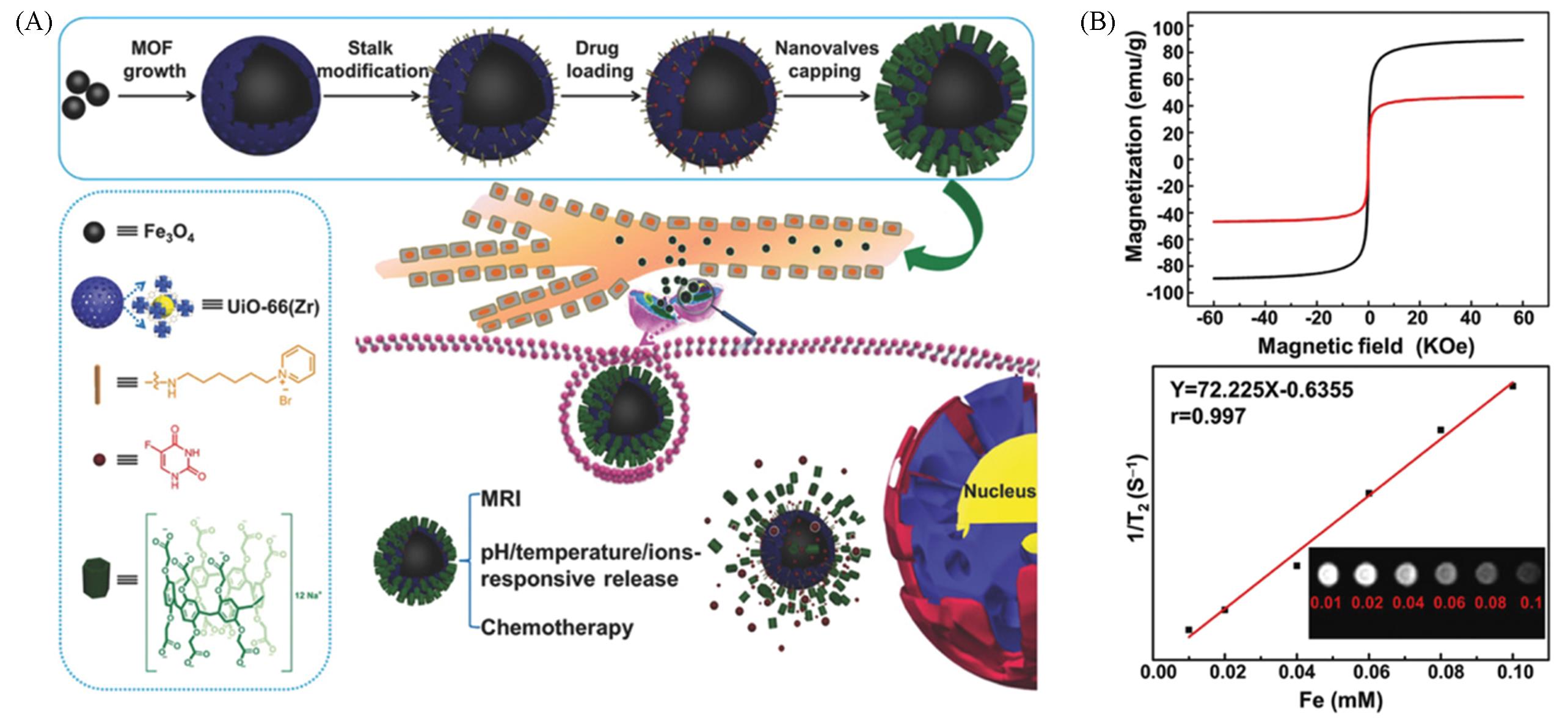
Fig.8 Synthesis, MRI imaging and chemotherapy properties of Fe3O4@UiO⁃66@WP6[71](A) Schematic description of the construction of Fe3O4@UiO-66@WP6 and the application for MRI and chemotherapy; (B) magnetic hysteresis curves of Fe3O4 NPs(black) and 5-Fu-loaded Fe3O4@UiO-66@WP6(red), and T2-weighted transverse relaxivity of Fe3O4@UiO-66@WP6 with a series of Fe concentrations.Copyright 2018, Wiley-VCH Verlag GmbH & Co. KGaA, Weinheim.
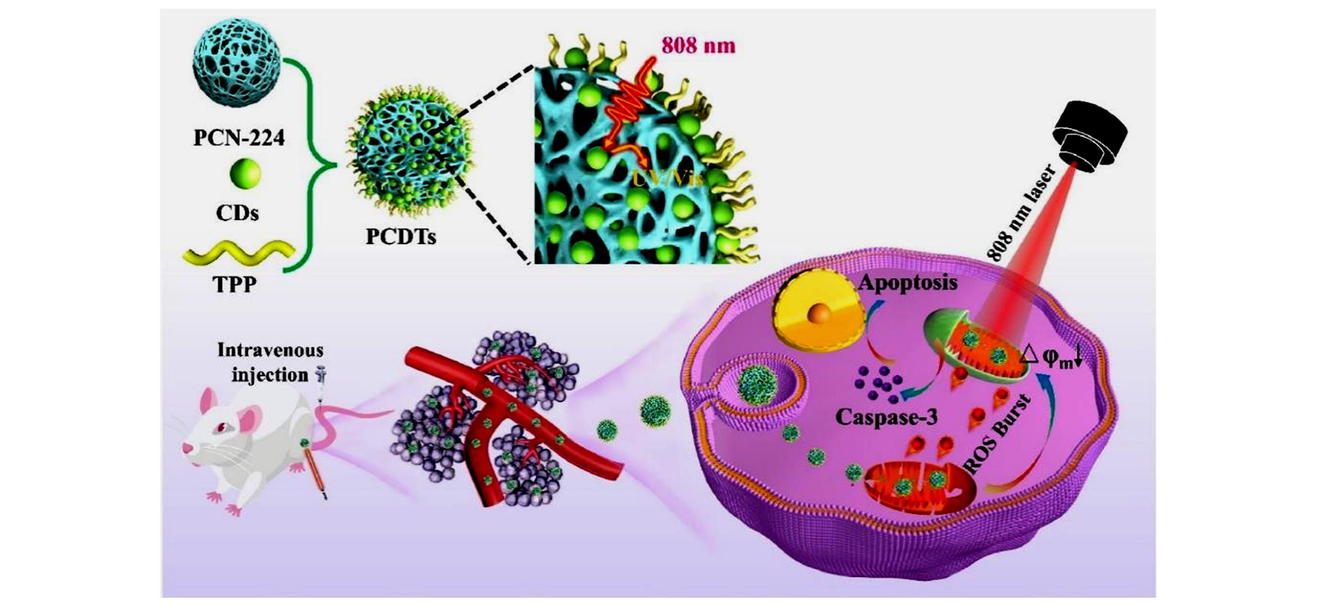
Fig.9 Schematic illustration of the structure of PCDTs and their application to 808 nm NIR light⁃activated and mitochondria⁃targeted PDT[78]Copyright 2022, Elsevier Ltd.
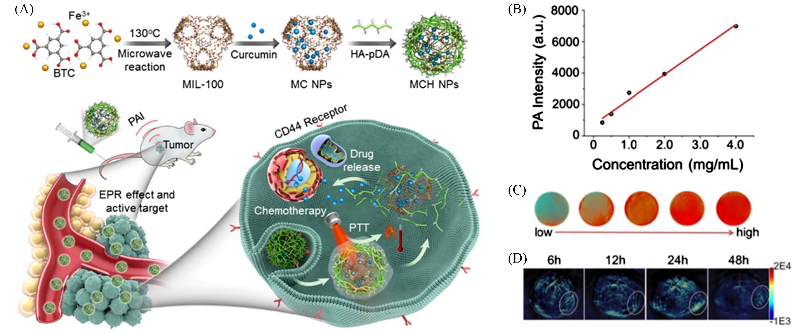
Fig.10 Synthesis, PA imaging and chemo/photothermal tumor therapy properties of MCH NPs[79](A) Schematic illustration for the construction and applications of MIL-100(Fe) for photoacoustic imaging-guided chemo-/photothermal combinational tumour therapy; (B) PA intensity of the nanoparticles at different concentrations; (C) corresponding in vitro PA images; (D) PA images of HeLa tumour-bearing mice at different time points after intravenous injection with the nanoparticles.Copyright 2018, American Chemical Society.
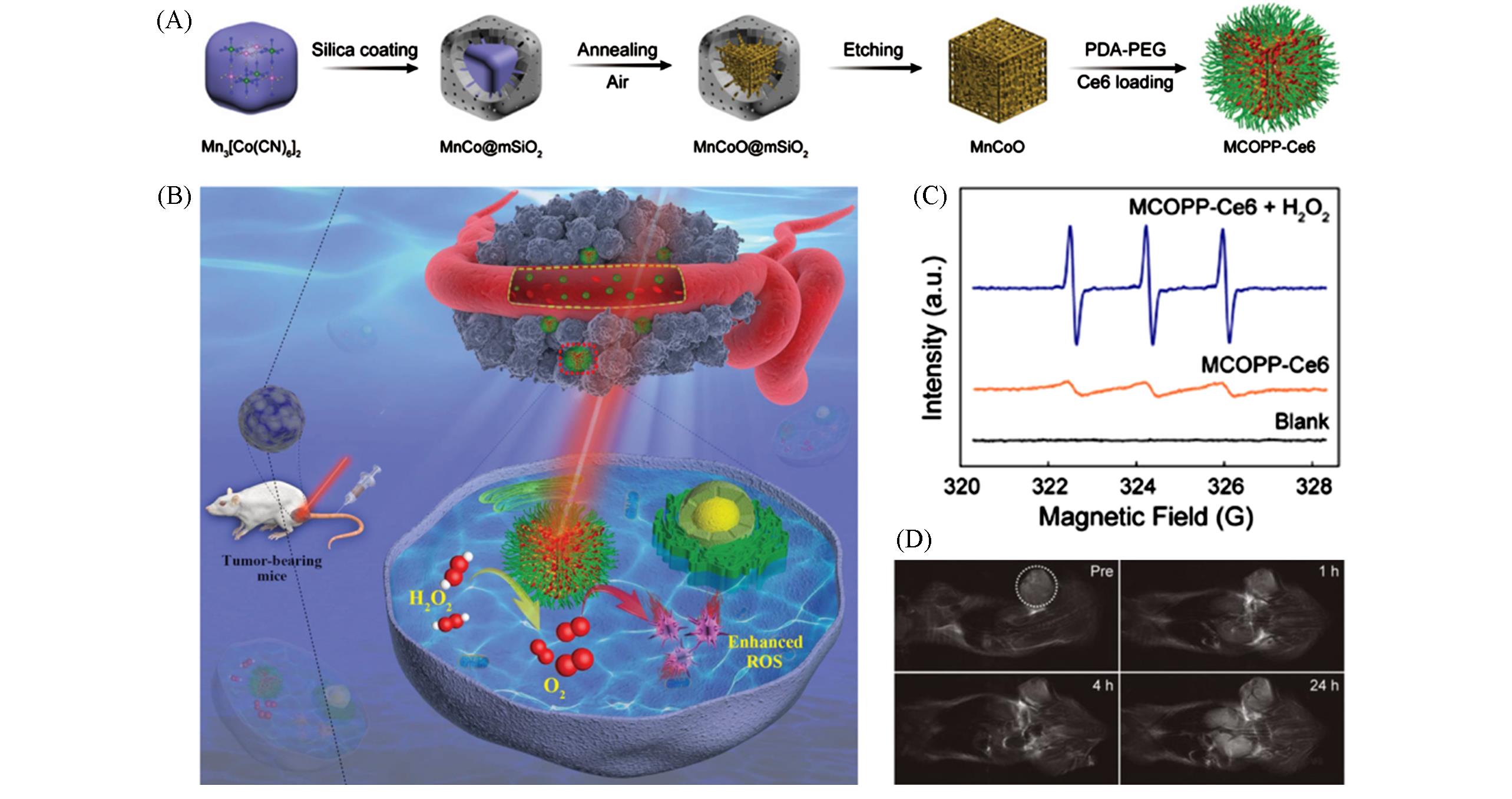
Fig.11 Synthesis, MRI imaging and PDT properties of MCOPP⁃Ce6[81](A) Synthesis procedure of MCOPP nanozyme and postloading of Ce6 photosensitizer; (B) schematic illustration of mesoporous MCOPP nanozyme for enhanced PDT of cancer; (C) ESR spectra of 1O2 trapped by TEMP after different treatments under near- infrared irradiation; (D) T2-weighted magnetic resonance imaging of murine breast tumor-bearing mouse after administration of MCOPP nanozyme. Tumor is marked with white dashed line.Copyright 2019, Wiley-VCH Verlag GmbH & Co. KGaA, Weinheim.
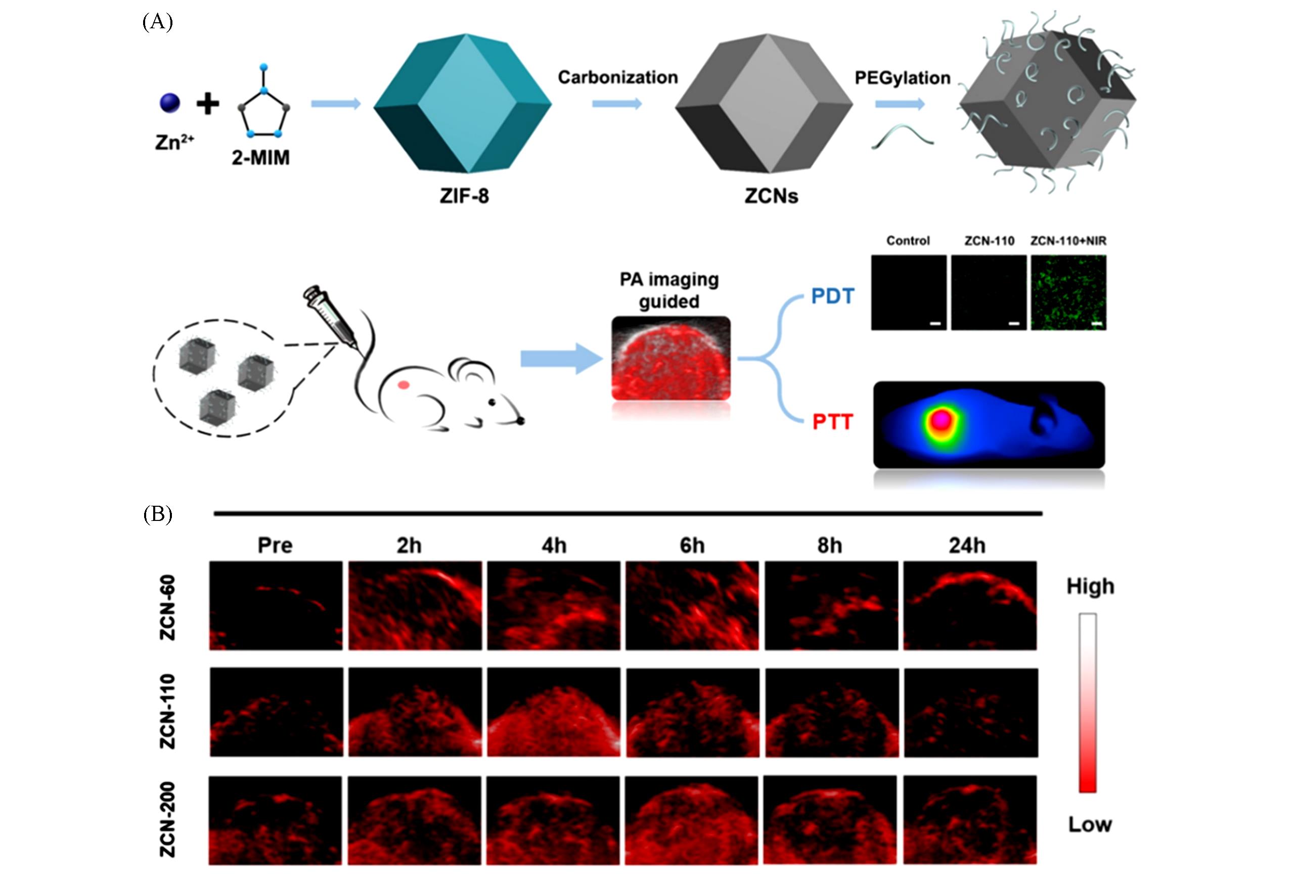
Fig.12 Synthesis, PA imaging and PDT/PTT tumor therapy properties of ZCNs[83](A) Schematic representation of the synthesis and applications of ZIF-8-derived carbon nanoparticles(ZCNs) for PAI-guided photothermal/photodynamic combined therapy; (B) PA imaging of A549 tumour-bearing mice at different times after intravenous injection with ZCNs. The images display the tumour region of the mice.Copyright 2018, American Chemical Society.
| 1 | Chen H. M., Zhang W. Z., Zhu G. Z., Xie J., Chen X. Y., Nat. Rev. Mater., 2017, 2(7), 1—18 |
| 2 | Luker G. D., Luker K. E., J. Nucl. Med., 2008, 49(1), 1—4 |
| 3 | Xu M. H., Wang L. H. V., Rev. Sci. Instrum., 2006, 77(4), 041101 |
| 4 | Jun Y. W., Huh Y. M., Choi J. S., Lee J. H., Song H. T., Kim S., Yoon S., Kim K. S., Shin J. S., Suh J. S., J. Am. Chem. Soc., 2005, 127(16), 5732—5733 |
| 5 | Mourtzakis M., Prado C. M. M., Lieffers J. R., Reiman T., McCargar L. J., Baracos V. E., Appl. Physiol. Nutr. Metab., 2008, 33(5), 997—1006 |
| 6 | Deng X. Y., Rong J., Wang L., Vasdev N., Zhang L., Josephson L., Liang S. H., Angew. Chem. Int. Ed., 2019, 58(9), 2580—2605 |
| 7 | Gerwing M., Herrmann K., Helfen A., Schliemann C., Berdel W. E., Eisenblätter M., Wildgruber M., Nat. Rev. Clin. Oncol., 2019, 16(7), 442—458 |
| 8 | Holohan C., van Schaeybroeck S., Longley D. B., Johnston P. G., Nat. Rev. Cancer, 2013, 13(10), 714—726 |
| 9 | Mallidi S., Anbil S., Bulin A. L., Obaid G., Ichikawa M., Hasan T., Theranostics, 2016, 6(13), 2458—2487 |
| 10 | Hompland T., Fjeldbo C. S., Lyng H., Cancers(Basel), 2021, 13(3), 499 |
| 11 | Nam J., Son S., Park K. S., Zou W. P., Shea L. D., Moon J. J., Nat. Rev. Mater., 2019, 4(6), 398—414 |
| 12 | Singh N., Son S., An J. S., Kim I., Choi M., Kong N., Tao W., Kim J. S., Chem. Soc. Rev., 2021, 50(23), 12883—12896 |
| 13 | Cai Y., Chen X. Y., Si J. X., Mou X. Z., Dong X. C., Small, 2021, 17(52), 2103072 |
| 14 | Johnson K. K., Koshy P., Yang J. L., Sorrell C. C., Adv. Funct. Mater., 2021, 31(43), 2104199 |
| 15 | Cheng L., Wang X. W., Gong F., Liu T., Liu Z., Adv. Mater., 2020, 32(13), 1902333 |
| 16 | Madamsetty V. S., Mukherjee A., Mukherjee S., Front. Pharmacol., 2019, 10, 1264 |
| 17 | Zhou H. C., Kitagawa S., Chem. Soc. Rev., 2014, 43(16), 5415—5418 |
| 18 | Qian Q. H., Asinger P. A., Lee M. J., Han G., Rodriguez K. M., Lin S., Benedetti F. M., Wu A. X., Chi W. S., Smith Z. P., Chem. Rev., 2020, 120(16), 8161—8266 |
| 19 | Li H. Y., Zhao S. N., Zang S. Q., Li J., Chem. Soc. Rev., 2020, 49(17), 6364—6401 |
| 20 | Wu C. D., Zhao M., Adv. Mater., 2017, 29(14), 1605446 |
| 21 | Lim D. W., Kitagawa H., Chem. Soc. Rev., 2021, 50(11), 6349—6368 |
| 22 | Lu K. D., Aung T., Guo N. N., Weichselbaum R., Lin W. B., Adv. Mater., 2018, 30(37), 1707634 |
| 23 | Liu J. T., Huang J., Zhang L., Lei J. P., Chem. Soc. Rev., 2021, 50(2), 1188—1218 |
| 24 | Wang F., Li Z., Zhang X. B., Luo R. A., Hou H. L., Lei J. P., Chem. Commun., 2021, 57(63), 7826—7829 |
| 25 | Rieter W. J., Taylor K. M. L., An H. Y., Lin W. L., Lin W. B., J. Am. Chem. Soc., 2006, 128(28), 9024—9025 |
| 26 | Giménez⁃Marqués M., Hidalgo T., Serre C., Horcajada P., Coord. Chem. Rev., 2016, 307, 342—360 |
| 27 | Cai Y., Wei Z., Song C. H., Tang C. C., Han W., Dong X. C., Chem. Soc. Rev., 2019, 48(1), 22—37 |
| 28 | Yang G. B., Phua S. Z. F., Bindra A. K., Zhao Y. L., Adv. Mater., 2019, 31(10), 1805730 |
| 29 | Zhao N. N., Yan L. M., Zhao X. Y., Chen X. Y., Li A. H., Zheng D., Zhou X., Dai X. G., Xu F. J., Chem. Rev., 2019, 119(3), 1666—1762 |
| 30 | Luo T. K., Nash G. T., Xu Z. W., Jiang X. M., Liu J. Q., Lin W. B., J. Am. Chem. Soc., 2021, 143(34), 13519—13524 |
| 31 | Zeng J. Y., Zhang M. K., Peng M. Y., Gong D., Zhang X. Z., Adv. Funct. Mater., 2018, 28(8), 1705451 |
| 32 | Li Y. L., Zhao P. R., Gong T., Wang H., Jiang X. W., Cheng H., Liu Y. Y., Wu Y. L., Bu W. B., Angew. Chem. Int. Ed., 2020, 59(50), 22537—22543 |
| 33 | Wu M. X., Yang Y. W., Adv. Mater., 2017, 29(23), 1606134 |
| 34 | Chowdhury M. A., J. Biomed. Mater. Res. Part A, 2017, 105(4), 1184—1194 |
| 35 | He F., Wen N. C., Xiao D. P., Yan J. H., Xiong H. J., Cai S. D., Liu Z. B., Liu Y. F., Curr. Med. Chem., 2020, 27(13), 2189—2219 |
| 36 | Zhou Z. J., Yang L. J., Gao J. H., Chen X. Y., Adv. Mater., 2019, 31(8), 1804567 |
| 37 | Pandey A., Dhas N., Deshmukh P., Caro C., Patil P., García⁃Martín M. L., Padya B., Nikam A., Mehta T., Mutalik S., Coord. Chem. Rev., 2020, 409, 213212 |
| 38 | Peller M., Böll K., Zimpel A., Wuttke S., Inorg. Chem. Front., 2018, 5(8), 1760—1779 |
| 39 | Hatakeyama W., Sanchez T. J., Rowe M. D., Serkova N. J., Liberatore M. W., Boyes S. G., ACS Appl. Mater. Interfaces, 2011, 3(5), 1502—1510 |
| 40 | Della Rocca J., Lin W. B., Eur. J. Inorg. Chem., 2010, 2010(24), 3725—3734 |
| 41 | Mao X. P., Xu J. D., Cui H. G., Wiley Interdiscip. Rev. Nanomed. Nanobiotechnol., 2016, 8(6), 814—841 |
| 42 | Rieter W. J., Pott K. M., Taylor K. M., Lin W. B., J. Am. Chem. Soc., 2008, 130(35), 11584—11585 |
| 43 | Qin L., Sun Z. Y., Cheng K., Liu S. W., Pang J. X., Xia L. M., Chen W. H., Cheng Z., Chen J. X., ACS Appl. Mater. Interfaces, 2017, 9(47), 41378—41386 |
| 44 | Liu C. H., Cao Y., Cheng Y. R., Wang D. D., Xu T. L., Su L., Zhang X. J., Dong H. F., Nat. Commun., 2020, 11(1), 1—9 |
| 45 | Yu S. J., Huang X. J., Xu C. F., Xu L. S., Sun Y., Shen Q. Y., Wang B., Zhu H. L., Lin W. X., Hu Q., J. Solid State Chem., 2022, 313, 123349 |
| 46 | Lee N., Hyeon T., Chem. Soc. Rev., 2012, 41(7), 2575—2589 |
| 47 | Horcajada P., Chalati T., Serre C., Gillet B., Sebrie C., Baati T., Eubank J. F., Heurtaux D., Clayette P., Kreuz C., Chang J. S., Hwang Y. K., Marsaud V., Bories P. N., Cynober L., Gil S., Ferey G., Couvreur P., Gref R., Nat. Mater., 2010, 9(2), 172—178 |
| 48 | Zhang Y., Liu C. Q., Wang F. M., Liu Z., Ren J. S., Qu X. G., Chem. Commun., 2017, 53(11), 1840—1843 |
| 49 | Horcajada P., Gref R., Baati T., Allan P. K., Maurin G., Couvreur P., Ferey G., Morris R. E., Serre C., Chem. Rev., 2012, 112(2), 1232—1268 |
| 50 | Yang J., Yang Y. W., View, 2020, 1(2), e28 |
| 51 | deKrafft K. E., Boyle W. S., Burk L. M., Zhou O. Z., Lin W. B., J. Mater. Chem., 2012, 22(35), 18139—18144 |
| 52 | Lu K. D., He C. B., Guo N. N., Chan C., Ni K. Y., Lan G. X., Tang H. D., Pelizzari C., Fu Y. X., Spiotto M. T., Weichselbaum R. R., Lin W. B., Nat. Biomed. Eng., 2018, 2(8), 600—610 |
| 53 | Zhang K., Meng X., Cao Y., Yang Z., Dong H. F., Zhang Y. D., Lu H. T., Shi Z. J., Zhang X. J., Adv. Funct. Mater., 2018, 28(42), 1804634 |
| 54 | Robison L., Zhang L., Drout R. J., Li P., Haney C. R., Brikha A., Noh H., Mehdi B. L., Browning N. D., Dravid V. P., Cui Q., Islamoglu T., Farha O. K., ACS Appl. Bio. Mater., 2019, 2(3), 1197—1203 |
| 55 | Zhang T., Wang L., Ma C., Wang W. Q., Ding J., Liu S., Zhang X. W., Xie Z. G., J. Mater. Chem. B, 2020, 8(48), 11107—11108 |
| 56 | Basu S., Alavi A., PET Clinics, 2016, 11(3), 203—207 |
| 57 | Duman F. D., Forgan R. S., J. Mater. Chem. B, 2021, 9(16), 3423—3449 |
| 58 | Chen D. Q., Yang D. Z., Dougherty C. A., Lu W. F., Wu H. W., He X. R., Cai T., Van Dort M. E., Ross B. D., Hong H., ACS Nano, 2017, 11(4), 4315—4327 |
| 59 | Abazari R., Ataei F., Morsali A., Slawin A. M., Z. Carpenter⁃Warren C. L., ACS Appl. Mater. Interfaces, 2019, 11(49), 45442—45454 |
| 60 | Wang W. Q., Wang L., Li Z. S., Xie Z. G., Chem. Commun., 2016, 52(31), 5402—5405 |
| 61 | Zhang H., Tian X. T., Shang Y., Li Y. H., Yin X. B., ACS Appl. Mater. Interfaces, 2018, 10(34), 28390—28398 |
| 62 | Liu Y., Gong C. S., Dai Y. L., Yang Z., Yu G. C., Liu Y. J., Zhang M. R., Lin L. S., Tang W., Zhou Z. J., Zhu G. Z., Chen J. J., Jacobson O., Kiesewetter D. O., Wang Z. T., Chen X. Y., Biomaterials, 2019, 218, 119365 |
| 63 | Li S. Y., Cheng H., Xie B. R., Qiu W. X., Zeng J. Y., Li C. X., Wan S. S., Zhang L., Liu W. L., Zhang X. Z., ACS Nano, 2017, 11(7), 7006—7018 |
| 64 | Mei J., Leung N. L. C., Kwok R. T. K., Lam J. W. Y., Tang B. Z., Chem. Rev., 2015, 115(21), 11718—11940 |
| 65 | Cai X. L., Liu B., Angew. Chem. Int. Ed., 2020, 59(25), 9868—9886 |
| 66 | Dong M. J., Li W. Q., Xiang Q., Tan Y., Xing X. T., Wu C. X., Dong H. F., Zhang X. J., ACS Appl. Mater. Interfaces, 2022, 14(26), 29599—29612 |
| 67 | Taylor⁃Pashow K. M. L., Della Rocca J., Xie Z. G., Tran S., Lin W. B., J. Am. Chem. Soc., 2009, 131(40), 14261—14263 |
| 68 | Emelianov S. Y., Li P. C., O’Donnell M., Phys. Today, 2009, 62(5), 34—39 |
| 69 | Zhou G. X., Wang Y. S., Jin Z. K., Zhao P. H., Zhang H., Wen Y. Y., He Q. J., Nanoscale Horiz., 2019, 4(5), 1185—1193 |
| 70 | Zhang K., Yu Z. F., Meng X. D., Zhao W. D., Shi Z. J., Yang Z., Dong H. F., Zhang X. J., Adv. Sci., 2019, 6(14), 1900530 |
| 71 | Wu M. X., Gao J., Wang F., Yang J., Song N., Jin X. Y., Mi P., Tian J., Luo J. Y., Liang F., Yang Y. W., Small, 2018, 14(17), 1704440 |
| 72 | Zhang H. P., Zhang Q., Liu C. S., Han B., Biomater. Sci., 2019, 7(4), 1696—1704 |
| 73 | Zhuang Y. X., Katayama Y., Ueda J., Tanabe S., Opt. Mater., 2014, 36(11), 1907—1912 |
| 74 | Lv Y., Ding D. D., Zhuang Y. X., Feng Y. S., Shi J. P., Zhang H. W., Zhou T. L., Chen H. M., Xie R. J., ACS Appl. Mater. Interfaces, 2019, 11(2), 1907—1916 |
| 75 | Duman F. D., Hocaoglu I., Ozturk D. G., Gozuacik D., Kiraz A., Acar H. Y., Nanoscale, 2015, 7(26), 11352—11362 |
| 76 | Aguilera⁃Sigalat J., Bradshaw D., Coord. Chem. Rev., 2016, 307, 267—291 |
| 77 | Zhao A. D., Chen Z. W., Zhao C. Q., Gao N., Ren J. S., Qu X. G., Carbon, 2015, 85, 309—327 |
| 78 | Xiang Q., Li W., Tan Y., Shi J. W., Dong M. J., Cheng J. L., Huang J. K., Zhang W. Y., Gong Y. C., Yang Q. Q., Yang L. Z., Dong H. F., Zhang X. J., Chem. Eng. J., 2022, 444, 136706 |
| 79 | Zhang Y., Wang L., Liu L., Lin L., Liu F., Xie Z. G., Tian H. Y., Chen X. S., ACS Appl. Mater. Interfaces, 2018, 10(48), 41035—41045 |
| 80 | Zhang K., Meng X. D., Yang Z., Dong H. F., Zhang X. J., Biomaterials, 2020, 258, 120278 |
| 81 | Wang D. D., Wu H. H., Lim W. Q., Phua S. Z. F., Xu P. P., Chen Q. W., Guo Z., Zhao Y. L., Adv. Mater., 2019, 31(27), 1901893 |
| 82 | Pan X. T., Wang W. W., Huang Z. J., Liu S., Guo J., Zhang F. R., Yuan H. J., Li X., Liu F. Y., Liu H. Y., Angew. Chem. Int. Ed., 2020, 132(32), 13659—13633 |
| 83 | Yang P., Tian Y., Men Y. Z., Guo R. R., Peng H. B., Jiang Q., Yang W. L., ACS Appl. Mater. Interfaces, 2018, 10(49), 42039—42049 |
| [1] | 冯丽, 邵兰兴, 李思骏, 全文选, 庄金亮. 超薄Sm-MOF纳米片的合成及可见光催化降解芥子气模拟剂性能[J]. 高等学校化学学报, 2022, 43(4): 20210867. |
| [2] | 储彬彬, 何耀. 硅基纳米探针用于眼部疾病的成像检测与治疗[J]. 高等学校化学学报, 2022, 43(12): 20220546. |
| [3] | 徐海东, 王睿, 梁高林. 自组装多肽探针在磁共振成像中的应用[J]. 高等学校化学学报, 2022, 43(12): 20220440. |
| [4] | 李奥, 李凌轩, 左翠翠, 陈传凯, 樊一凡, 步逸凡, 林泓域, 高锦豪. 基于硼酸酯的19F磁共振分子探针的设计合成及活体深组织活性氧物种的激活响应成像[J]. 高等学校化学学报, 2022, 43(12): 20220545. |
| [5] | 叶卓, 吉墨轩, 刘定斌. 动脉粥样硬化光学成像探针研究进展[J]. 高等学校化学学报, 2022, 43(12): 20220556. |
| [6] | 汪诗琪, 罗博文, 俞计成, 顾臻. 近红外二区活体荧光成像在肿瘤诊疗中的应用[J]. 高等学校化学学报, 2022, 43(12): 20220577. |
| [7] | 卢美如, 张宏宇, 石百媚, 孙茂忠, 徐丽广, 胥传来, 匡华. 手性纳米材料: 生物成像、 生物传感与治疗[J]. 高等学校化学学报, 2022, 43(12): 20220683. |
| [8] | 常通航, 程震. 整合荧光成像和化疗的有机小分子诊疗探针的研究进展[J]. 高等学校化学学报, 2022, 43(12): 20220430. |
| [9] | 张庆鹏, 关国强, 刘慧怡, 陆畅, 周颖, 宋国胜. 磁粒子成像示踪剂的研究进展[J]. 高等学校化学学报, 2022, 43(12): 20220375. |
| [10] | 徐心昱, 张乐天, 曹晖, 马原, 刘柳卉, 宋国胜, 张晓兵. 脂质响应型探针用于动脉粥样硬化成像及治疗的研究进展[J]. 高等学校化学学报, 2022, 43(12): 20220383. |
| [11] | 马小飞, 胡山, 李俊彬, 杨盛, 谌委菊, 卿志和, 周怡波, 杨荣华. 细胞内源性分子辅助荧光信号放大策略及细胞成像[J]. 高等学校化学学报, 2022, 43(12): 20220320. |
| [12] | 陈尚钰, 沈清明, 孙鹏飞, 范曲立. 小分子基温敏聚合物纳米粒子的制备及在近红外二区荧光成像与光热治疗中的应用[J]. 高等学校化学学报, 2022, 43(12): 20220392. |
| [13] | 汤乔伟, 蔡小青, 李江, 诸颖, 王丽华, 田阳, 樊春海, 胡钧. 同步辐射X射线成像技术在脑成像研究中的应用[J]. 高等学校化学学报, 2022, 43(12): 20220379. |
| [14] | 赵恒智, 余方志, 李翔菲, 李乐乐. 基于DNA与上转换纳米颗粒相结合的生物传感与成像研究进展[J]. 高等学校化学学报, 2022, 43(12): 20220626. |
| [15] | 赵雪琪, 赵越, 薛静, 白敏, 陈锋, 孙颖, 宋大千, 赵永席. 单细胞核酸编码扩增成像分析[J]. 高等学校化学学报, 2022, 43(12): 20220572. |
| 阅读次数 | ||||||
|
全文 |
|
|||||
|
摘要 |
|
|||||