

Chem. J. Chinese Universities ›› 2020, Vol. 41 ›› Issue (6): 1252.doi: 10.7503/cjcu20200026
• Analytical Chemistry • Previous Articles Next Articles
BAI Cuiting,YUE Renye,LUO Liegao,MA Nan*( )
)
Received:2020-01-13
Online:2020-06-10
Published:2020-02-26
Contact:
Nan MA
E-mail:nan.ma@suda.edu.cn
Supported by:CLC Number:
TrendMD:
BAI Cuiting, YUE Renye, LUO Liegao, MA Nan. Quantitative Analysis of MicroRNA Content by Fluorescence Imaging in Cancer Cells Using Dual-color Fluorescence Nanosensor †[J]. Chem. J. Chinese Universities, 2020, 41(6): 1252.
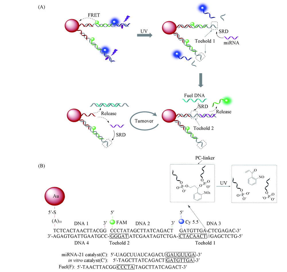
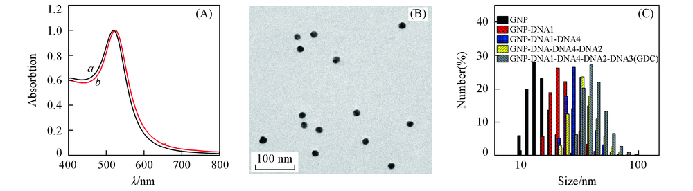
| Name | DNA Sequence(5'→3') |
|---|---|
| DNA1(thiolated DNA) | SH-AAAAAAAAAATCTCACTAACTTACGG |
| DNA2(FAM) | FAM-CCCTATAGCTTATCAGACT |
| DNA3(Cy5.5) | Cy5.5-GATGTTGACTCGAGAC |
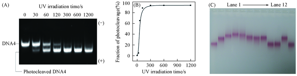
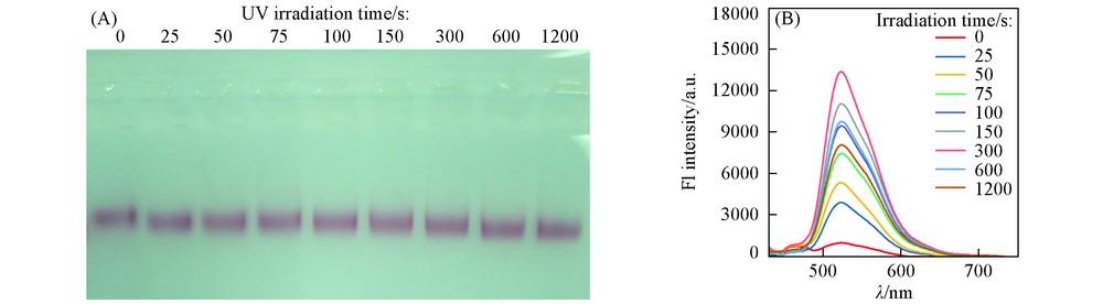
| DNA4(PC Linker DNA) | GTCTCGAG//TCAACATCAGTCTGATAAGCTATAGGGCCGTAAGTTAGTGAGA |
| Fuel DNA(F) | CTAACTTACGGCCCTATAGCTTATCAGACT |
| Catalyst DNA(C') | TAGCTTATCAGACTGATGTTGA |
| MiRNA-21(C) | UAGCUUAUCAGACUGAUGUUGA |
| FAM-DNA1 | SH-AAAAAAAAAATCTCACTAACTTACGG-FAM |
| FAM-DNA4 | GTCTCGAGTCAACATCAGTCTGATAAGCTATAGGGCCGTAAGTTAGTGAGA-FAM |
| 1-Mismatch[C'(mis-1)] | TAGCTTATCAGACTGATCTTGA |
| 2-Mismatches[C'(mis-2)] | TAGCTTATCAGTCTGATCTTGA |
| 3-Mismatches[C'(mis-3)] | TAGCTAATCAGTCTGATCTTGA |
| Name | DNA Sequence(5'→3') |
|---|---|
| DNA1(thiolated DNA) | SH-AAAAAAAAAATCTCACTAACTTACGG |
| DNA2(FAM) | FAM-CCCTATAGCTTATCAGACT |
| DNA3(Cy5.5) | Cy5.5-GATGTTGACTCGAGAC |
| DNA4(PC Linker DNA) | GTCTCGAG//TCAACATCAGTCTGATAAGCTATAGGGCCGTAAGTTAGTGAGA |
| Fuel DNA(F) | CTAACTTACGGCCCTATAGCTTATCAGACT |
| Catalyst DNA(C') | TAGCTTATCAGACTGATGTTGA |
| MiRNA-21(C) | UAGCUUAUCAGACUGAUGUUGA |
| FAM-DNA1 | SH-AAAAAAAAAATCTCACTAACTTACGG-FAM |
| FAM-DNA4 | GTCTCGAGTCAACATCAGTCTGATAAGCTATAGGGCCGTAAGTTAGTGAGA-FAM |
| 1-Mismatch[C'(mis-1)] | TAGCTTATCAGACTGATCTTGA |
| 2-Mismatches[C'(mis-2)] | TAGCTTATCAGTCTGATCTTGA |
| 3-Mismatches[C'(mis-3)] | TAGCTAATCAGTCTGATCTTGA |
| 103 C'/L molar ratio | FL intensity/a.u. | 103 C'/L molar ratio | FL intensity/a.u. | ||
|---|---|---|---|---|---|
| FAM | Cy5.5 | FAM | Cy5.5 | ||
| 0 | 1298 | 20635 | 15 | 26138 | 23284 |
| 4 | 7351 | 24341 | 20 | 33258 | 22946 |
| 10 | 16737 | 23975 | |||
| 103 C'/L molar ratio | FL intensity/a.u. | 103 C'/L molar ratio | FL intensity/a.u. | ||
|---|---|---|---|---|---|
| FAM | Cy5.5 | FAM | Cy5.5 | ||
| 0 | 1298 | 20635 | 15 | 26138 | 23284 |
| 4 | 7351 | 24341 | 20 | 33258 | 22946 |
| 10 | 16737 | 23975 | |||
| [1] |
Lim L. P., Lau N. C., Garrettengele P., Grimson A., Schelter J. M., Castle J., Bartel D. P., Linsley P. S., Johnson J. M., Nature, 2005, 433, 769—773
doi: 10.1038/nature03315 URL |
| [2] |
He L., Hannon G. J., Nat. Rev. Genet., 2004, 5, 522—531
doi: 10.1038/nrg1379 URL |
| [3] |
Ambros V., Nature, 2004, 431, 350—355
doi: 10.1038/nature02871 URL |
| [4] |
Liu H. Y., Bei X. Q., Xia Q. T., Fu Y., Zhang S., Liu M. C., Fan K., Zhang M. Z., Yang Y., Microchimica Acta, 2015, 183(1), 297—304
doi: 10.1007/s00604-015-1636-z URL |
| [5] |
Rajesha R., Frank. J. S., Nat. Rev. Drug Discov., 2017, 16(3), 203—222
doi: 10.1038/nrd.2016.246 URL |
| [6] |
Chen Y. X., Huang K. J., Niu K. X., Biosens. Bioelectron., 2018, 99, 612—624
doi: 10.1016/j.bios.2017.08.036 URL |
| [7] |
Cullen B. R., Nature, 2009, 457(7228), 421—425
doi: 10.1038/nature07757 URL |
| [8] |
Matsumura T., Sugimachi K., Iinuma H., Takahashi Y., Kurashige J., Sawada G., Ueda M., Uchi R., Ueo H., Takano Y., Shinden Y., Eguchi H., Yamamoto H., Doki Y., Mori M., Ochiya T., Mimori K., Br. J. Cancer, 2015, 113(2), 275—281
doi: 10.1038/bjc.2015.201 URL |
| [9] |
Nelson P. T., Baldwin D. A., Scearce L. M., Oberholtzer J. C., Tobias J. W., Mourelatos Z., Nat. Methods, 2004, 1, 155—161
doi: 10.1038/nmeth717 URL |
| [10] |
Markou A., Tsaroucha E. G., Kaklamanis L., Fotinou M., Georgoulias V., Lianidou E. S., Clin. Chem., 2008, 54, 1696—1704
doi: 10.1373/clinchem.2007.101741 URL |
| [11] |
Varkonyi-Gasic E., Wu R., Wood M., Walton E. F., Hellens R. P., Plant Methods, 2007, 3, 12
doi: 10.1186/1746-4811-3-12 URL |
| [12] |
Cheng Y., Lei J. P., Chen Y. L., Ju H. X., Biosens. Bioelectron., 2014, 51, 431—436
doi: 10.1016/j.bios.2013.08.014 URL |
| [13] |
Zhang P., Wu X. Y., Yuan R., Chai Y. Q., Anal. Chem., 2015, 87, 3202—3207
doi: 10.1021/ac504455z URL |
| [14] |
Zhang D. C., Yan Y. R., Cheng W., Zhang W., Li Y. H., Ju H. X., Ding S. J., Microchim. Acta., 2013, 180, 397—403
doi: 10.1007/s00604-013-0945-3 URL |
| [15] |
Gao X. F., Xu H., Baloda M., Gurung A. S., Xu L. P., Wang T., Biosens. Bioelectron., 2014, 54, 578—584
doi: 10.1016/j.bios.2013.10.055 URL |
| [16] |
Liu L. Z., Jiang S. T., Wang L., Zhang Z., Xie G. M., Microchim. Acta, 2015, 182, 77—84
doi: 10.1007/s00604-014-1273-y URL |
| [17] |
Sztandera K., Gorzkiewicz M., Klajnert-Maculewicz B., Mol. Pharm., 2019, 16(1), 1—23
doi: 10.1021/acs.molpharmaceut.8b00810 URL |
| [18] |
Reinhard B. M., Siu M., Agarwal H., Alivisatos A. P., Liphardt J., Nano Lett., 2005, 5(22), 2246—2252
doi: 10.1021/nl051592s URL |
| [19] |
Yun C. S., Javier A., Jennings T., Fisher M., Hira S., Peterson S., Hopkins B., Reich N. O., Strouse G. F., J. Am. Chem. Soc., 2005, 127(23), 3115—3119
doi: 10.1021/ja043940i URL |
| [20] |
Gaylord B. S., Heeger A. J., Bazan G. C., Proc. Natl. Acad. Sci., 2002, 99, 10954—10957
doi: 10.1073/pnas.162375999 URL |
| [21] |
Niikura K., Matsunaga T., Suzuki T., Kobayashi S., Yamaguchi H., Orba Y., Kawaguchi A., Hasegawa H., Kajino K., Ninomiya T., Ijiro K., Sawa H., ACS Nano, 2013, 7(5), 3926—3938
doi: 10.1021/nn3057005 URL |
| [22] |
Jain P. K., Lee K. S., El-Sayed I. H., El-Sayed M. A., J. Phys. Chem. B, 2006, 110(14), 7238—7248
doi: 10.1021/jp057170o URL |
| [23] |
Huang K. J., Liu Y. J., Wang H. B., Wang Y. Y., Liu Y. M., Biosens. Bioelectron., 2014, 55, 195—202
doi: 10.1016/j.bios.2013.11.061 URL |
| [24] |
Huang K. J., Liu Y. J., Shi G. W., Yang X. R., Liu Y. M., Sens. Actuators B: Chem., 2014, 201, 579—585
doi: 10.1016/j.snb.2014.05.055 URL |
| [25] |
Komarala E. P., Tyagi H., Thiyagatajan S., Pradhan L., Aslam M., Bahadur D., J. Mater. Chem. B, 2017, 5(21), 3852—3861
doi: 10.1039/C7TB00015D URL |
| [26] |
Zhang J., Li C., Zhang X., Huo S., Jin S., An F. F., Wang X., Xue X., Okeke C. I., Duan G., Guo F., Zhang X., Hao J., Wang P. C., Zhang J., Liang X. J., Biomaterials, 2015, 42, 103—111
doi: 10.1016/j.biomaterials.2014.11.053 URL |
| [27] | Jin X. T., Liu G., Li J. Z., Sun L. L., Wang J. R., Li J. F., Li P., Chen W. Q., Wang Q., Tong T., Chem. J. Chinese Universities, 2016, 37(2), 224—231 |
| 金新天, 刘刚, 李君哲, 孙丽丽, 王俊荣, 李俊锋, 李沛, 陈文庆, 王强, 佟倜 . 高等学校化学学报, 2016, 37(2), 224—231) | |
| [28] |
Wang J., Wang D. X., Tang A. N., Kong D. M., Anal. Chem., 2019, 91, 5244—5251
doi: 10.1021/acs.analchem.9b00007 URL |
| [29] |
Jiang L., Zhou Q., Mu K., Xie H., Zhu Y., Zhu W., Zhao Y., Xu H., Yang X., Biomaterials, 2013, 34(30), 7418—7428
doi: 10.1016/j.biomaterials.2013.05.078 URL |
| [30] |
Dong H. F., Zhang J., Ju H. X., Lu H. T., Wang S. Y., Jin S., Hao K. H., Du H. W., Zhang X. J., Anal. Chem., 2012, 84, 4587—4593
doi: 10.1021/ac300721u URL |
| [31] |
Dong H. F., Hao K. H., Tian Y. P., Jin S., Lu H. T., Zhou S. F., Zhang X. J., Biosens. Bioelectron., 2014, 53, 377—383
doi: 10.1016/j.bios.2013.09.061 URL |
| [32] | Wang Z. Y., Liu M., Zhang C. Y. , Chem. J. Chinese Universities 2017, 38(1), 1—11 |
| ( 王子月, 刘萌, 张春阳 . 高等学校化学学报, 2017, 38(1) 1—11) | |
| [33] |
Yang J. M., Dou B. T., Yuan R., Xiang Y., Anal. Chem., 2017, 89, 5138—5143
doi: 10.1021/acs.analchem.7b00827 URL |
| [34] |
Liu J. T., Du P., Zhang J., Shen H., Lei J. P., Chem. Commun., 2018, 54(20), 2550—2553
doi: 10.1039/C7CC09579A URL |
| [35] |
Zhang D. Y., Turberfield A. J., Yurke B., Winfree E., Science, 2007, 318(5853), 1121—1125
doi: 10.1126/science.1148532 URL |
| [36] |
He X. W., Zeng T., Li Z., Wang G. L., Ma N., Angew. Chem. Int. Ed., 2016, 55, 3073—3076
doi: 10.1002/anie.201509726 URL |
| [37] | Luo X. C., Li Z., Wang G. L., He X. W., Shen X. Q., Sun Q. H., Wang L., Yue R. Y., Ma N., ACS Appl. Mater. Interfaces, 2017, 10, 1021 |
| [38] |
Shen Y., Li Z., Wang G. L., Ma N., ACS Sens., 2018, 3, 494—503
doi: 10.1021/acssensors.7b00922 URL |
| [39] |
Jin R., Wu G., Li Z., Mirkin C. A., Schatz G. C., J. Am. Chem. Soc., 2003, 125, 1643—1654
doi: 10.1021/ja021096v URL |
| [1] | LIU Miao, LIU Ruibo, LIU Badi, QIAN Ying. Synthesis, Two-photon Fluorescence Imaging and Photodynamic Therapy of Lysosome-targeted Indole-BODIPY Photosensitizer [J]. Chem. J. Chinese Universities, 2022, 43(10): 20220326. |
| [2] | CHEN Hongda, ZHANG Hua, WANG Zhenxin. Development of Small Animals in vivo Fluorescence-photothermal Dual Mode Imaging System [J]. Chem. J. Chinese Universities, 2021, 42(3): 725. |
| [3] | LIANG Yuxin, ZHAO Rong, LIANG Xinyue, FANG Xiaohong. Single-molecule Imaging and Analysis of Signal Transduction Proteins on Cell Membranes † [J]. Chem. J. Chinese Universities, 2020, 41(6): 1127. |
| [4] | SHAO Wei, LEE Jiyoung, LI Fangyuan, LING Daishun. Organic Small Molecule Nanoparticles for Phototheranostics [J]. Chem. J. Chinese Universities, 2020, 41(11): 2356. |
| [5] | ZHANG Yimeng, ZHANG Huixin, LIU Yang. Recent Advances of Exosomes Bioanalysis and Their Clinic Applications [J]. Chem. J. Chinese Universities, 2020, 41(11): 2306. |
| [6] | WANG Tingting, LI Yuan, YANG Lili, BAO Changhao, CHENG Han. In vivo Dynamic Detection of Aloe Polysaccharides Using Carbon Fiber Microelectrodes Modified with Gold Nanoparticles † [J]. Chem. J. Chinese Universities, 2020, 41(1): 87. |
| [7] | ZHU Qinfu,HU Kezhen,LI Xiaojie,CHEN Mingqing. Preparation of Dendrimer-gold Nanoparticle Composites for pH Responsive Drug Delivery† [J]. Chem. J. Chinese Universities, 2019, 40(5): 1065. |
| [8] | Yong ZHANG,Cheng SHEN,Zhirong XING,Guiqi CHEN,Zi LU,Zhibing HOU,Xuemei CHEN. Benzimidazole-Derived Fluorescence Enhancement Probe for Visual Detection of HClO † [J]. Chem. J. Chinese Universities, 2019, 40(12): 2480. |
| [9] | Siqi SUN,Ying WANG,Chuanyin SUN,Runwei WANG,Zhendong ZHANG,Zongtao ZHANG,Shilun QIU. Preparation and Catalytic Performance of Bowl-shaped Amphiphilic ZSM-5 Zeolites Supported Gold Nanoparticles † [J]. Chem. J. Chinese Universities, 2019, 40(12): 2436. |
| [10] | HE Caimei,ZHENG Jingyi,LI Xiaoxia. MoS2-Gold Nanoparticles and Thionine-gold Nanoparticle Based Signal-enhanced Electrochemical Aptasensor for the Detection of 17β-Estradiol † [J]. Chem. J. Chinese Universities, 2019, 40(10): 2090. |
| [11] | FENG Wei,WANG Bowei,ZHENG Yan,JIANG Yang. Preparation and Surface-enhanced Raman Scattering(SERS) of Single Au Nanodot† [J]. Chem. J. Chinese Universities, 2018, 39(9): 1875. |
| [12] | LI Aiju, WANG Yuxi, LU Shaoyong, LIU Kun. Ligand Exchange of Gold Nanoparticles with Thiol-terminated Polystyrene† [J]. Chem. J. Chinese Universities, 2018, 39(3): 552. |
| [13] | WANG Li, LI Zhi, SHEN Xiaoqin, MA Nan. Programming Single Quantum Dot Valencies via DNA Caging† [J]. Chem. J. Chinese Universities, 2018, 39(1): 32. |
| [14] | ZHANG Chanchan, ZHANG Fanghui, DING Lei, NI Zhenjie, JIANG Lang, DONG Huanli, ZHANG Xiaotao, LI Rongjin, HU Wenping. Organic Phototransistor Based on Surface Plasmon Resonance Effect† [J]. Chem. J. Chinese Universities, 2018, 39(1): 102. |
| [15] | HUANG Haiping, YUE Yafeng, XU Liang, LÜ Lianlian, HU Yongmei. Glucose Biosensor Based on Dy2(MoO4)3-AuNPs Composite Nanomaterial† [J]. Chem. J. Chinese Universities, 2017, 38(4): 554. |
| Viewed | ||||||
|
Full text |
|
|||||
|
Abstract |
|
|||||