

高等学校化学学报 ›› 2026, Vol. 47 ›› Issue (8): 20260080.doi: 10.7503/cjcu20260080
李欣岩1, 孙诗涵1, 苗滋伟2, 耿文倩1, 蒋彬1, 孙志英1, 李婉明1( )
)
收稿日期:2026-02-13
出版日期:2026-08-10
发布日期:2026-05-09
通讯作者:
李婉明
E-mail:wmli@cmu.edu.cn
基金资助:
LI Xinyan1, SUN Shihan1, MIAO Ziwei2, GENG Wenqian1, JIANG Bin1, SUN Zhiying1, LI Wanming1()
Received:2026-02-13
Online:2026-08-10
Published:2026-05-09
Contact:
LI Wanming
E-mail:wmli@cmu.edu.cn
Supported by:摘要:
研究了核酸适体L4的靶向结合特性, 并探讨了其作为分子探针对临床结直肠癌患者组织的靶向成像能力, 旨在为转移性结直肠癌的靶向诊断提供潜在的分子工具. 采用流式细胞术分析了核酸适体L4的结合特异性、 亲和性、 温度稳定性和细胞选择性; 通过酶处理实验探讨了核酸适体L4结合靶向分子的性质; 基于生物素-链霉亲和素的结合原理, 将核酸适体L4与量子点(Quantum dots, QDs)偶联形成了L4-QD探针, 对临床结直肠癌患者组织进行了靶向成像, 并分析了组织靶向性及与患者病理特征的相关性. 结果表明, 核酸适体L4可高特异性和高亲和力地结合靶细胞HCT116, 解离常数(Kd)值为(10.4±1.7) nmol/L, 且具有良好的温度稳定性. 临床组织成像结果显示, L4-QD探针能够对结直肠癌组织实现靶向成像, 且荧光强度与患者美国癌症联合委员会(American joint committee on cancer, AJCC)临床分期和生存预后不良呈显著正相关. 核酸适体L4对转移性结直肠癌细胞具有高特异性和高亲和力, 与量子点偶联可实现结直肠癌组织的靶向成像, 有望成为评估结直肠癌进展程度的潜在分子探针及转移性结直肠癌靶向诊断的新型工具.
中图分类号:
TrendMD:
李欣岩, 孙诗涵, 苗滋伟, 耿文倩, 蒋彬, 孙志英, 李婉明. 基于核酸适体L4的转移性结直肠癌的靶向成像. 高等学校化学学报, 2026, 47(8): 20260080.
LI Xinyan, SUN Shihan, MIAO Ziwei, GENG Wenqian, JIANG Bin, SUN Zhiying, LI Wanming. Targeted Imaging of Metastatic Colorectal Cancer Based on Aptamer L4. Chem. J. Chinese Universities, 2026, 47(8): 20260080.
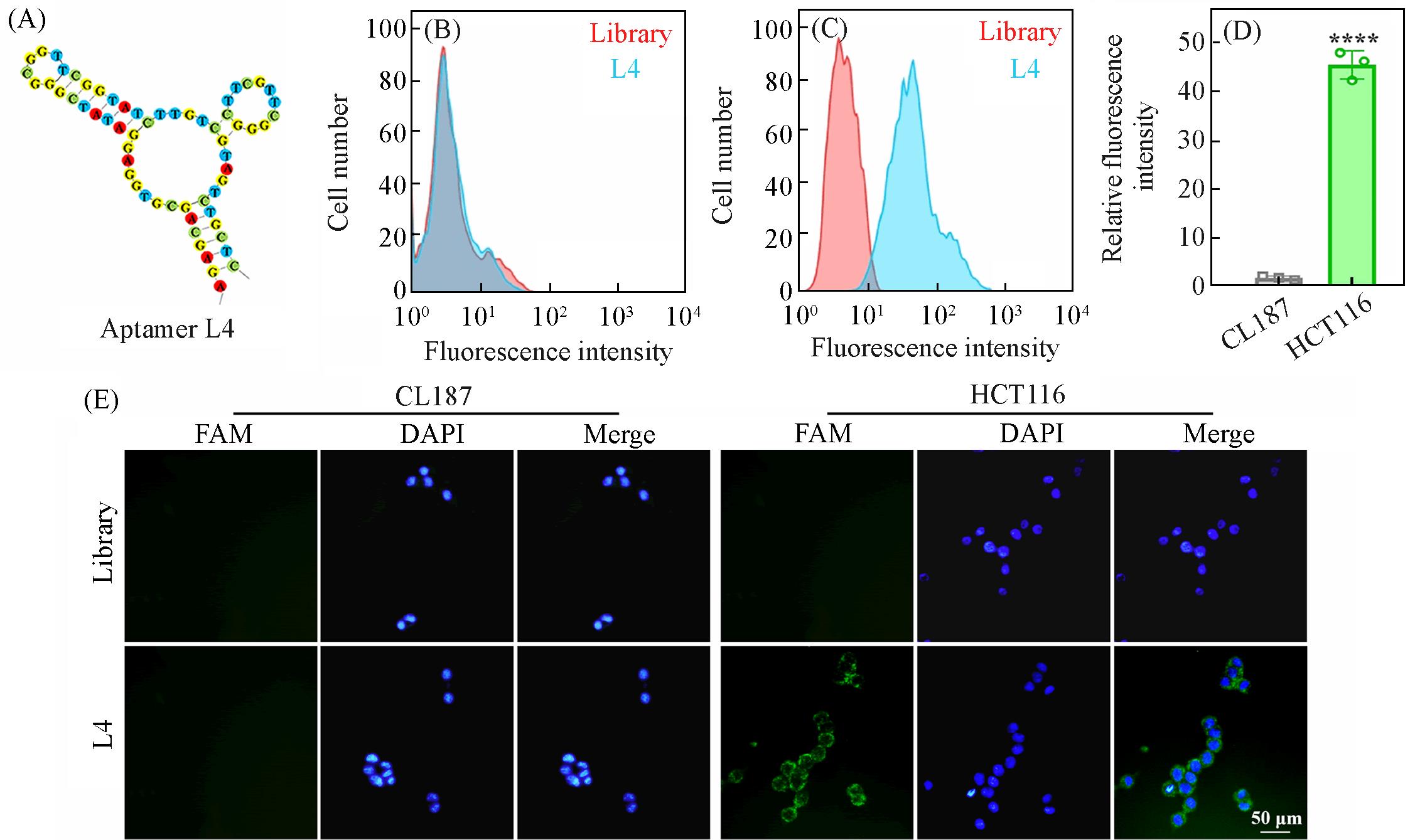
Fig.1 Analysis of binding specificity of aptamer L4 to the target HCT116 cells(A) Secondary structure of aptamer L4; (B, C) flow cytometry analysis of aptamer L4 binding to HCT116(B) and CL187(C) cells; (D)quantitative analysis of mean fluorescence intensity, ****P<0.0001, n=3; (E) fluorescence microscopy images of aptamer L4 binding to the target HCT116 or non-target CL187 cells.
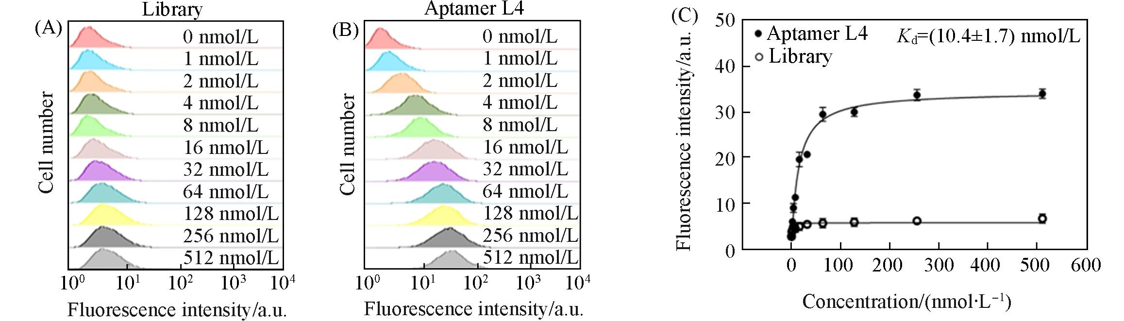
Fig.2 Affinity analysis of aptamer L4 binding to HCT116 cells(A, B) Flow cytometry histograms showing the binding of different concentrations(0—512 nmol/L) of aptamer Library(A) or aptamer L4(B) to HCT116 cells; (C) saturation binding curve of aptamer L4 to HCT116 cells with increasing concentrations of aptamer L4, and calculated Kd value, data are presented as mean±SD(n=3 independent experiments).
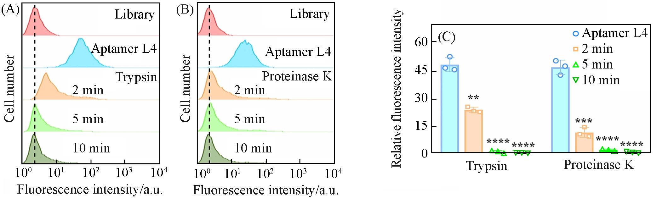
Fig.3 Flow cytometry analysis of aptamer L4 binding to HCT116 cells before and after trypsin/proteinase K treatmentHistograms of trypsin(A) and proteinase K(B) treated cells with 2, 5 and 10 min; (C) quantitative analysis of mean fluorescence intensity(MFI) showing the relative binding of aptamer L4 after enzyme treatments; data are presented as mean±SD, **P<0.01, ***P<0.001, ****P<0.0001, n=3.
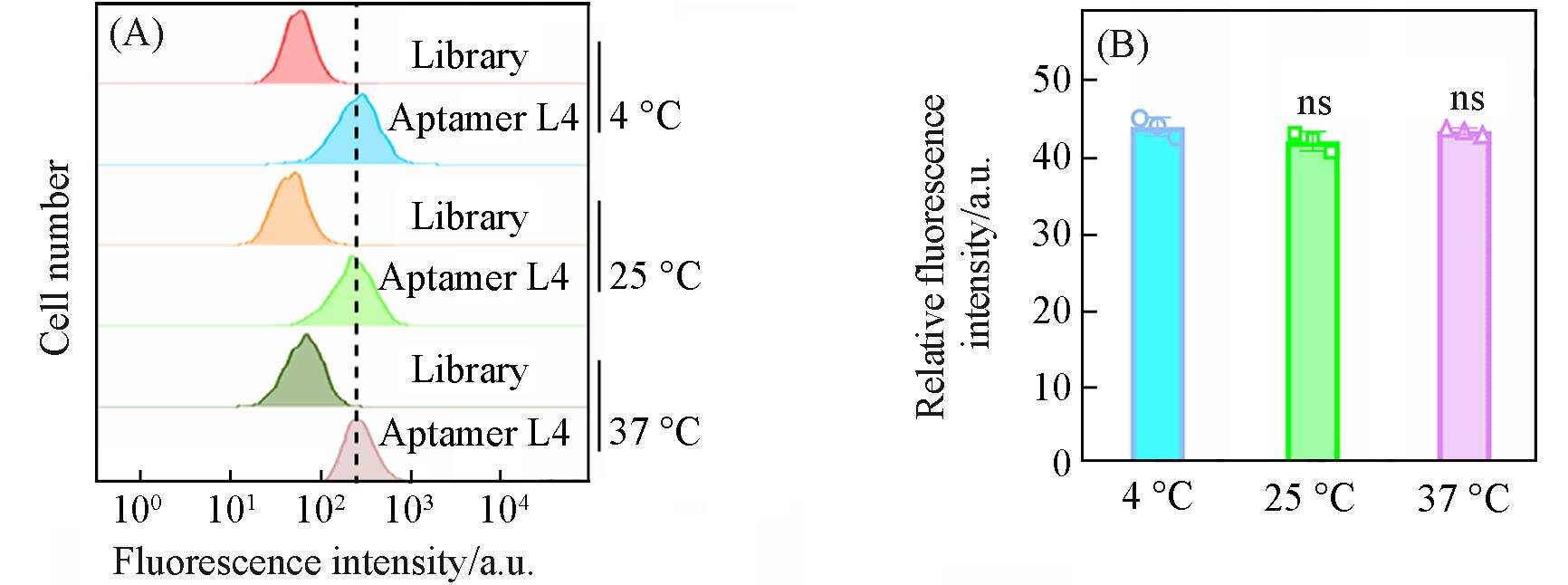
Fig.4 Flow cytometry analysis of aptamer L4 binding to HCT116 cells at different temperatures(A) Histograms; (B) quantitative analysis of mean fluorescence intensity, nsP>0.05, n=3.
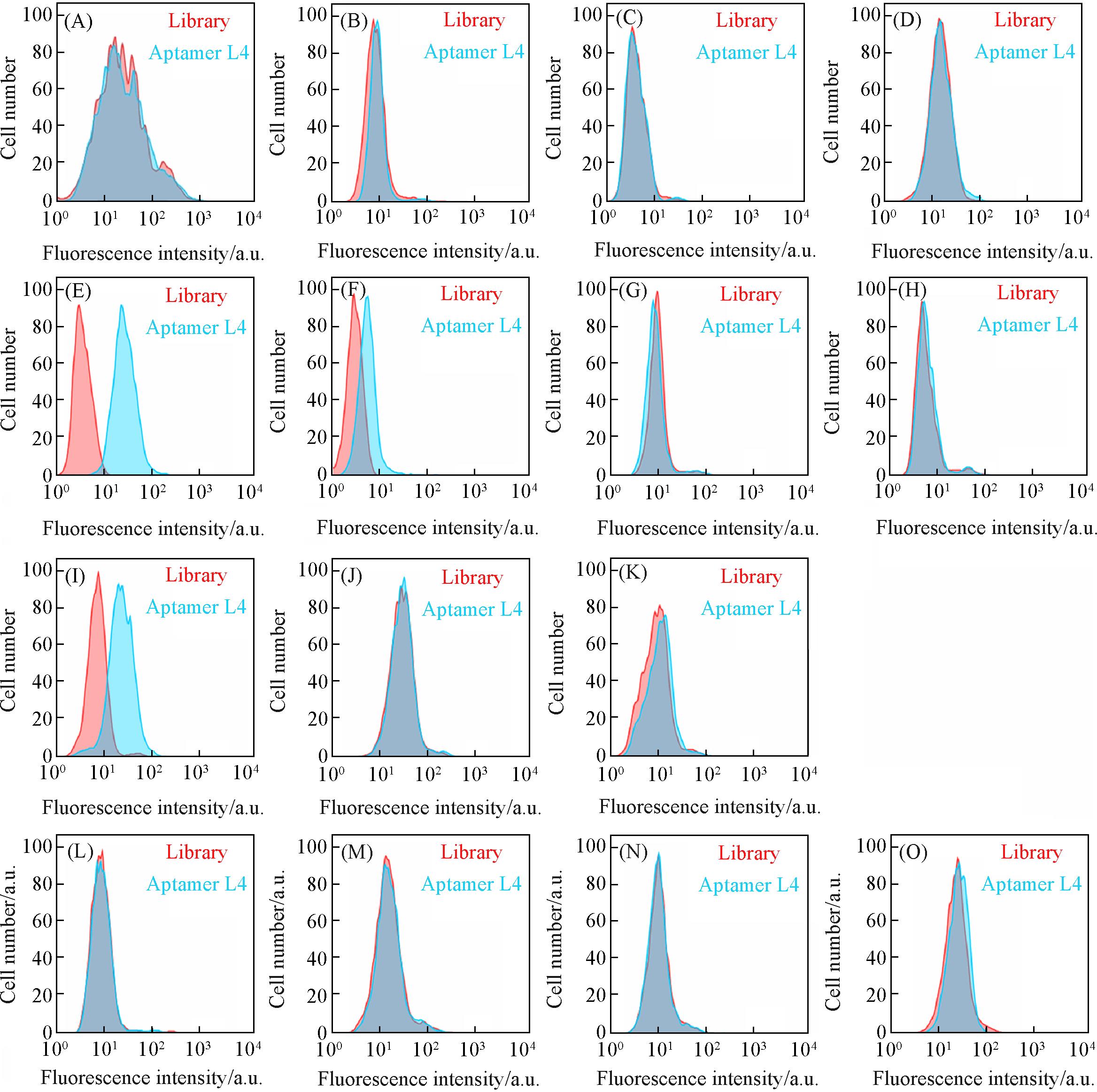
Fig.5 Flow cytometry analysis of aptamer L4 binding to various normal(A—D) and tumor(E—R) cell lines(A) COS-7; (B) GES-1; (C) CHO; (D) HEK293; (E) CHT116; (F) SW620; (G) SW480; (H) HT29; (I) COLO 205; (J) HCT-8; (K) CloneA; (L) MCF-7; (M) BGC-823; (N) SMMC7721; (O) A549.
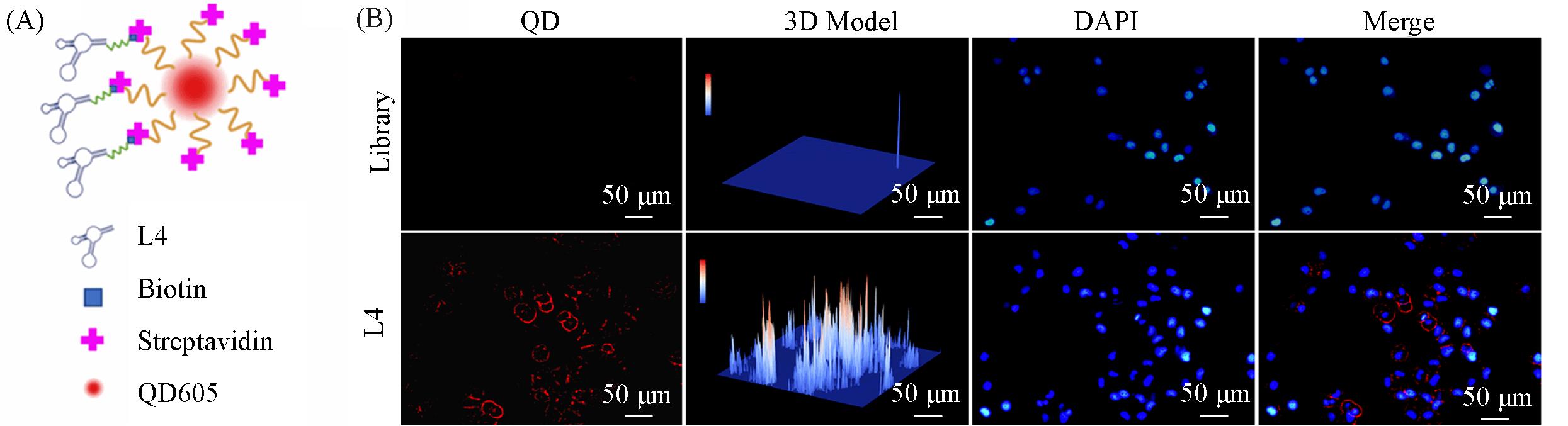
Fig.6 Targeted imaging of HCT116 cells by L4⁃QD probe(A) Schematic diagram of L4-QD probe; (B) fluorescence images of HCT116 cells attained by L4-QD probe.
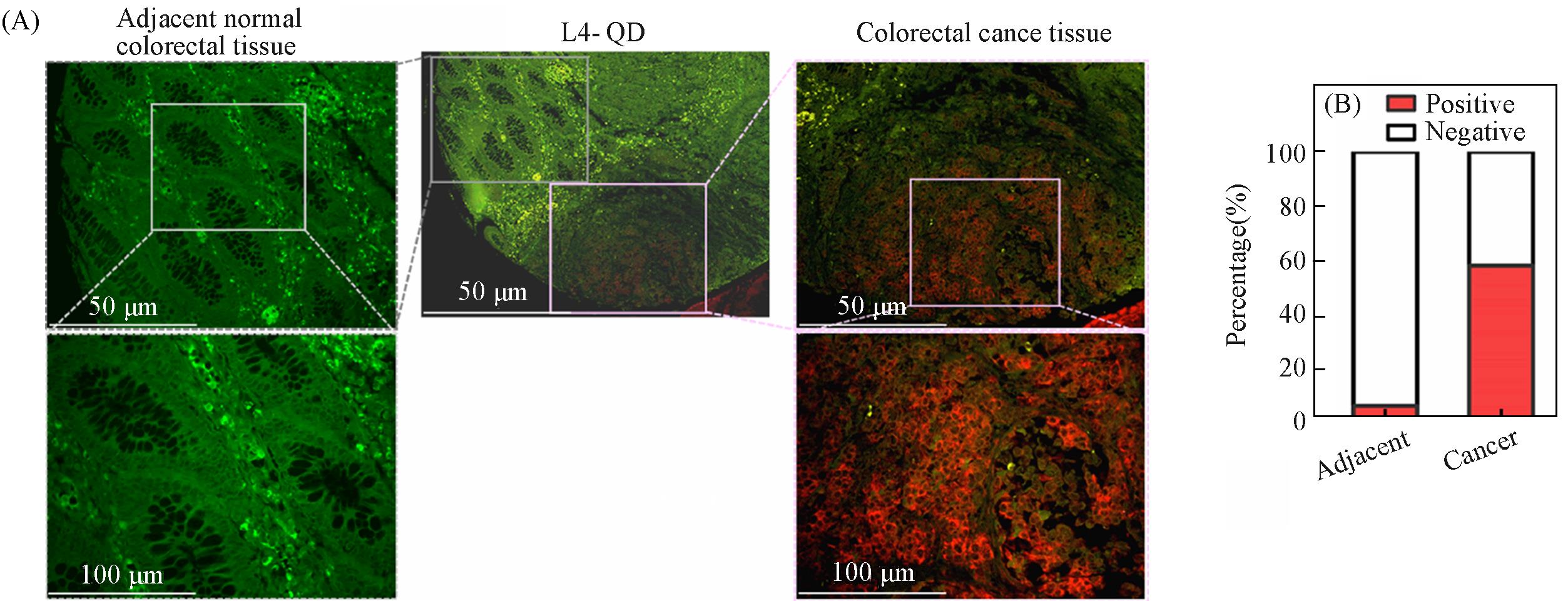
Fig.7 Imaging analysis of colorectal cancer tissues and adjacent normal tissues by L4⁃QD probe(A) Fluorescence imaging of L4-QD; (B) comparison of L4-QD positivity rates.
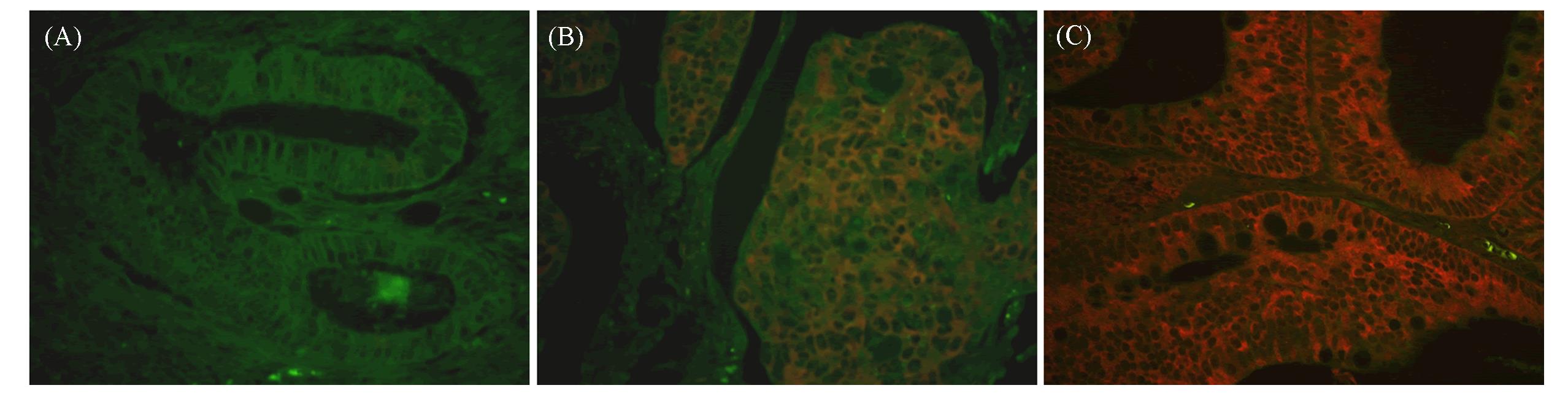
Fig.8 Representative immunofluorescence images of L4⁃QD probe staining in colorectal cancer tissues(A) Negative; (B) weak-moderate positive; (C) strong positive.
| Feature | L4⁃QD fluorescence intensity, n(%) | Spearman’s correlation | ||||
|---|---|---|---|---|---|---|
| Negative | Weak⁃moderate | Strong | r | P⁃value | ||
| 34(37.8%) | 34(37.8%) | 22(24.4%) | ||||
| Age | ≤70 | 22(39.29) | 19(33.93) | 15(26.78) | -0.02248 | 0.8353 |
| >70 | 10(31.25) | 15(46.88) | 7 (21.87) | |||
| Gender | Male | 26(44.8) | 21(36.2) | 11(19) | 0.2170 | 0.0400 |
| Female | 8(25) | 13(40.6) | 11(34.4) | |||
| Tumor size | ≤5 cm | 21(38.2) | 19(34.5) | 15(27.3) | -0.03909 | 0.7145 |
| >5 cm | 13(37.1) | 15(42.9) | 7(20) | |||
| AJCC clinical stage | I | 7(77.8) | 1(11.1) | 1(11.1) | 0.5675 | <0.0001 |
| II | 23(61.5) | 13(30.8) | 3(7.7) | |||
| III | 4(10.5) | 20(52.6) | 14(36.9) | |||
| IV | 0(0) | 0(0) | 4(100) | |||
Table 1 Correlation analysis of clinical and pathological characteristics of 90 patients with colorectal cancer and fluorescence intensity of L4-QD probe*
| Feature | L4⁃QD fluorescence intensity, n(%) | Spearman’s correlation | ||||
|---|---|---|---|---|---|---|
| Negative | Weak⁃moderate | Strong | r | P⁃value | ||
| 34(37.8%) | 34(37.8%) | 22(24.4%) | ||||
| Age | ≤70 | 22(39.29) | 19(33.93) | 15(26.78) | -0.02248 | 0.8353 |
| >70 | 10(31.25) | 15(46.88) | 7 (21.87) | |||
| Gender | Male | 26(44.8) | 21(36.2) | 11(19) | 0.2170 | 0.0400 |
| Female | 8(25) | 13(40.6) | 11(34.4) | |||
| Tumor size | ≤5 cm | 21(38.2) | 19(34.5) | 15(27.3) | -0.03909 | 0.7145 |
| >5 cm | 13(37.1) | 15(42.9) | 7(20) | |||
| AJCC clinical stage | I | 7(77.8) | 1(11.1) | 1(11.1) | 0.5675 | <0.0001 |
| II | 23(61.5) | 13(30.8) | 3(7.7) | |||
| III | 4(10.5) | 20(52.6) | 14(36.9) | |||
| IV | 0(0) | 0(0) | 4(100) | |||
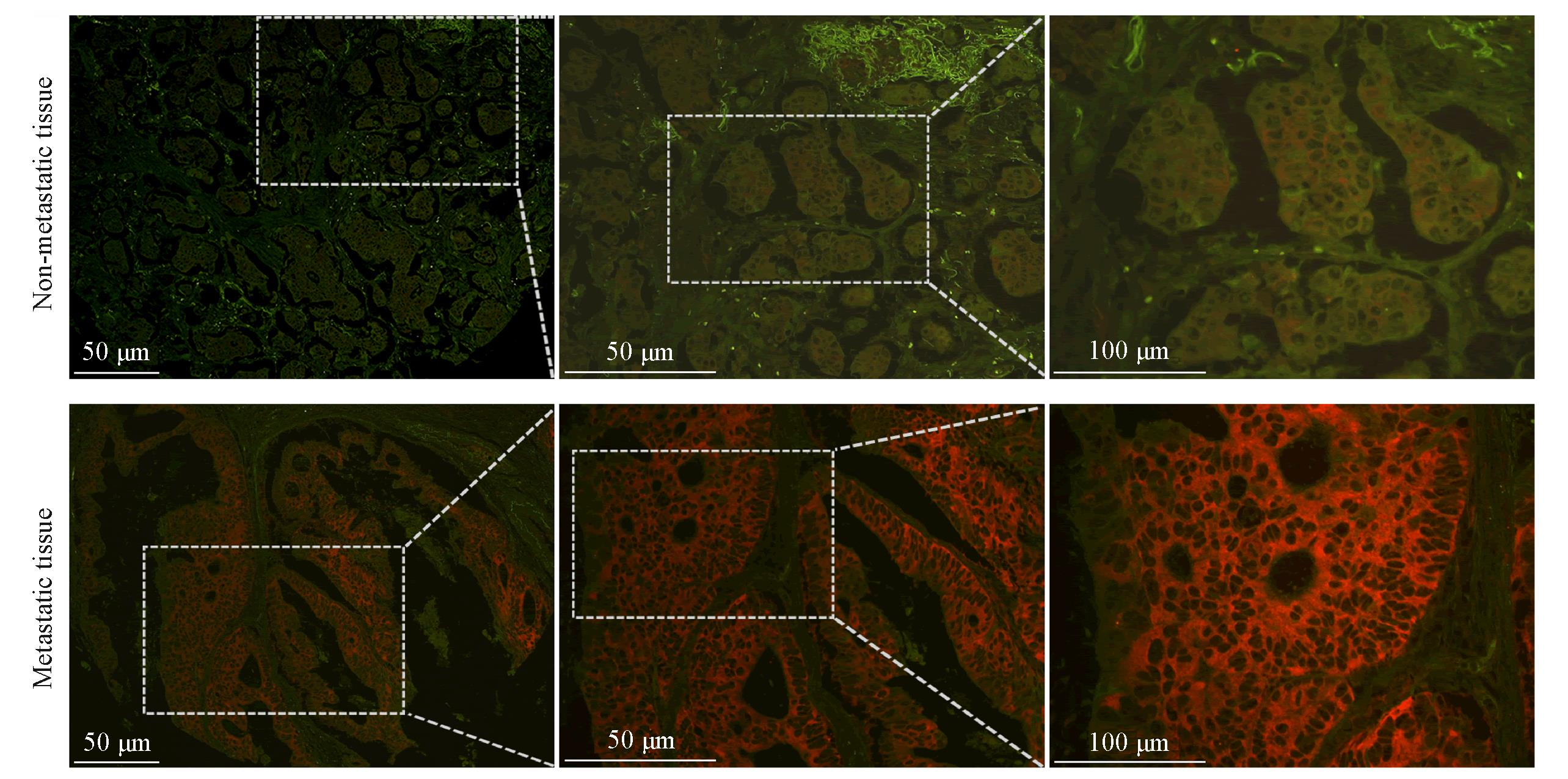
Fig.9 Representative immunofluorescence images of L4⁃QD probe staining in non⁃metastatic and metastatic colorectal cancer tissues at different magnifications
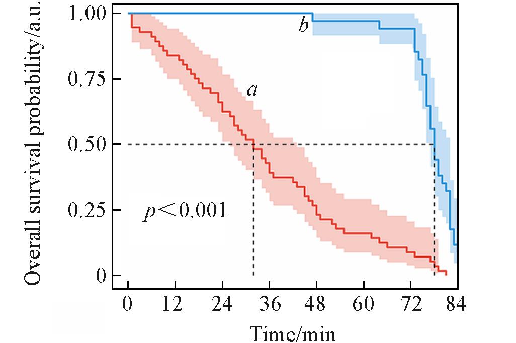
Fig.10 Overall survival curves of colorectal cancer patients with high(a) and low(b) aptamer L4 fluorescence intensities
| [1] | Cooray P. D., Cooper N. J., Med. Oncol., 2025, 42(4), 95 |
| [2] | Kiri S., Ryba T., Mol. Cancer, 2024, 23(1), 154 |
| [3] | Ciracì P., Studiale V., Taravella A., Antoniotti C., Cremolini C., Nat. Rev. Clin. Oncol., 2025, 22(1), 28—45 |
| [4] | André T., Shiu K. K., Kim T. W., Jensen B. V., Jensen L. H., Punt C. J. A., Smith D., Garcia⁃Carbonero R., Alcaide⁃Garcia J., Gibbs P., de la Fouchardiere C., Rivera F., Elez E., Le D. T., Yoshino T., Zuo Y., Fogelman D., Adelberg D., Diaz L. A., Ann. Oncol., 2025, 36(3), 277—284 |
| [5] | Strickler J. H., Bekaii⁃Saab T., Cercek A., Heinemann V., Nakamura Y., Raghav K., Siena S., Tabernero J., van Cutsem E., Yoshino T., Ramos J., Guan X. S., Andre T., Future Oncol., 2025, 21(3), 303—311 |
| [6] | Yaqub S., Longva A., Angelsen J. H., Brudvik K. W., Carling U., Edwin B., Fretland Å. A., Grünhagen D. J., Line P. D., Røsok B. I., Smedman T. M., Sparrelid E., Syversveen T., Villanger O., Lassen K., Scand. J. Surg., 2025, 114(2), 126—134 |
| [7] | Zhai F. H., Yun B. F., Ming J., Yu T. Y., Li B. H., Liu X., Wang X. S., Chen Z. H., Song C. F., Zhao M. Y., Li W. L., Liu Z. B., Liang A. B., Li J. Y., Zhang F., Adv. Mater., 2025, 37(18), 2501378 |
| [8] | Tonini V., Zanni M., World J. Gastroenterol., 2024, 30(3), 211—224 |
| [9] | Zygulska A. L., Pierzchalski P., Int. J. Mol. Sci., 2022, 23(2), 852 |
| [10] | Sur D. K. C., Brown P. C., Am. Fam. Physician, 2025, 112(3), 278—283 |
| [11] | Dong Q., Li Z. Q., Peng T. H., Chen Z., Tan W. H., Chem. J. Chinese Universities, 2020, 41(12), 2648—2657 |
| 董倩, 李兆倩, 彭天欢, 陈卓, 谭蔚泓. 高等学校化学学报, 2020, 41(12), 2648—2657 | |
| [12] | Jin Z., Li Y., Yi H., Wang M. H., Wang C. F., Du S. K., Zeng W. J., Zong Z., Int. J. Oncol., 2025, 66(3), 22 |
| [13] | Zhang Y., Wang Y., Zhang B. Q., Li P. F., Zhao Y., Biomed. Pharmacother., 2023, 163, 114786 |
| [14] | Wu Z. W., Li Y., Zhang Y. B., Hu H., Wu T. W., Liu S. Y., Chen W. Q., Xie S. G., Lu Z. X., Technol. Cancer Res. Treat., 2020, 19, 1533033820980426 |
| [15] | Liu S. C., Zhang H., World J. Gastroenterol., 2024, 30(33), 3818—3822 |
| [16] | Li W. M., Fang J., Chem. J. Chinese Universities, 2016, 37(7), 1262—1268 |
| 李婉明, 方瑾. 高等学校化学学报, 2016, 37(7), 1262—1268 | |
| [17] | Tang J. L., Shi H., He X. X., Wang K. M., Li D., Yan L. A., Lei Y. L., Liu J. B., Chem. J. Chinese Universities, 2014, 35(10), 2093—2100 |
| 汤进录, 石慧, 何晓晓, 王柯敏, 李朵, 颜律安, 雷艳丽, 刘剑波. 高等学校化学学报, 2014, 35(10), 2093—2100 | |
| [18] | Lin N. Q., Wu L., Xu X., Wu Q. Y., Wang Y. Z., Shen H. C., Song Y. L., Wang H. Y., Zhu Z., Kang D. Z., Yang C. Y., ACS Appl. Mater. Interfaces, 2021, 13(8), 9306—9315 |
| [19] | Loskutova A., Seitkali A., Aliyev D., Bukasov R., Int. J. Mol. Sci., 2025, 26(14), 6674 |
| [20] | Liang H. X., Wang M., Zhang Y. C., Bai L., Li X. M., Yu F. B., Cheng Z. Y., Zhao L. L., Chem. J. Chinese Universities, 2025, 46(1), 20240154 |
| 梁惠闲, 王淼, 张雨璨, 白丽, 李雪妹, 于法标, 程子译, 赵琳璐. 高等学校化学学报, 2025, 46(1), 20240154 | |
| [21] | Bao Z. H., Lu L. X., Wang J. Y., Zhu H. T., Dang X. L., Chen Y. C., Yin X. Z., Song Y. H., Wen X. J., J. Fluoresc., 2025, 36, 785—816 |
| [22] | Stephens C., Goodey N. M., Gubler U., Anal. Biochem., 2025, 703, 115890 |
| [23] | Feng L. J., Sun Y., Jia W. S., Yu Y., Liu C., Yang J., Luan Y. X., Chen J., Wang F. C., Biomolecules, 2025, 15(6), 818 |
| [24] | Chen C., Zhou S., Cai Y. Q., Tang F. Q., NPJ Precis. Oncol., 2017, 1, 37 |
| [25] | Li W. M., Bing T., Wang Q., Li Y. X., Sun S. H., Li X. Y., Ma Y. N., Wang R., Ba W., Li X. Y., Shangguan D. H., Fang J., Bioorg. Chem., 2025, 165, 108989 |
| [26] | Zhu G. Z., Chen X. Y., Adv. Drug Deliv. Rev., 2018, 134, 65—78 |
| [27] | Ji C. L., Wei J. Y., Zhang L., Hou X. R., Tan J., Yuan Q., Tan W. H., Chem. Rev., 2023, 123(22), 12471—12506 |
| [28] | Ochoa S., Milam V. T., ACS Synth. Biol., 2025, 14(8), 3049—3064 |
| [29] | Ishizu K., Sunose N., Yamazaki K., Tsuruo T., Sadahiro S., Makuuchi H., Yamori T., Biol. Pharm. Bull., 2007, 30(9), 1779—1783 |
| [30] | Yang Z., Wu D. J., Chen Y. S., Min Z. J., Quan Y. J., Cancer Biol. Ther., 2019, 20(9), 1195—1205 |
| [31] | Vincent Z., Urakami K., Maruyama K., Yamaguchi K., Kusuhara M., Genes Cancer, 2014, 5(7⁃8), 250—60 |
| [32] | Ji D. Y., Feng H. X., Liew S. W., Kwok C. K., Trends Biotechnol., 2023, 41(11), 1360—1384 |
| [33] | Kordasht H. K., Hasanzadeh M., Talanta, 2020, 220, 121436 |
| [34] | Chen S. Y., Cui J. L., Chen H. Y., Yu B., Long S. H., Eur. J. Med. Chem., 2023, 262, 115911 |
| [1] | 陈卓然, 龚玲玉, 艾立丽, 张鹏, 周艳华. 基于高内吞效率的核酸适体构建LYTACs嵌合体以增强白血病细胞膜蛋白的降解[J]. 高等学校化学学报, 2026, 47(7): 20260017. |
| [2] | 梁惠闲, 王淼, 张雨璨, 白丽, 李雪妹, 于法标, 程子译, 赵琳璐. 基于Ag2S量子点的光热治疗协同药物治疗在动脉粥样硬化中的应用[J]. 高等学校化学学报, 2025, 46(1): 20240154. |
| [3] | 张丽媛, 王查斯娜, 胡井香, 詹传郎. 离子掺杂调控双钙钛矿量子点发光性能的研究进展[J]. 高等学校化学学报, 2024, 45(9): 20240126. |
| [4] | 张慧莲, 杨新杰, 李军, 李泉, 张福娟, 张艳丽, 王红斌, 杨文荣, 庞鹏飞. 基于N掺杂Ti3C2 MXene量子点的荧光探针用于Hg2+和S2-的传感检测[J]. 高等学校化学学报, 2024, 45(5): 20230504. |
| [5] | 王瑞娜, 孙瑞粉, 钟添华, 池毓务. 大尺寸石墨烯量子点组装体的制备及电化学发光性能[J]. 高等学校化学学报, 2022, 43(8): 20220161. |
| [6] | 夏雾, 任颖异, 刘京, 王锋. 壳聚糖包裹CdSe量子点组装体的水相可见光催化CO2还原[J]. 高等学校化学学报, 2022, 43(7): 20220192. |
| [7] | 黄珊, 姚建东, 宁淦, 肖琦, 刘义. 石墨烯量子点荧光探针对碱性磷酸酶活性的高效检测[J]. 高等学校化学学报, 2021, 42(8): 2412. |
| [8] | 周洁琼, 黄艳, 张志凌, 庞代文, 田智全. 水溶性近红外Ⅱ区荧光Ag2Te量子点的合成[J]. 高等学校化学学报, 2021, 42(6): 2072. |
| [9] | 舒馨, 王迪, 袁春玲, 覃秀, 王益林. 基于碳量子点过氧化物模拟酶的肌氨酸比色测定[J]. 高等学校化学学报, 2021, 42(6): 1761. |
| [10] | 刘珂, 靳宇, 梁建功, 吴园. 化学修饰提高核酸适体结合亲和力的研究进展[J]. 高等学校化学学报, 2021, 42(11): 3477. |
| [11] | 解忱, 陈娜, 杨雁冰, 袁荃. 核酸适体功能化的二维材料场效应晶体管传感器研究进展[J]. 高等学校化学学报, 2021, 42(11): 3406. |
| [12] | 赵卓, 王雪强. 核酸适体偶联药物的生物偶联构建技术与应用[J]. 高等学校化学学报, 2021, 42(11): 3367. |
| [13] | 刘学娇, 杨帆, 刘爽, 张春娟, 刘巧玲. 核酸适体靶向的膜蛋白识别与功能调控研究进展[J]. 高等学校化学学报, 2021, 42(11): 3277. |
| [14] | 张晓荣, 陈岚岚, 胡善文. 基于分子识别的细菌检测研究进展[J]. 高等学校化学学报, 2021, 42(11): 3468. |
| [15] | 吉采灵, 程兴, 谈洁, 袁荃. 功能化核酸适体的筛选及分子识别应用[J]. 高等学校化学学报, 2021, 42(11): 3457. |
| 阅读次数 | ||||||
|
全文 |
|
|||||
|
摘要 |
|
|||||
