

高等学校化学学报 ›› 2026, Vol. 47 ›› Issue (9): 20260103.doi: 10.7503/cjcu20260103
刘雨庆1,2,3, 程杰1,2,3, 王越4, 焦凯3, 诸颖3, 李江3, 郭琳洁3( )
)
收稿日期:2026-03-13
出版日期:2026-09-10
发布日期:2026-04-22
通讯作者:
郭琳洁
E-mail:guolinjie@shu.edu.cn
基金资助:
LIU Yuqing1,2,3, CHENG Jie1,2,3, WANG Yue4, JIAO Kai3, ZHU Ying3, LI Jiang3, GUO Linjie3()
Received:2026-03-13
Online:2026-09-10
Published:2026-04-22
Contact:
GUO Linjie
E-mail:guolinjie@shu.edu.cn
Supported by:摘要:
一维DNA纳米材料在诸多领域具有广泛的应用前景, 但其自组装效率的定量表征仍面临挑战. 本文以六螺旋束(six-helix bundle, 6HB) DNA纤维为模型体系, 基于已有的图像分析方法, 针对高长径比一维纤维易交叉缠绕、 难以进行单颗粒识别与计数的问题进行整合与参数化处理, 建立了一套基于原子力显微镜(AFM)图像分析的自组装效率可视化测量方法. 该方法以面积覆盖率和轮廓长度密度为量化指标, 实现了对一维DNA纤维网络结构的定量表征. 利用该方法考察了缓冲液pH(5~11)和Mg2+浓度(6.25~87.5 mmol/L)对6HB DNA纤维组装行为的影响. 结果表明, 6HB DNA纤维的有效组装存在明确的条件窗口: pH在6~8范围内有利于高产率形成长纤维, 其中pH=8时自组装效率达到峰值(面积覆盖率约36%, 轮廓长度密度约8 μm/μm²). 当pH偏离该范围时, 连续的一维网络逐渐转变为碎片化或无序聚集结构, 两项参数接近背景水平. 同时, 纤维自组装对Mg2+浓度表现出明显的阈值响应特征, 浓度高于25 mmol/L时进入快速增长区, 在50 mmol/L以上时形成稳定的长纤维产物(面积覆盖率> 20%, 轮廓长度密度> 4 μm/μm²). 这些测量结果通过可视化和定量分析, 验证了DNA纳米结构自组装对pH值和离子强度等环境条件的高度依赖性. 该研究框架有望为一维DNA纳米材料的设计、 制备与应用提供有力的定量分析工具.
中图分类号:
TrendMD:
刘雨庆, 程杰, 王越, 焦凯, 诸颖, 李江, 郭琳洁. 一维DNA纳米材料自组装效率的可视化测量. 高等学校化学学报, 2026, 47(9): 20260103.
LIU Yuqing, CHENG Jie, WANG Yue, JIAO Kai, ZHU Ying, LI Jiang, GUO Linjie. Visualized Measurement of Self-assembly Efficiency in One-dimensional DNA Nanomaterials. Chem. J. Chinese Universities, 2026, 47(9): 20260103.
| Name | Sequence(5′⁃3′) |
|---|---|
| X⁃1 | TACTGACCTCGTGCGTTCGTACAAGAACCATTGACCTGCGCG |
| X⁃2 | CGAACCTGATTAGAGACCCTGATGGCGGTGGAGACACTCACG |
| X⁃3 | CTCGCGCTTCCCATTGGTCTCCTTCTATTGCTAGACCGGAGT |
| X⁃4 | AGAAAGCCATACGCGCCTTCTTGCGGAACTGGTAGCCATGAG |
| X⁃5 | GATTAGGTGGGCCGAAATAGACAACCTACGAACTAGCACCCA |
| X⁃6 | TAATCGACGCTTTACGACAAGGCCGATTGATCACGAGTTCCG |
| Y⁃1 | GCAATAGAAGCGGAACTCGTGTATGGCTTTCTTACGAACGCA |
| Y⁃2 | ATGGTTCTTGCTCATGGCTACCCCACCTAATCCAGGGTCTCT |
| Y⁃3 | CCACCGCCATTGGGTGCTAGTAGCGTCGATTAGAGACCAATG |
| Y⁃4 | ATCAATCGGCACTCCGGTCTACGAGGTCAGTAAGAAGGCGCG |
| Y⁃5 | CAGTTCCGCACGCGCAGGTCAAATCAGGTTCGTCTATTTCGG |
| Y⁃6 | TCGTAGGTTGCGTGAGTGTCTGGAAGCGCGAGCTTGTCGTAA |
Table 1 DNA sequences used for the assembly of 6HB DNA fibers
| Name | Sequence(5′⁃3′) |
|---|---|
| X⁃1 | TACTGACCTCGTGCGTTCGTACAAGAACCATTGACCTGCGCG |
| X⁃2 | CGAACCTGATTAGAGACCCTGATGGCGGTGGAGACACTCACG |
| X⁃3 | CTCGCGCTTCCCATTGGTCTCCTTCTATTGCTAGACCGGAGT |
| X⁃4 | AGAAAGCCATACGCGCCTTCTTGCGGAACTGGTAGCCATGAG |
| X⁃5 | GATTAGGTGGGCCGAAATAGACAACCTACGAACTAGCACCCA |
| X⁃6 | TAATCGACGCTTTACGACAAGGCCGATTGATCACGAGTTCCG |
| Y⁃1 | GCAATAGAAGCGGAACTCGTGTATGGCTTTCTTACGAACGCA |
| Y⁃2 | ATGGTTCTTGCTCATGGCTACCCCACCTAATCCAGGGTCTCT |
| Y⁃3 | CCACCGCCATTGGGTGCTAGTAGCGTCGATTAGAGACCAATG |
| Y⁃4 | ATCAATCGGCACTCCGGTCTACGAGGTCAGTAAGAAGGCGCG |
| Y⁃5 | CAGTTCCGCACGCGCAGGTCAAATCAGGTTCGTCTATTTCGG |
| Y⁃6 | TCGTAGGTTGCGTGAGTGTCTGGAAGCGCGAGCTTGTCGTAA |

Fig.1 Workflow of AFM image processing used in the fiber network quantification method(A) Raw AFM topography image; (B) image after background flattening and line correction; (C) high-contrast binarized image obtained by threshold segmentation for area coverage ratio calculation; (D) single-pixel-width fiber skeleton extracted via skeletonization for contour length density calculation.
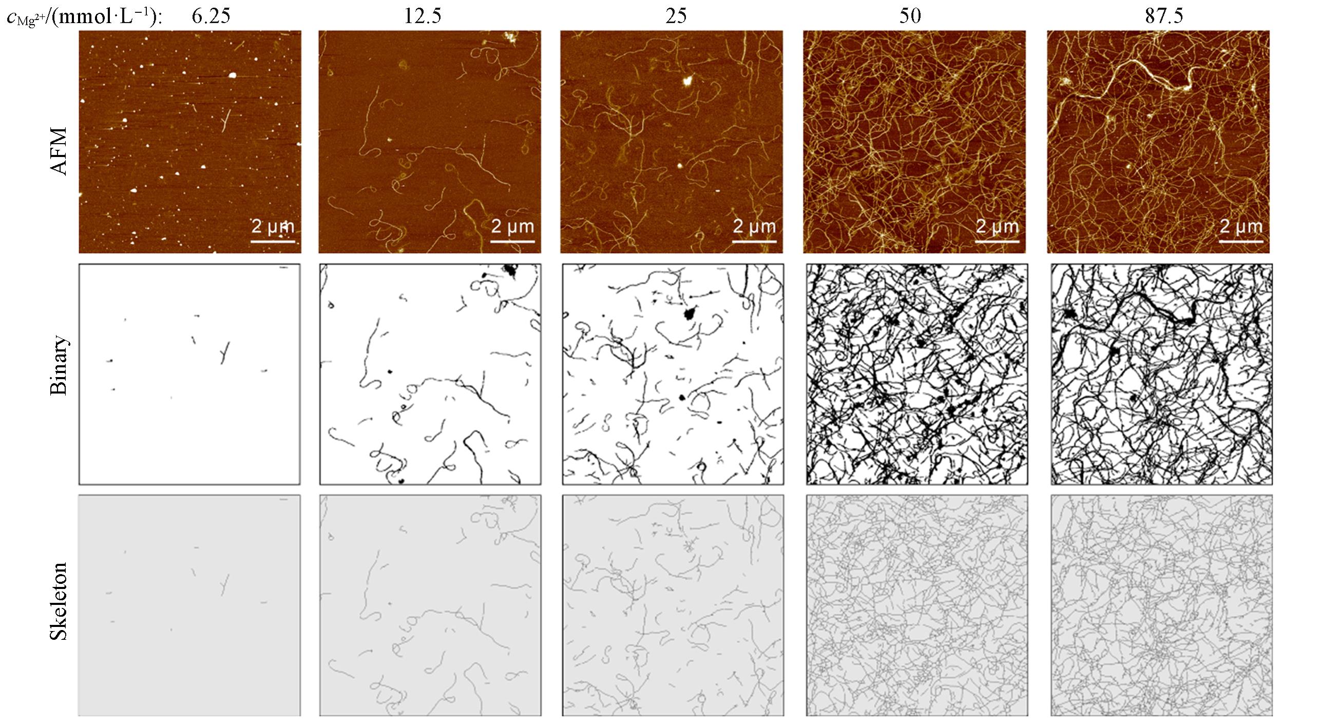
Fig.2 AFM images of 6HB DNA fibers assembled with different Mg2+ concentrationsTop row: raw AFM images; middle row: binarized images; bottom row: skeletonized images.
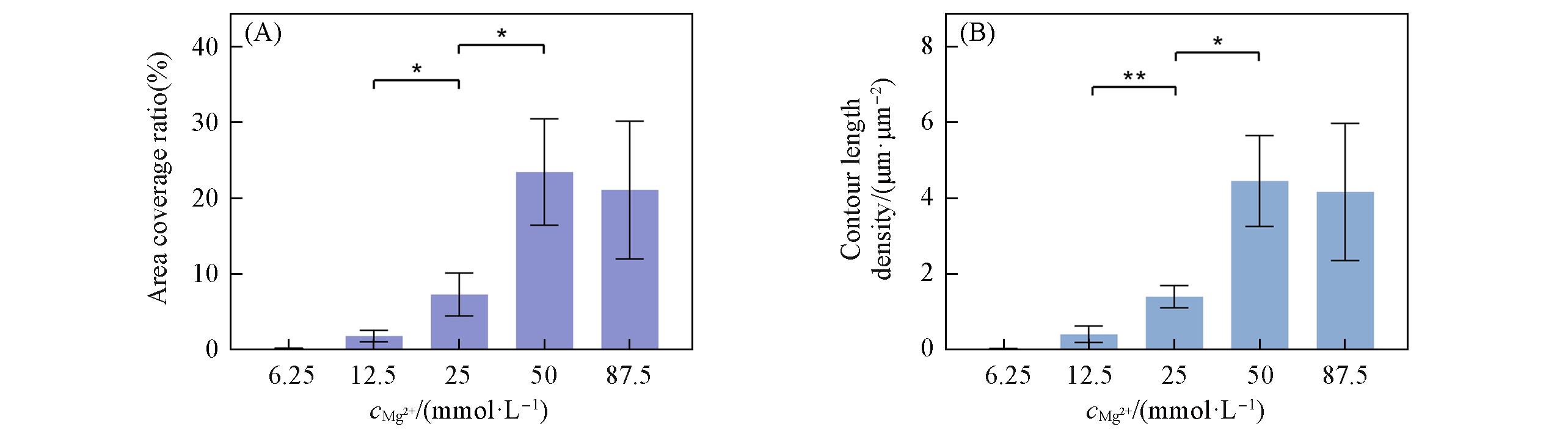
Fig.3 Quantitative analysis of 6HB DNA fibers assembled with different Mg2+ concentrations(A) Area coverage ratio(%); (B) contour length density(μm/μm²). Data are presented as mean±SD(n=3, independent samples). Statistical analysis was performed using one-way ANOVA(*P < 0.05, **P < 0.01).
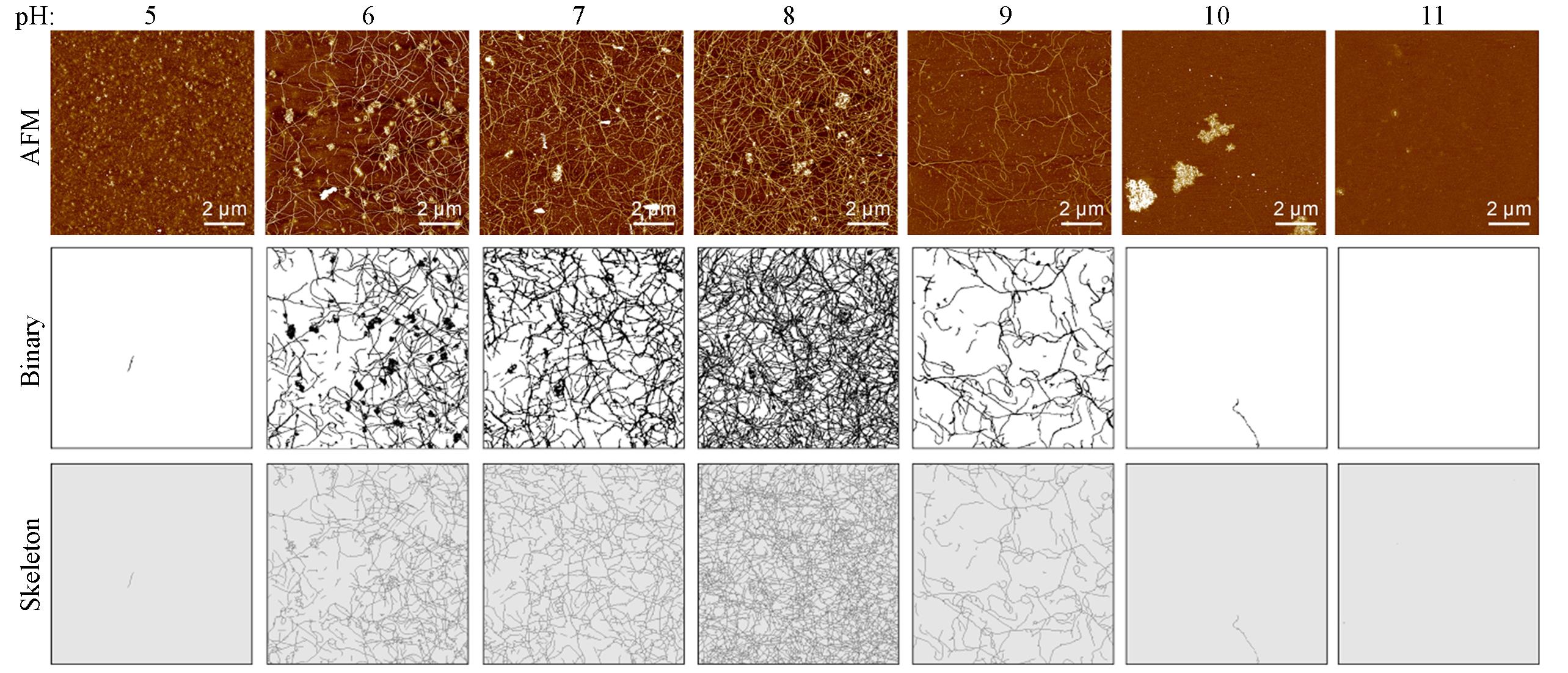
Fig.4 AFM images of 6HB DNA fibers assembled with different pH valuesTop row: raw AFM topography; middle row: binarized images; bottom row: skeletonized images.
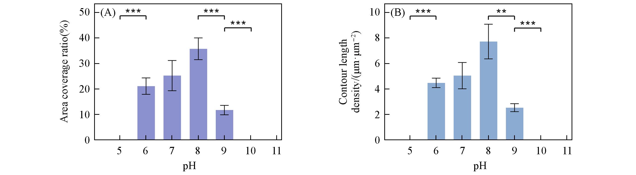
Fig.5 Quantitative analysis of 6HB DNA fibers assembled with different pH values(A) Area coverage ratio(%); (B) Contour length density(μm·μm⁻²). Data are presented as mean ± SD(n=3, independent samples). Statistical analysis was performed using one-way ANOVA(*P < 0.05, **P < 0.01, ***P < 0.001).
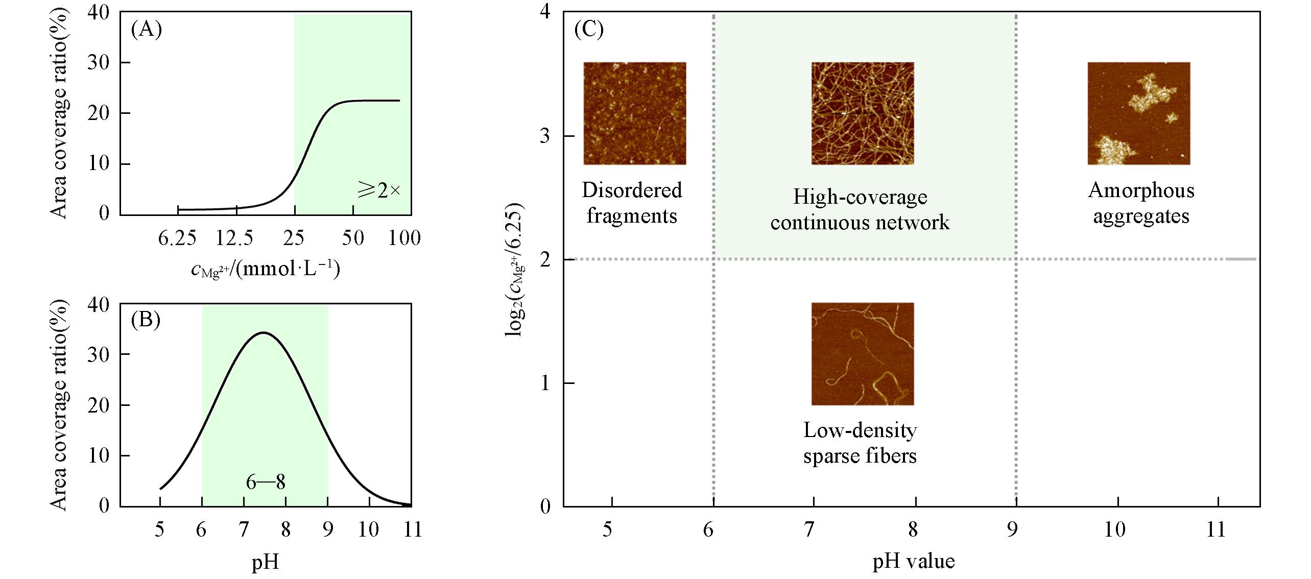
Fig.6 Quantitative response analysis and structural state mapping of 6HB DNA fibers assembly in the pH⁃Mg2+ parameter space(A) Dependence of area coverage ratio on Mg2+ concentration at constant pH. The data were empirically fitted with a sigmoidal function; (B) Dependence of area coverage ratio on pH at constant Mg2+ concentration. The data were empirically fitted with a Gaussian function; (C) Structural state diagram of 6HB DNA fibers in the pH-Mg²⁺ space. The Mg²⁺ concentrations on the Y-axis are expressed as log2- transformed values relative to the lowest tested concentration(6.25 mmol·L-1). Representative AFM images show distinct morphologies. Dashed lines indicate boundaries between different structural states. Shaded regions denote the effective assembly windows.
| [1] | Seeman N. C., Sleiman H. F., Nat. Rev. Mater., 2017, 3(1), 17068 |
| [2] | Rothemund P. W. K., Nature, 2006, 440(7082), 297—302 |
| [3] | Keren K., Berman R. S., Buchstab E., Sivan U., Braun E., Science, 2003, 302(5649), 1380—1382 |
| [4] | Fu J., Yang Y. R., Johnson⁃Buck A., Liu M., Liu Y., Walter N. G., Woodbury N. W., Yan H., Nat. Nanotechnol., 2014, 9(7), 531—536 |
| [5] | Um S. H., Lee J. B., Park N., Kwon S. Y., Umbach C. C., Luo D., Nat. Mater., 2006, 5(10), 797—801 |
| [6] | Elblová P., Anthi J., Liu M., Lunova M., Jirsa M., Stephanopoulos N., Lunov O., JACS Au, 2025, 5(4), 1591—1616 |
| [7] | Fang X., Zheng J., Gao W., Liu Z., Li X., Li C., Gao T., Zhang R., Coord. Chem. Rev., 2026, 552, 217525 |
| [8] | Green L. N., Subramanian H. K. K., Mardanlou V., Kim J., Hariadi R. F., Franco E., Nat. Chem., 2019, 11(6), 510—520 |
| [9] | Ong L. L., Hanikel N., Yaghi O. K., Grun C., Strauss M. T., Bron P., Lai⁃Kee⁃Him J., Schueder F., Wang B., Wang P., Kishi J. Y., Myhrvold C., Zhu A., Jungmann R., Bellot G., Ke Y., Yin P., Nature, 2017, 552(7683), 72—77 |
| [10] | Jones M. R., Seeman N. C., Mirkin C. A., Science, 2015, 347(6224), 1260901 |
| [11] | Hong F., Zhang F., Liu Y., Yan H., Chem. Rev., 2017, 117(20), 12584—12640 |
| [12] | Castro C. E., Kilchherr F., Kim D. N., Shiao E. L., Wauer T., Wortmann P., Bathe M., Dietz H., Nat. Methods, 2011, 8(3), 221—229 |
| [13] | Zhang F., Jiang S., Wu S., Li Y., Mao C., Liu Y., Yan H., Nat. Nanotechnol., 2015, 10(9), 779—784 |
| [14] | Hansma H. G., Hoh J. H., Annu. Rev. Biophys. Biomol. Struct., 1994, 23, 115—140 |
| [15] | Saleem S. S., Ravichandran S., Ravichandran S., Chinnadurai K. R., Saravanan A., Vickram S., ACS Appl. Bio Mater., 2025, 8(10), 8467—8512 |
| [16] | Högberg B., Liedl T., Shih W. M., J. Am. Chem. Soc., 2009, 131(26), 9154—9155 |
| [17] | Fang W., Xie M., Hou X., Liu X., Zuo X., Chao J., Wang L., Fan C., Liu H., Wang L., J. Am. Chem. Soc., 2020, 142(19), 8782—8789 |
| [18] | Zhang S., Zhang H., Li M., Song H., Sun J., Fan C., Xie Y., Wang H., Ge Z., Nano Lett., 2024, 24(42), 13438—13446 |
| [19] | Wang J., Wei Y., Zhang P., Wang Y., Xia Q., Liu X., Luo S., Shi J., Hu J., Fan C., Li B., Wang L., Zhou X., Li J., Nano Lett., 2022, 22(17), 7173—7179 |
| [20] | Yin P., Hariadi R. F., Sahu S., Choi H. M. T., Park S. H., Labean T. H., Reif J. H., Science, 2008, 321(5890), 824—826 |
| [21] | Stein A. M., Vader D. A., Jawerth L. M., Weitz D. A., Sander L. M., J. Microscopy, 2008, 232(3), 463—475 |
| [24] | Engel A., Müller D. J., Nat. Struct. Biol., 2000, 7(9), 715—718 |
| [25] | Hansma H. G., Laney D. E., Biophys. J., 1996, 70(4), 1933—1939 |
| [26] | Schindelin J., Arganda⁃Carreras I., Frise E., Kaynig V., Longair M., Pietzsch T., Preibisch S., Rueden C., Saalfeld S., Schmid B., Tinevez J. Y., White D. J., Hartenstein V., Eliceiri K., Tomancak P., Cardona A., Nat. Methods, 2012, 9(7), 676—682 |
| [27] | Sezgin M., Sankur B., J. Electron. Imaging, 2004, 13, 146—168 |
| [28] | Otsu N., IEEE Trans. Syst. Man Cybern., 1979, 9(1), 62—66 |
| [29] | Saha P. K., Borgefors G., Sanniti Di Baja G., Pattern Recognition Letters, 2016, 76, 3—12 |
| [30] | Zhang T. Y., Suen C. Y., Commun. ACM, 1984, 27(3), 236–239 |
| [31] | Woodson S. A., Curr. Opin. Chem. Biol., 2005, 9(2), 104—109 |
| [32] | Bloomfield V. A., Biopolymers, 1997, 44(3), 269—282 |
| [33] | Ke Y., Ong L. L., Shih W. M., Yin P., Science, 2012, 338(6111), 1177—1183 |
| [34] | Tao Q., Chen Q., Bian X. J., Liu G., Yan J., Chin. J. Anal. Chem., 2021, 49(5), 743—751 |
| 陶晴, 陈谦, 卞晓军, 刘刚, 颜娟. 分析化学, 2021, 49(5), 743—751 | |
| [35] | Ramakrishnan S., Ijäs H., Linko V., Keller A., Comput. Struct. Biotechnol. J., 2018, 16, 342—349 |
| [36] | Fang W. N., Fan C. H., Liu H. J., Acta Polym. Sin., 2017, (12), 1993—2000 |
| 方维娜, 樊春海, 柳华杰. 高分子学报, 2017, (12), 1993—2000 |
| [1] | 刘少芳, 谢奎. 宏观尺寸单晶多孔材料[J]. 高等学校化学学报, 2026, 47(9): 20260187. |
| [2] | 翟晓璐, 吕云, 李会, 黄科科, 王艳红, 卢红成. 甲酸-草酸共配体低维磁性镱配合物的构筑及磁性[J]. 高等学校化学学报, 2026, 47(9): 20260193. |
| [3] | 燕霞, 赵嘉仪, 文浩, 齐延歌, 刘博洋, 杜江锋, 宋建波. 氧化铪增强兔原位肝癌放疗疗效并减轻肺部转移负荷[J]. 高等学校化学学报, 2026, 47(8): 20250391. |
| [4] | 宋志国, 王鑫, 朱志慧, 徐一铭, 王敏. 含萘二磺酸类配体单核Cd(II)配合物传感器对Fe3+的高选择性检测[J]. 高等学校化学学报, 2026, 47(8): 20260062. |
| [5] | 林明穗, 王启龙, 梁晴晴, 陈乔, 赖惠文, 陈雪莹, 郭新颖, 马战文, 赖安群, 潘中华, 肖旺钏. 过渡金属多核配合物Cu4的合成及电催化水氧化性能[J]. 高等学校化学学报, 2026, 47(8): 20250361. |
| [6] | 师凯, 刘子荐, 张潇, 赵立彦, 姚同杰. 基于偶氮苯衍生物构筑的Y-MOF的合成、 表征及对硝基化合物的荧光检测性能[J]. 高等学校化学学报, 2026, 47(7): 20250365. |
| [7] | 周海刚, 程孟, 张月, 魏徐兵, 李晓伟, 胡松青. 面向污染物识别的荧光噻吩基MOFs的构筑与性能[J]. 高等学校化学学报, 2026, 47(6): 20250274. |
| [8] | 魏颖真, 冯婧怡, 王若瑜, 宋海涛, 闫文付. 同轴3D打印核壳结构复合催化剂研究进展[J]. 高等学校化学学报, 2026, 47(6): 20260082. |
| [9] | 张应鹏 刘娟娟 秦凡凡 杨云裳 胡赜太. 铁/锰比例调控的双金属纳米酶用于有机磷农药比色传感[J]. 高等学校化学学报, 0, (): 20260095. |
| [10] | 李宜霏 线尚兰 蒋洲洋 唐怀军 夏溶蔓 王威贤 吴一冰 方秀英 李香 姜艾晶. 一种阳离子型铱(III)配合物与硅胶复合纳米发光材料的制备及其在潜指纹检测中的应用[J]. 高等学校化学学报, 0, (): 20260137. |
| [11] | 张凯为, 秦安军. 聚集诱导发光镧系配合物[J]. 高等学校化学学报, 2026, 47(5): 20260013. |
| [12] | 韩雨宸, 张蒙, 杨琪, 彭秋晨, 李恺. 一种金铜簇配位聚合物的制备与力致变色性能[J]. 高等学校化学学报, 2026, 47(4): 20260007. |
| [13] | 张捷, 霍志鹏, 钟国强. 亚微米球Eu2O3/B4C/HDPE复合材料的制备及中子/伽马辐射屏蔽性能[J]. 高等学校化学学报, 2026, 47(3): 20250298. |
| [14] | 黄馨怡, 阙茂梅, 李瑶, 雷珊, 林思琪, 王力. Dawson型多金属氧酸盐-VC复合物对 α-葡萄糖苷酶的抑制作用及机制研究[J]. 高等学校化学学报, 2026, 47(3): 20250236. |
| [15] | 程文婷, 李智婷, 翟颖. 三水合碳酸镁在限域旋混反应器中的结晶优化[J]. 高等学校化学学报, 2026, 47(3): 20250217. |
| 阅读次数 | ||||||
|
全文 |
|
|||||
|
摘要 |
|
|||||
