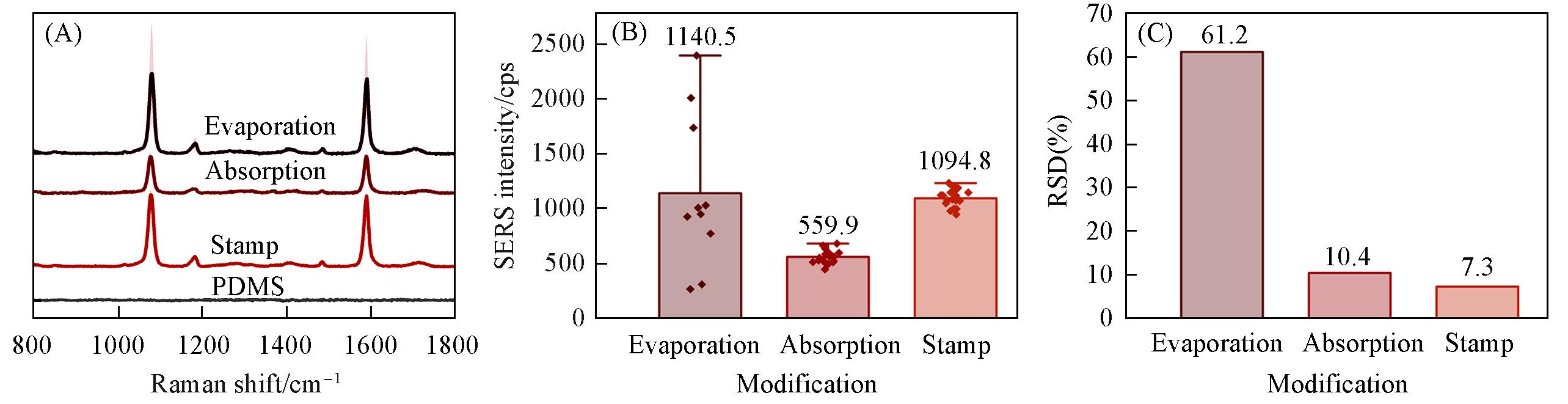
| 1 |
Ma H., Pan S. Q., Wang W. L., Yue X., Xi X. H., Yan S., Wu D. Y., Wang X., Liu G., Ren B., ACS Nano, 2024, 18(22), 14000—14019
|
| 2 |
Itoh T., Prochazka M., Dong Z. C., Ji W., Yamamoto Y. S., Zhang Y., Ozaki Y., Chem. Rev., 2023, 123(4), 1552—1634
|
| 3 |
Liebel M., Pazos⁃Perez N., van Hulst N. F., Alvarez⁃Puebla R. A., Nat. Nanotechnol., 2020, 15(12), 1005—1011
|
| 4 |
He X., Wei W., Duan X., Nanotechnol. Precis. Eng., 2023, 6(4), 045002
|
| 5 |
Chen S. Y., Zhang X. Y., Liu B., Tian D., Li Q., Chen F., Hu C. L., Chen J., Chem. J. Chinese Universities, 2021, 42(8), 2381—2392
|
|
陈韶云, 张行颖, 刘奔, 田杜, 李奇, 陈芳, 胡成龙, 陈建. 高等学校化学学报, 2021, 42(8), 2381—2392
|
| 6 |
Haldavnekar R., Vijayakumar S. C., Venkatakrishnan K., Tan B., ACS Nano, 2020, 14(11), 15468—15491
|
| 7 |
Xia D. C., Zhou R., Tu B., Cai Z. W., Gao N., Ji X. X., Chang G., Ren X. M., He Y. B., Chem. J. Chinese Universities, 2022, 43(3), 67—77
|
|
夏大成, 周瑞, 涂博, 蔡志伟, 高难, 姬晓旭, 常钢, 任小明, 何云斌. 高等学校化学学报, 2022, 43(3), 67—77
|
| 8 |
Deng K., Luo Z., Tan L., Quan Z., Chem. Soc. Rev., 2020, 49(16), 6002—6038
|
| 9 |
Wei W. B., Bai F., Fan H. Y., Angew. Chem. Int. Ed., 2019, 58(35), 11956—11966
|
| 10 |
Xu L., Li X. Y., Wang X., Zou Z. M., Nanotechnology, 2021, 32(13), 135601
|
| 11 |
Chen X. Y., Cui A. R., He M. Y., Yan M., Zhang X. C., Ruan J., Yang S. K., Nano Lett., 2023, 23(14), 6736—6743
|
| 12 |
Song L., Xu B. B., Cheng Q., Wang X., Luo X., Chen X., Chen T., Huang Y., Sci. Adv., 2021, 7(52), eabk2852
|
| 13 |
Liu Y., Kim M., Cho S. H., Jung Y. S., Nano Today, 2021, 37, 101063
|
| 14 |
Kim M., Panagiotakopoulou M., Chen C., Ruiz S. B., Ganesh K., Tammela T., Heller D. A., Nature Rev. Cancer, 2023, 23(9), 581—599
|
| 15 |
Cheng Y., Pang S. W., Microsystems & Nanoengineering, 2023, 9(1), 6
|
| 16 |
Zhang R. Y., Duan X. X., Zhang S. H., Guo W. L., Sun C., Han Z. Y., Nanotechnol. Precis. Eng., 2023, 6(2), 023003
|
| 17 |
Kang S., Wu X., Qi H., Yang K., Feng R., Guo W., Sun C., Duan X., Wang Y., Small Structures, 2024, 5(5), 2300432
|
| 18 |
Nemec S., Kilian K. A., Nat. Rev. Mater., 2021, 6(1), 69—83
|
| 19 |
Yang L. T., Conley B. M., Rathnam C., Cho H. Y., Pongkulapa T., Conklin B., Lee K. B., ACS Nano, 2022, 16(4), 5577—5586
|
| 20 |
He S., Pang W., Wu X. Y., Yang Y., Li W. J., Qi H., Sun C. L., Duan X. X., Wang Y. Y., Nanoscale, 2022, 14(41), 15281—15290
|
| 21 |
Zhu F., Liu Z., Wu X., Xu D., Li Q., Chen X., Pang W., Duan X., Wang Y., Ultrasonics Sonochemistry, 2023, 100, 106618
|
| 22 |
Guo Q., Wu X., Duan X., He S., Pang W., Wang Y., J. Colloid Interface Sci., 2021, 586, 67—74
|
| 23 |
Yang K., Kang S., Wu X., Liu B., Sun C., Wang Y., Appl. Phys. Lett., 2023, 123(14), 143505
|
| 24 |
Wang S., Wang Z., Tang N., Liu C., He S., Liu B., Qu H., Duan X., Pang W., Wang Y., Nanotechnology, 2019, 30(17), 175302
|
| 25 |
Meitl M. A., Zhu Z. T., Kumar V., Lee K. J., Feng X., Huang Y. Y., Adesida I., Nuzzo R. G., Rogers J. A., Nature Materials, 2006, 5(1), 33—38
|
| 26 |
He S., Pang W., Wu X. Y., Yang Y., Li W. J., Qi H., Yang K., Duan X. X., Wang Y. Y., ACS Nano, 2022, 16(5), 8427—8439
|
| 27 |
Wu X., Yang K., He S., Zhu F., Kang S., Liu B., Sun C., Pang W., Wang Y., J. Colloid Interface Sci., 2023, 647, 429—437
|
| 28 |
Kesharwani P., Ma R., Sang L., Fatima M., Sheikh A., Abourehab M. A. S., Gupta N., Chen Z. S., Zhou Y., Molecular Cancer, 2023, 22(1), 98
|
| 29 |
Cao J., Sun T., Grattan K. T. V., Sensor. Actuat. B: Chem., 2014, 195, 332—351
|
| 30 |
Wang T., Liang G., Appl. Therm. Eng., 2023, 226, 120310
|
| 31 |
Wang S., Wang Y., Chinese J. Anal. Lab., 2019, 38(8), 891—896
|
|
王爽, 王艳艳. 分析试验室, 2019, 38(8), 891—896
|
| 32 |
Zhang C., Qi G., Kong J., Diao X., Ju X., Wang J., Dong S., Jin Y., Anal. Chem., 2023, 95(48), 17716—17725
|
| 33 |
Wang W., Vikesland P. J., Anal. Chem., 2023, 95(49), 18055—18064
|
| 34 |
Lee G., Cho Y., Kim E. H., Choi J. M., Chae S. S., Lee M. G., Kim J., Choi W. J., Kwon J., Han E. H., Kim S. H., Park S., Chung Y. H., Chi S. G., Jung B. H., Shin J. H., Lee J. O., ACS Appl. Mater. Interfaces, 2022, 14(1), 20—31
|
| 35 |
Higgins S. G., Becce M., Belessiotis⁃Richards A., Seong H., Sero J. E., Stevens M. M., Adv. Mater., 2020, 32(9), e1903862
|
| 36 |
Xie W., Wei X., Kang H., Jiang H., Chu Z., Lin Y., Hou Y., Wei Q., Adv. Sci., 2023, 10(9), 2204594
|
 ), WANG Yanyan(