

高等学校化学学报 ›› 2015, Vol. 36 ›› Issue (11): 2134.doi: 10.7503/cjcu20150744
付翠翠, 梁丽佳, 齐国华, 徐抒平, 徐蔚青( )
)
收稿日期:2015-09-22
出版日期:2015-11-10
发布日期:2015-10-23
作者简介:联系人简介: 徐蔚青, 男, 博士, 教授, 博士生导师, 主要从事纳米结构与材料光谱学研究. E-mail:基金资助:
FU Cuicui, LIANG Lijia, QI Guohua, XU Shuping, XU Weiqing*()
Received:2015-09-22
Online:2015-11-10
Published:2015-10-23
Contact:
XU Weiqing
E-mail:xuwq@jlu.edu.cn
Supported by:摘要:
表面增强拉曼散射(SERS)生物传感技术是以生物成分为敏感元件或探测对象, 研究生物分子间相互作用的重要工具之一, 被广泛应用于环境监测、 食品安全、 临床检验及疾病诊断等众多领域. 本文总结了耦合增强SERS生物传感技术方面的进展及其在分析检测和癌症诊断方面的应用. 主要包括基于耦合增强 SERS 生物传感技术方法、 高灵敏度的SERS传感芯片的制备及其应用和新型SERS技术研究癌细胞及组织.
中图分类号:
TrendMD:
付翠翠, 梁丽佳, 齐国华, 徐抒平, 徐蔚青. SERS生物传感技术及其应用进展. 高等学校化学学报, 2015, 36(11): 2134.
FU Cuicui, LIANG Lijia, QI Guohua, XU Shuping, XU Weiqing. Biomolecule-assisted Surface-enhanced Raman Scattering(SERS) Technology and SERS Biosensing†. Chem. J. Chinese Universities, 2015, 36(11): 2134.
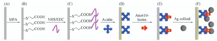
Fig.1 SPR-SERS spectroscopy for detecting bio-identification under silver colloid enhancement[18] Copyright from the Royal Society of Chemistry.
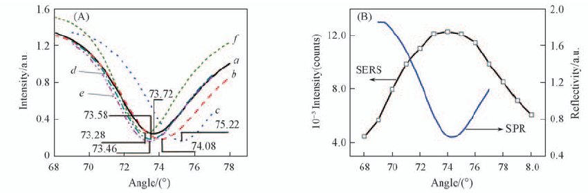
Fig.2 SPR curves of the layer-by-layer assembly of avidin and Atto610-biotin under silver colloid enhancement(A) and SPR curve and the plot of SERS intensities of Atto610-biotin(2.0 mg/mL) captured by avidin on silver film at different incident angles with silver colloid enhancement(B)[18] Curves a—f correspond to the process(A)—(F) in Fig.1. Copyright from the Royal Society of Chemistry.
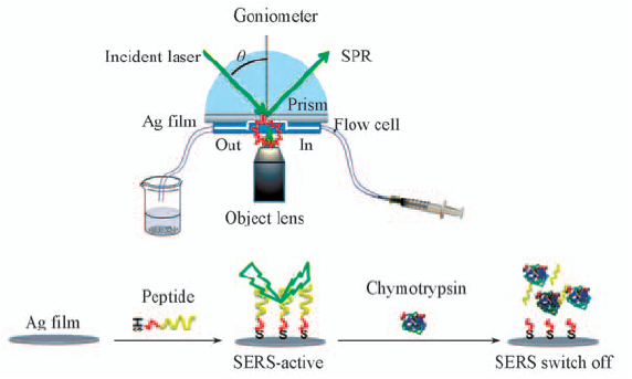
Fig.3 SPR-SERS spectroscopy for detecting chymotrypsin-catalyzed reaction[19] Copyright from the Royal Society of Chemistry.
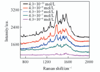
Fig.4 SERS spectra of the peptide reacting with different concentrations of chymotrypsin for 90 min[19] Copyright from the Royal Society of Chemistry.
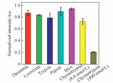
Fig.5 Analysis of the specificity of the SPR-SERS biosensor[19] Copyright from the Royal Society of Chemistry.
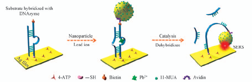
Fig.6 Schematic Diagram of the Fabrication of the DNAzyme-Based Plasmonic Nanomechine and Its Mechanism for the Detection of Pb2+ [29] Copyright from the American Chemical Society.
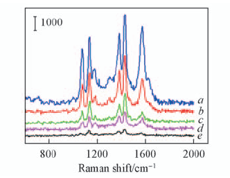
Fig.7 SERS spectra of 4-ATP(1.0 × 10⁻⁴ mol/L) with different concentrations of Pb2+ ions[29] Concentration of Pb2+/(mol·L-1): a. 1.0 × 10⁻⁶; b. 1.0 × 10⁻⁷; c. 1.0× 10⁻⁸; d. 1.0 × 10⁻⁹; e. 0. Copyright from the American Chemical Society.
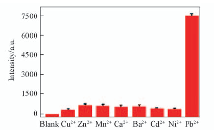
Fig.8 Selectivity of the DNAzyme-based SERS biosensor for Pb2+detection [29] Copyright from the American Chemical Society.
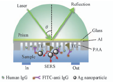
Fig.9 Schematic illustration for the SERS detection based on the nanoporous waveguide(PAA) film[34] Copyright from the Elsevier.
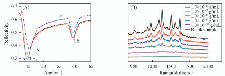
Fig.10 Typical reflection spectra(A) of the PAA waveguide layer before(a) and after the adsorptions of human IgG(b), FITC-anti IgG(c) and SERS detection of FITC-anti IgG with different concentrations of IgG through bioidentification by waveguide-assisted SERS method(B)[34] Copyright from the Elsevier.
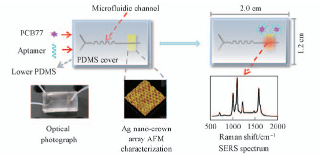
Fig.11 Schematic diagram of the fabrication of the aptamer-based SERS microfluidic sensor for the detection of PCB77[44] Copyright from the American Chemical Society.
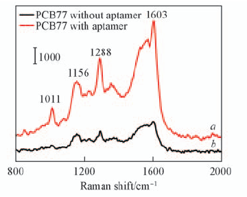
Fig.12 Comparison of SERS spectra of PCB77(1.0×10⁻⁷ mol/L) in microfluidic sensor with(a) and without(b) the aptamer[44] Copyright from the American Chemical Society.
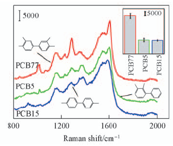
Fig.13 Specificity test for the aptamer-based SERS microfluidic sensor with 1.0×10⁻⁶ mol/L PCB[44] Insert compares the SERS intensities of PCB77, PCB5, and PCB15 according to the 1288 cm-1 peak[44]. Copyright from the American Chemical Society.
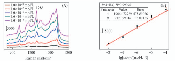
Fig.14 SERS spectra of different concentrations of PCB77(A) and SERS intensity at 1288 cm-1 as a function of concentration of PCB77(B)[44] Copyright from the American Chemical Society.
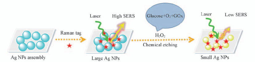
Fig.15 Glucose-sensing mechanism based on a “turn-off” SERS response
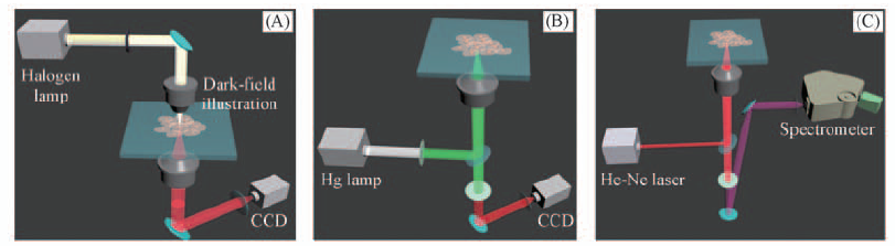
Fig.16 Schematic diagram of our Raman platform with three working modes[64] (A) Dark-field imaging; (B) fluorescence imaging; (C) Raman detection. Copyright from the american chemical society.
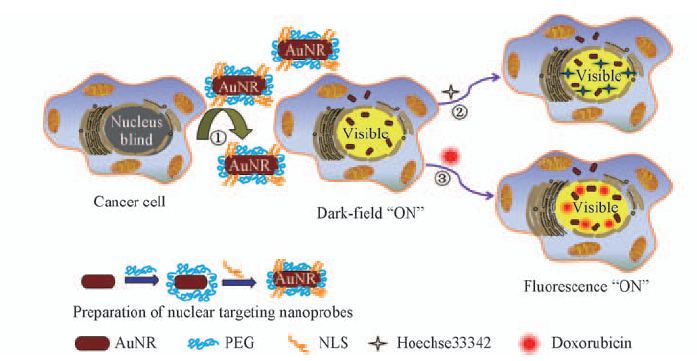
Fig.17 Illustration of SGC-7901 cell treated with nuclear targeting nanoprobes and two model drugs[64] Copyright from the American Chemical Society.
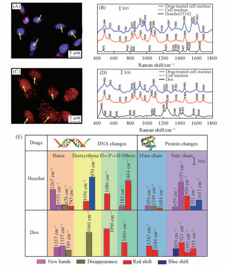
Fig.18 Major changes of SERS spectra in the nucleus before and after drug action[64] (A), (C) Dark-field/fluorescence coimages of cancer cells incubated with NLS-PEG-AuNRs and drugs(Hoechst and Dox); (B), (D) mean SERS spectra of cancer cells before and after treatment with drugs, and SERS spectra of Hoechst and Dox; (E) major changes of SERS spectra in the nucleus after drug action. Copyright from the American Chemical Society.
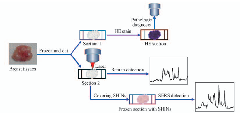
Fig.19 Raman spectra and SHINERS spectrum detection of breast tissue[70] Copyright from the Springer.
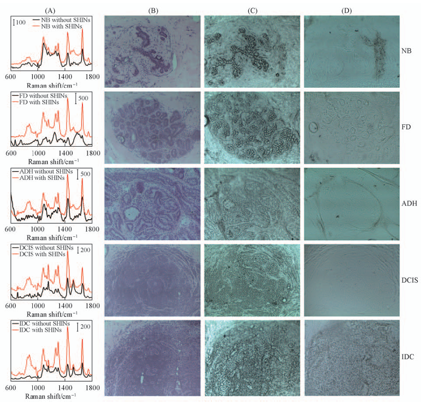
Fig.20 Mean spectra of breast tissue(A), optical images of HE tissue sections(B), Raman spectrum detection of breast tissue(C) and optical images of the breast tissue detected by SHINERS spectroscopy(D)[70] The photo shows an area of 0.8 mm×0.6 mm. Copyright from the Springer.
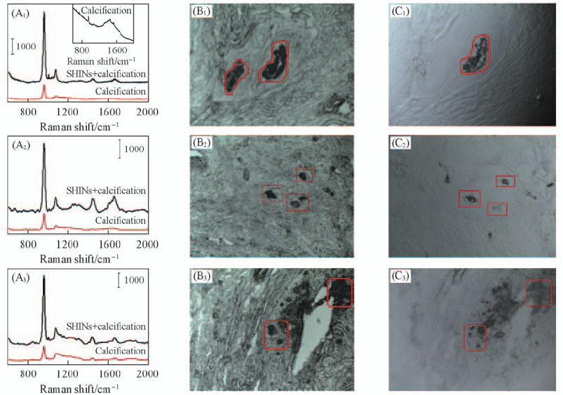
Fig.21 Raman and SERS spectra(A1—A3) of FD, ADH and DCIS containing calcified fibroadenomas, precancerous lesions and in situ duct and optical images(B, C) [71] (B1)—(B3) Breast tissue with calcification in Raman spectra; (C1)—(C3) breast tissue with calcification in SHINERS spectra. The photo shows an area of 0.8 mm×0.6 mm. Copyright from the Elsevier.
| [1] | Kirsch J., Siltanen C., Zhou Q., Chem. Soc. Rev., 2013, 42(22), 8733—8768 |
| [2] | Raman C. V., Krishnan K. S., Nature, 1928, 121(3048), 501—504 |
| [3] | Fleischmann M., Hendra P. J., McQuillan A. J., Chem. Phys. Lett., 1974, 26(2), 163—166 |
| [4] | Jeanmaire D. L., Van Duyne R. P., J. Electroanal. Chem., 1977, 84(1), 1—20 |
| [5] | Wu Z. T., Liu Y. Z., Zhou X. D., Shen A. G., Hu J. M., J. Anal. Sci., 2014, 30(6), 829—839 |
| (伍子同, 刘翼振, 周晓东, 沈爱国, 胡继明. 分析科学学报, 2014, 30(6), 829—839) | |
| [6] | Zheng J. W., Li X. W., Xu H. Y., Zhou Y. G., Gu R. A., Spectrosc. Spect. Anal., 2003, 23(2), 294—296 |
| (郑军伟, 李晓伟, 徐浩元, 周耀国, 顾仁敖. 光谱学与光谱分析, 2003, 23(2), 294—296) | |
| [7] | Ge M., Yao J. L., Cui Y., Jiang Y., Gu R. A., Chem. J. Chinese Universities, 2007, 28(8), 1464—1468 |
| (葛明, 姚建林, 崔颜, 蒋芸, 顾仁敖. 高等学校化学学报, 2007, 28(8), 1464—1468) | |
| [8] | Bao F., Yao J. L., Gu R. A., Langmuir, 2009, 25(18), 10782—10787 |
| [9] | Chen S., Yuan Y. X., Yao J. L., Han S. Y., Gu R. A., Chem. Commun., 2011, 47(14), 4225—4227 |
| [10] | Cui Y., Ren B., Tian Z. Q., J. Southeast Univ.(MedSci. Edi.), 2011, 30(1), 254—262 |
| (崔颜, 任斌, 田中群. 东南大学学报(医学版), 2011, 30(1), 254—262) | |
| [11] | Huang J., Guo M., Ke H. T., Zong C., Ren B., Liu G., Shen H., Ma Y. F., Wang X. Y., Zhang H. L., Deng Z. W., Chen H. B., Zhang Z. J., Adv. Mater., 2015, 27(34), 5049—5056 |
| [12] | Bantz K. C., Meyer A. F., Wittenberg N. J., Im H., Kurtulus O., Lee S. H., Lindquist N. C., Oh S. H., Haynes C. L., Phys. Chem. Chem. Phys., 2011, 13, 11551—11567 |
| [13] | Futamata M., Keim E., Bruckbauer A., Schumacher D., Otto A., Appl. Surf. Sci., 1996, 100/101, 60—63 |
| [14] | Liu D. L., Zhao Q., Lu D. F., Qi Z. M., Chem. J. Chinese Universities, 2014, 35(10), 2207—2213 |
| (刘德龙, 赵乔, 逯丹凤, 祁志美. 高等学校化学学报, 2014, 35(10), 2207—2213) | |
| [15] | Yih J. N., Chen S. J., Huang K. T., Su Y. T., Lin G. Y., Proc SPIE., 2004, 5327, 5—9 |
| [16] | Liu Y., Xu S. P., Tang B., Wang Y., Zhou J., Zheng X. L., Zhao B., Xu W. Q., Rev. Sci. Instrum., 2010, 81(3), 036105 |
| [17] | Liu Y., Xu S. P., Li H. B., Jian X. G., Xu W. Q., Chem. Commun., 2011, 47(13), 3784—3786 |
| [18] | Fu C. C., Hu C. X., Liu Y., Xu S. P., Xu W. Q., Anal. Method, 2012, 4(10), 3107—3110 |
| [19] | Fu C. C., Xu W. Q., Chen G., Xu S. P., Analyst, 2013, 138(21), 6282—6286 |
| [20] | Fang Y., Seong N., Dlott D. D., Science, 2008, 321(5887), 388—392 |
| [21] | Xu H., Aizpurua J., Käll M., Apell P., Phys. Rev. E, 2000, 62(3), 4318—4324 |
| [22] | Wang X., Li M., Meng L., Lin K., Feng J., Huang T., Yang Z., Ren B., ACS Nano, 2014, 8(1), 528—536 |
| [23] | Stöckle R. M., Suh Y. D., Deckert V., Zenobi R., Chem. Phys. Lett., 2000, 318(1—3), 131—136 |
| [24] | Halas N. J., Lal S., Chang W. S., Link S., Nordlander P., Chem. Rev., 2011, 111, 3913—3961 |
| [25] | Xuan X. Y., Xu S. P., Liu Y., Li H. B., Xu W. Q., Lombardi John R., J. Phys. Chem. Lett., 2012, 3(19), 2773—2778 |
| [26] | Hermann T., Patel D. J., Science, 2000, 287(5454), 820—825 |
| [27] | Kim N. H., Lee S. J., Moskovits M., Adv. Mater., 2011, 23(36), 4152—4156 |
| [28] | Zheng J., Jiao A., Yang R., Li H., Li J., Shi M., Ma C., Jiang Y., Deng L., Tan W., J. Am. Chem. Soc., 2012, 134(49), 19957—19960 |
| [29] | Fu C. C., Xu W. Q., Wang H. L., Ding H., Liang L. J., Cong M., Xu S. P., Anal. Chem., 2014, 86(23), 11494—11497 |
| [30] | McKee K. J., Meyer M. W., Smith E. A., Anal. Chem., 2012, 84(21), 9049—9055 |
| [31] | Meyer M. W., McKee K. J., Nguyen V. T., Smith E. A., J. Phys. Chem. C, 2012, 116(47), 24987—24992 |
| [32] | Hu D. B., Qi Z. M., J. Phys. Chem. C, 2013, 117(31), 16175—16181 |
| [33] | Gu Y. J., Xu S. P., Li H. B., Wang S. Y., Cong M., Lombardi J. R., Xu W. Q., J. Phys. Chem. Lett., 2013, 4(18), 3153—3157 |
| [34] | Fu C. C., Gu Y. J., Wu Z. Y., Wang Y. Y., Xu S. P., Xu W. Q., Sens. Actuators B, 2014, 201, 173—176 |
| [35] | Manz A., Graber N., Widmer H. M., Sens. Actuators B, 1990, 1(1—6), 244—248 |
| [36] | Thorsen T., Maerkl S. J., Quake S. R., Science, 2002, 298(5593), 580—584 |
| [37] | Whitesides G. M., Nature, 2006, 442(7101), 368—373 |
| [38] | Ríos Á., Zougagh M., Avilaa M., Anal. Chim. Acta, 2012, 740, 1—11 |
| [39] | Chen G., Wang Y. Y., Wang H. L., Cong M., Chen L., Yang Y. A., Geng Y. J., Li H. B., Xu S. P., Xu W. Q., RSC Adv., 2014, 4(97), 54434—54440 |
| [40] | Choi N., Lee K., Lim D. W., Lee E. K., Chang S. I., Oh K. W., Choo J., Lab Chip, 2012, 12(24), 5160—5167 |
| [41] | Lee M., Lee K., Kim K. H., Oh K. W., Choo J., Lab Chip, 2012, 12(19), 3720—3727 |
| [42] | Lu X., Samuelson D. R., Xu Y., Zhang H., Wang S., Rasco B. A., Xu J., Konkel M. E., Anal. Chem., 2013, 85(4), 2320—2327 |
| [43] | Malfatti L., Falcaro P., Marmiroli B., Amenitsch H., Piccinini M., Falqui A., Innocenzi P., Nanoscale, 2011, 3(9), 3760—3766 |
| [44] | Fu C. C., Wang Y., Chen G., Yang L. Y., Xu S. P., Xu W. Q., Anal. Chem., 2015, 87(19), 9555—9558 |
| [45] | Wang X., Xu S., Cong M., Li H., Gu Y., Xu W., Small, 2012, 8(7), 972—976 |
| [46] | Wang Y., Wang Y., Wang H., Cong M., Xu W., Xu S., Phys. Chem. Chem. Phys., 2015, 17(2), 1173—1179 |
| [47] | Shafer-Peltier K. E., Haynes C. L., Glucksberg M. R., Van Duyne R. P., J. Am. Chem. Soc., 2003, 125(2), 588—593 |
| [48] | Haynes C. L, Yonzon C. R, Zhang X., Van Duyne R. P., J. Raman Spectrosc., 2005, 36, 471—484 |
| [49] | Yonzon C. R., Haynes C. L., Zhang X., Walsh J. T., Van Duyne R. P., Anal. Chem., 2004, 76(1), 78—85 |
| [50] | Yonzon C. R., Stuart D. A., Zhang X., McFarland A. D., Haynes C. L., Van Duyne R. P., Talanta, 2005, 67(3), 438—448 |
| [51] | Lyandres O., Shah N. C., Yonzon C. R., Walsh J. T., Glucksberg M. R., Van Duyne R. P., Anal Chem., 2005, 77(19), 6134—6139 |
| [52] | Yang X., Yu Y., Gao Z., ACS Nano, 2014, 8(5), 4902—4907 |
| [53] | Xia Y., Ye J., Tan K., Wang J., Yang G., Anal. Chem., 2013, 85(13), 6241—6247 |
| [54] | Kong K. V., Lam Z., Lau W. K. O., Leong W. K., Olivo M., J. Am. Chem. Soc., 2013, 135(48), 18028—18031 |
| [55] | Yehezkeli O., Tel—Vered R., Raichlin S., Willner I., ACS Nano, 2011, 5(3), 2385—2391 |
| [56] | Qi G. H., Jia K. Q., Fu C. C., Xu S. P., Xu W. Q., J. Opt., 2015, 17, 114020-1—6 |
| [57] | Qian X., Peng X. H., Ansari D. O., YinGoen Q., Chen G. Z., Shin D. M., Yang L., Young A. N., Wang M. D., Nie S., Nat. Biotechnol., 2008, 26(1), 83—90 |
| [58] | Raj L., Ide T., Gurkar A. U., Foley M., Schenone M., Li X., Tolliday N. J., Golub T. R., Carr S. A., Shamji A. F., Stern A. M., Mandinova A., Schreiber S. L., Lee S. W., Nature, 2012, 481(7382), 534—534 |
| [59] | Bair J. S., Palchaudhuri R., Hergenrother P. J., J. Am. Chem. Soc., 2010, 132(15), 5469—5478 |
| [60] | Panikkanvalappil S. R., Mackey M. A., El-Sayed M. A., J. Am. Chem. Soc., 2013, 135(12), 4815—4821 |
| [61] | Panikkanvalappil S. R., Mahmoud M. A., Mackey M. A., El-Sayed M. A., ACS Nano, 2013, 7(9), 7524—7533 |
| [62] | Kang B., Austin L. A., El-Sayed M. A., ACS Nano, 2014, 8(5), 4883—4892 |
| [63] | Li H., Wang H., Huang D., Liang L., Gu Y., Liang C., Xu S., Xu W., Rev. Sci. Instrum., 2014, 85(5), 056109-1—3 |
| [64] | Liang L., Huang D., Wang H., Li H., Xu S., Chang Y., Li H., Yang Y., Liang C., Xu W., Anal. Chem., 2015, 87(4), 2504—2510 |
| [65] | Chen W. Q., Zheng R. S., Zhang S. W., Zhao P., Li G. L., Wu L. Y., He J., Chin J. Cancer Res., 2013, 25(1), 10—21 |
| [66] | Hu C. X., Zheng C., Zhang H. P., Bi L. R., Xu S. P., Fan Z. M., Han B., Xu W. Q., Chem. J. Chinese Universities, 2013, 34(12), 2721—2727 |
| (胡成旭, 郑超, 张海鹏, 毕丽荣, 徐抒平, 范志民, 韩冰, 徐蔚青. 高等学校化学学报, 2013, 34(12), 2721—2727) | |
| [67] | Zheng C., Zhang H. P., Han B., Liang L. J., Zou Y. B., Xu S. P., Lin H. P., Xu W. Q., Fan Z. M., Chem. J. Chinese Universities, 2015, 36(1), 74—78 |
| (郑超, 张海鹏, 韩冰, 梁丽佳, 邹亚斌, 徐抒平, 林和平, 徐蔚青, 范志民. 高等学校化学学报, 2015, 36(1), 74—78) | |
| [68] | Anema J. R., Li J. F., Yang Z. L., Ren B., Tian Z. Q., Annu. Rev. Anal. Chem., 2011, 4, 129—150 |
| [69] | Li J. F., Huang Y. F., Ding Y., Yang Z. L., Li S. B., Zhou X. S., Fan F. R., Zhang W., Zhou Z. Y., Wu Y., Ren B., Wang Z. L., Tian Z. Q., Nature, 2010, 464(7287), 392—395 |
| [70] | Zheng C., Liang L., Xu S., Zhang H., Hu C., Bi L., Fan Z., Han B., Xu W., Anal. Bioanal.Chem., 2014, 406(22), 5425—5432 |
| [71] | Liang L. J., Zheng C., Zhang H. P., Xu S. P., Zhang Z., Hu C. X., Bi L. R., Fan Z. M., Han B., Xu W. Q., Spectrochim Acta Part A, 2014, 132, 397—402 |
| [1] | 江博文, 陈敬轩, 成永华, 桑微, 寇宗魁. 单原子材料在电化学生物传感中的研究进展[J]. 高等学校化学学报, 2022, 43(9): 20220334. |
| [2] | 王君旸, 刘争, 张茜, 孙春燕, 李红霞. DNA银纳米簇在功能核酸荧光生物传感器中的应用[J]. 高等学校化学学报, 2022, 43(6): 20220010. |
| [3] | 夏大成, 周瑞, 涂博, 蔡志伟, 高难, 姬晓旭, 常钢, 任小明, 何云斌. 银/金纳米线阵列表面增强拉曼基底的制备及对孔雀石绿的高灵敏度检测[J]. 高等学校化学学报, 2022, 43(3): 20210731. |
| [4] | 陈韶云, 张行颖, 刘奔, 田杜, 李奇, 陈芳, 胡成龙, 陈建. Ag纳米粒子在TiO2四棱柱阵列上的可控生长及其SERS效应[J]. 高等学校化学学报, 2021, 42(8): 2381. |
| [5] | 徐梦祎, 黄雪雯, 李小杰, 魏玮, 刘晓亚. “串珠状”复合纳米组装体修饰丝网印刷电极构建的生物传感器[J]. 高等学校化学学报, 2021, 42(6): 1768. |
| [6] | 李奇, 田杜, 陈韶云, 钟敏, 胡成龙, 陈建. 银纳米片在无纺布纤维上的可控组装及其SERS效应[J]. 高等学校化学学报, 2021, 42(3): 736. |
| [7] | 王博东, 潘美辰, 卓颖. 二氧化硅纳米颗粒表面原位还原银纳米簇电化学发光传感界面的构建与分子识别[J]. 高等学校化学学报, 2021, 42(11): 3519. |
| [8] | 王庆, 何雨秋, 王富安. 多功能脱氧核酶用于生物医学分析的研究进展[J]. 高等学校化学学报, 2021, 42(11): 3334. |
| [9] | 赵卓, 王雪强. 核酸适体偶联药物的生物偶联构建技术与应用[J]. 高等学校化学学报, 2021, 42(11): 3367. |
| [10] | 席京, 陈娜, 杨雁冰, 袁荃. 长余辉纳米材料的控制合成及在疾病诊断中的应用[J]. 高等学校化学学报, 2021, 42(11): 3247. |
| [11] | 柯梦婷, 袁江培, 张恒, 方煜. 多孔配位聚合物靶向亚细胞器用于生物成像和诊疗[J]. 高等学校化学学报, 2021, 42(11): 3295. |
| [12] | 张嘉懿, 丁臻尧, 王丹丹, 陈礼平, 封心建. 基于多孔金结构的三相界面酶电极的制备及高效电化学酶传感性能[J]. 高等学校化学学报, 2021, 42(10): 3167. |
| [13] | 唐文涛, 李圣凯, 王昚, 陈龙, 陈卓. 基于激光介导富集的表面增强拉曼分析[J]. 高等学校化学学报, 2021, 42(10): 3054. |
| [14] | 张梦瑶, 余仁鹏, 韩梅, 刘建芳, 李末霞, 胡家文, 田中群. 盐诱导金纳米粒子自组装和沉降制备具有宽波段吸收特性的黑金[J]. 高等学校化学学报, 2020, 41(8): 1903. |
| [15] | 袁中文, 贺利贞, 陈填烽. 单原子催化剂的生物医学应用[J]. 高等学校化学学报, 2020, 41(12): 2690. |
| 阅读次数 | ||||||
|
全文 |
|
|||||
|
摘要 |
|
|||||