

高等学校化学学报 ›› 2021, Vol. 42 ›› Issue (9): 2701.doi: 10.7503/cjcu20210118
朱兆田1, 李圣凯1, 宋明慧1, 蔡芯琪1, 宋志灵2, 陈龙3, 陈卓1( )
)
收稿日期:2021-02-06
出版日期:2021-09-10
发布日期:2021-09-08
通讯作者:
陈卓
E-mail:zhuochen@hnu.edu.cn
基金资助:
ZHU Zhaotian1, LI Shengkai1, SONG Minghui1, CAI Xinqi1, SONG Zhiling2, CHEN Long3, CHEN Zhuo1()
Received:2021-02-06
Online:2021-09-10
Published:2021-09-08
Contact:
CHEN Zhuo
E-mail:zhuochen@hnu.edu.cn
Supported by:摘要:
多功能金属石墨纳米囊由于其良好的稳定性和独特的理化性质, 在生物医学领域受到了广泛关注. 利用石墨烯外壳独特的拉曼散射特征峰作为拉曼标签或者内标, 结合等离子体纳米核优异的表面增强拉曼散射(SERS)和双光子发光(TPL)性能, 可实现SERS生物分析以及肿瘤细胞或组织的Raman/TPL双模成像. 利用表面积大的石墨烯外壳作为药物负载平台, 结合等离子体纳米核的近红外光吸收能力, 可实现光介导的病原菌杀灭以及肿瘤细胞或实体瘤的热疗与化疗的协同治疗. 此外, 利用石墨烯外壳优异的荧光猝灭性能, 还实现了生物分子的荧光检测; 利用磁性纳米核独特的磁学性能, 可实现生物样品的分离和富集、 细菌的原位磁共振成像检测以及磁靶向胃部口服药物的递送. 本综述首先介绍了金属石墨纳米囊的制备、 分类和性质, 然后概述了它们在生物检测、 生物成像和治疗3个方面的应用进展, 并进一步总结了它们的发展现状包括生物毒性和生物医学应用的优缺点, 最后对其在生物医学领域的发展方向做出了展望. 我们期望多功能的金属石墨纳米囊能够为今后的临床生物医学应用提供可靠的纳米平台.
中图分类号:
TrendMD:
朱兆田, 李圣凯, 宋明慧, 蔡芯琪, 宋志灵, 陈龙, 陈卓. 多功能金属石墨纳米囊的生物医学应用进展. 高等学校化学学报, 2021, 42(9): 2701.
ZHU Zhaotian, LI Shengkai, SONG Minghui, CAI Xinqi, SONG Zhiling, CHEN Long, CHEN Zhuo. Recent Progress of Versatile Metal Graphitic Nanocapsules in Biomedical Applications. Chem. J. Chinese Universities, 2021, 42(9): 2701.
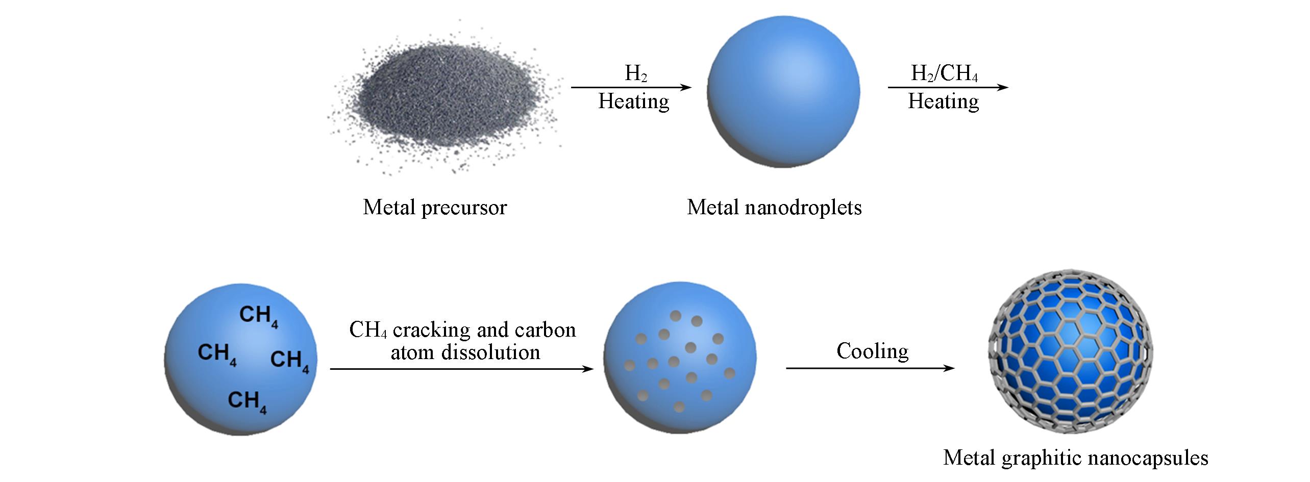
Scheme 1 Mechanism and synthesis process of metal graphitic nanocapsules by CVD method
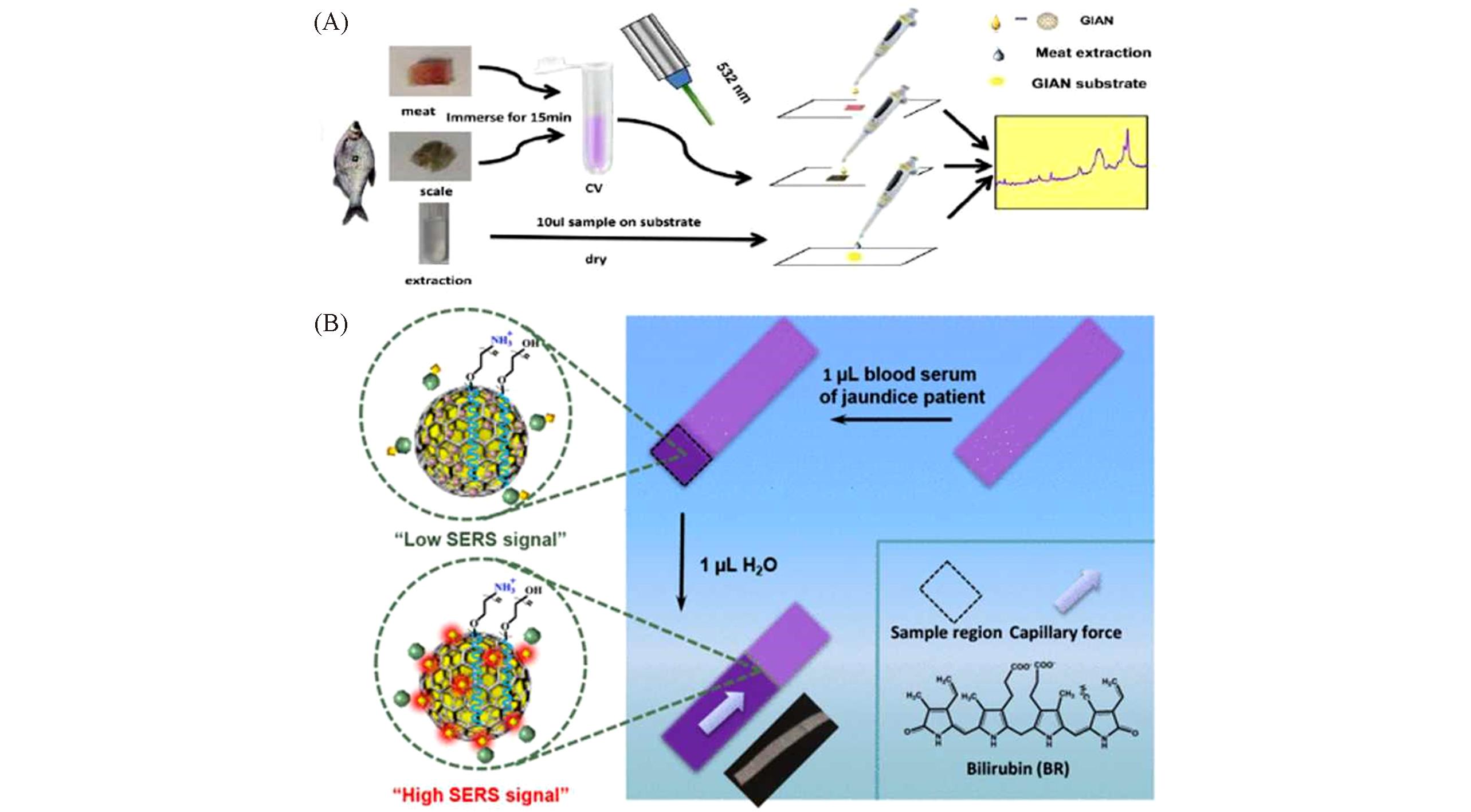
Fig.1 GIANs for sensitive SERS bioanalysis(A) Schematic illustration of CV detection on site. Adapted with permission from Ref.[18]. Copyright 2016, American Chemical Society.(B) Adsorption process of BR occurred on the surface of GIANs in fetal bovine serum(FBS) matrix. Inset image is the digital photo of CS@GIANs dripped with BR(FBS) aqueous after separation. Adapted with permission from Ref.[20]. Copyright 2018, American Chemical Society.
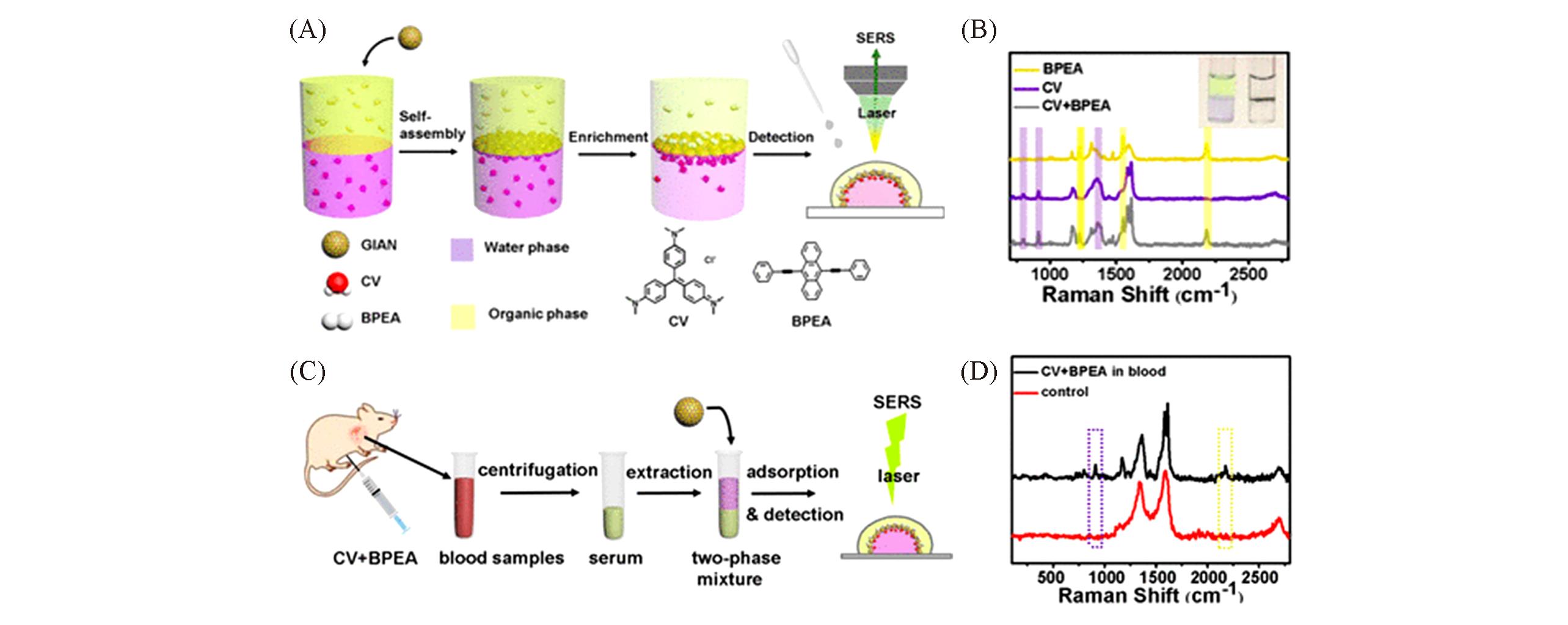
Fig.2 GIANs for simultaneous multiphase SERS analysis(A) Schematic diagram of multiphase Raman detectionwith GIANs; (B) SERS spectra for the multiplex interfacial detection of CV and BPEA; (C) schematic illustration of Raman analysis of CV and BPEA in mice; (D) blood analysis of mice injected with CV and BPEA(black line). Insets of (B) are photographs of the solutions corresponding to the spectra. Adapted with permission from Ref.[58]. Copyright 2018, American Chemical Society.
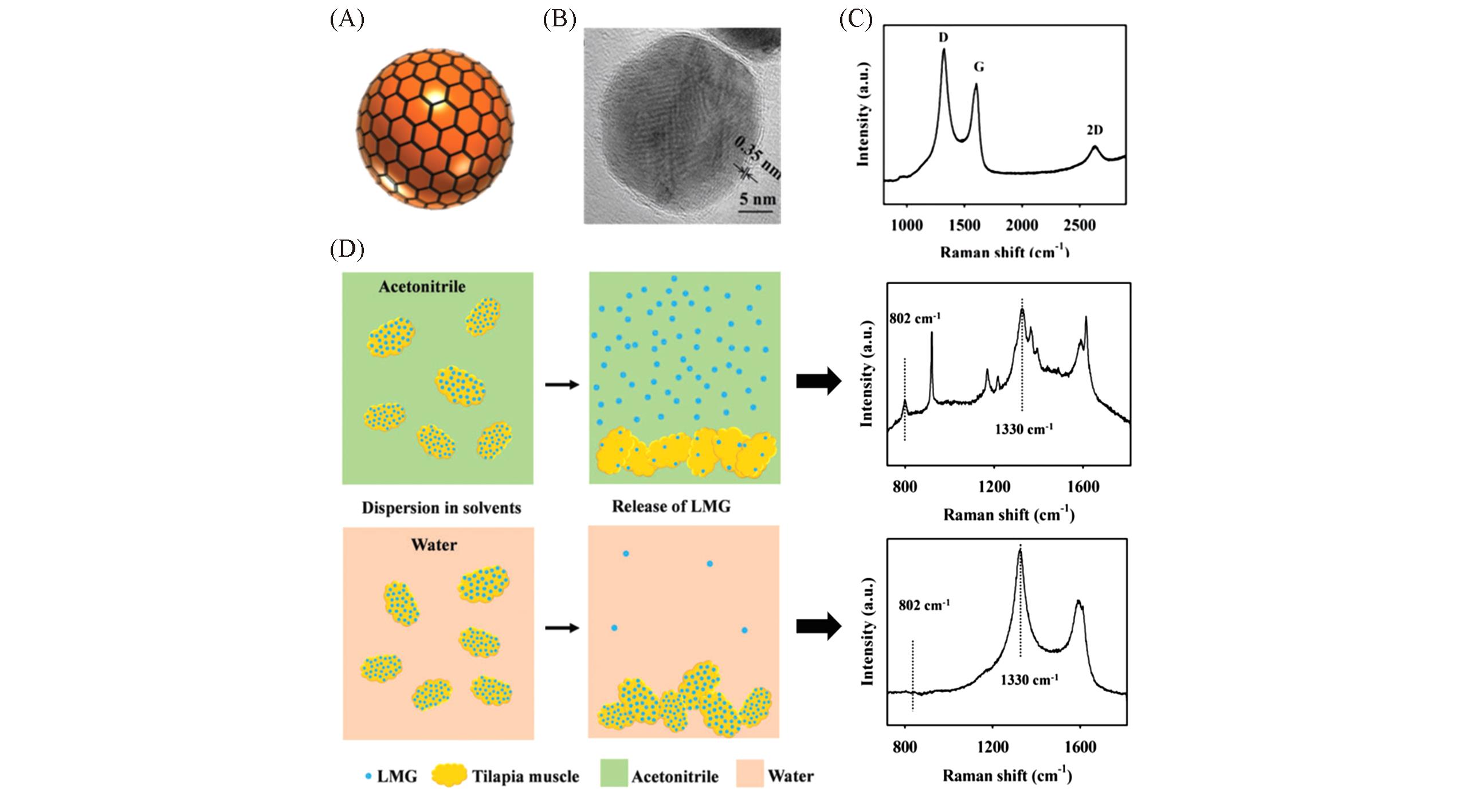
Fig.3 AgCu@G for sensitive SERS bioanalysisSchematic diagram(A), TEM image(B) and Raman spectrum(C) of AgCu@G, (D) SERS detection of LMG in contaminated fish muscle washed with different solvents. Adapted with permission from Ref.[44]. Copyright 2018, Royal Society of Chemistry.
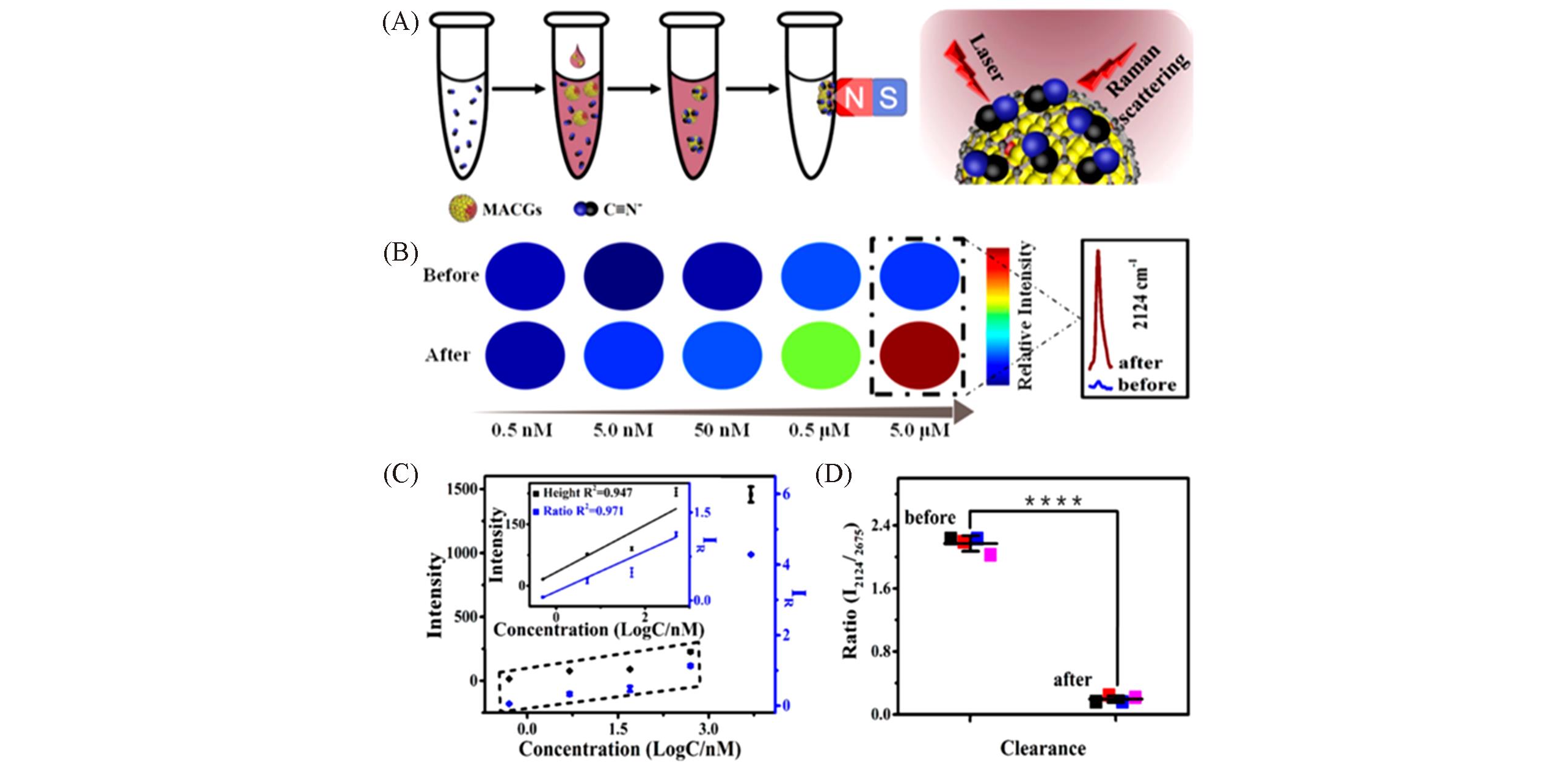
Fig.4 AgCu@G for sensitive SERS bioanalysis(A) Schematic diagram of enrichment processes; (B) hot map of different CN- concentrations before and after enrichment; (C) change of logarithmic CN- concentration without(black) and with(blue) IS(inset is the linear fitting of the logarithmic CN- concentration); (D) corresponding data analysis to demonstrate the cyanide clearance ability of MACGs in the water sample(height means intensity at 2124 cm-1, and ratio is I2124/I2675. P values were calculated by the Student’s t test: **** P<0.0001). Adapted with permission from Ref.[28]. Copyright 2019, American Chemical Society.
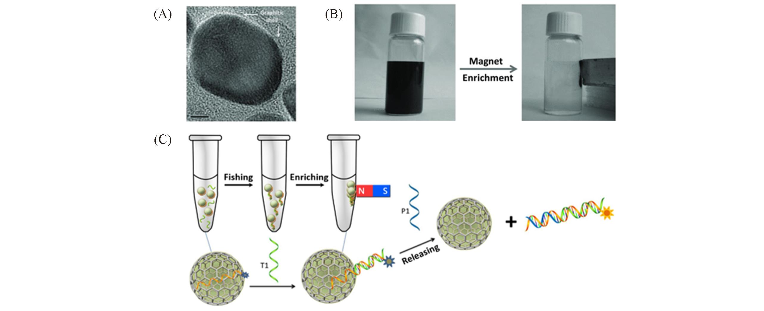
Fig.5 Co@G for fluorescence detection of DNA(A) TEM image of Co@G; (B) suspensions of Co@G aqueous solution before(left) and after(right) magnetic enrichment; (C) schematic diagram of Co@G for fluorescence detection of DNA. Adapted with permission from Ref.[26]. Copyright 2017, Wiley-VCH.
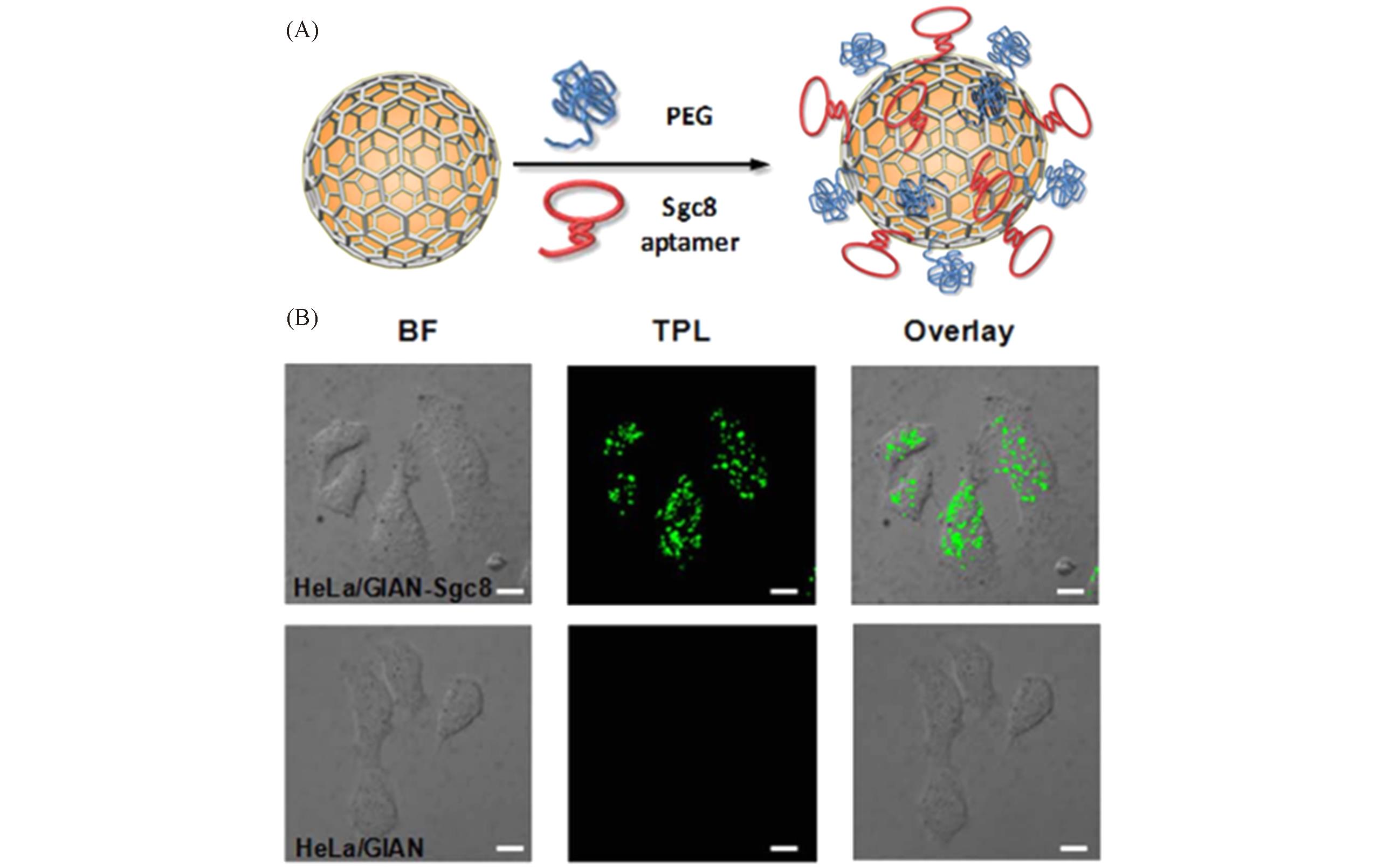
Fig.6 Targeted cell imaging with GIANs(A) Schematic illustration of Sgc-8 aptamer?functionalized GIAN; (B) TPL confocal images of HeLa incubated with GIAN and GIAN-Sgc8. BF: bright field, scale bar: 10 mm. Adapted with permission from Ref.[23]. Copyright 2014, Nature Publishing Group.
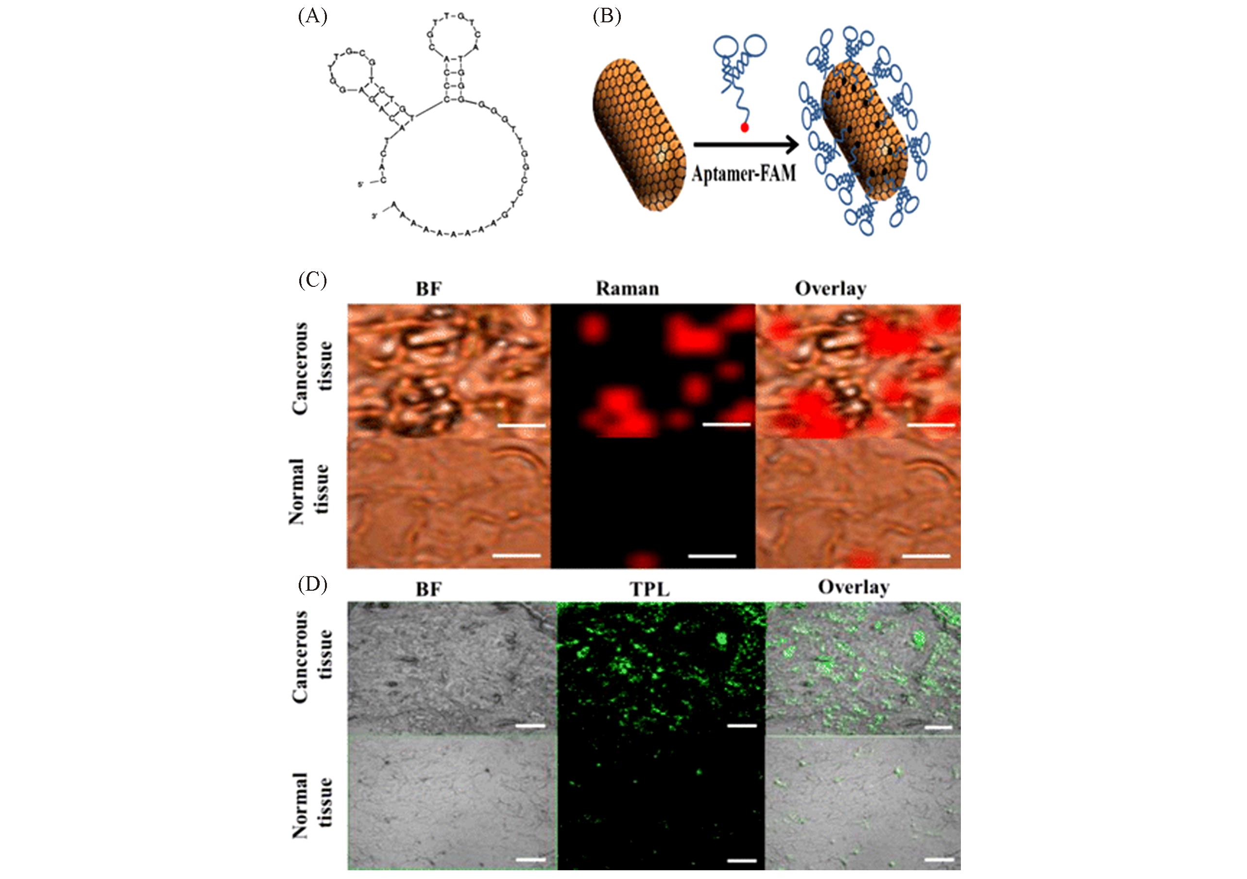
Fig.7 Targeted tissue imaging with AuNR@G(A) Structure of SYL3C aptamer; (B) schematic illustration of SYL3C aptamer?functionalized AuNR@G; (C) targeted Raman imaging of cancerous breast and normal liver tissues of rat treated with aptamer-AuNR@G nanocapsules at room temperature for 40 min with 1.0 s integration time per pixel(BF: bright-field; scale bar: 10 μm); (D) TPL confocal images of cancerous breast and normal liver tissues of rat treated with aptamer-AuNR@G nanocapsules(scale bar: 50 μm). Adapted with permission from Ref.[22]. Copyright 2016, American Chemical Society.
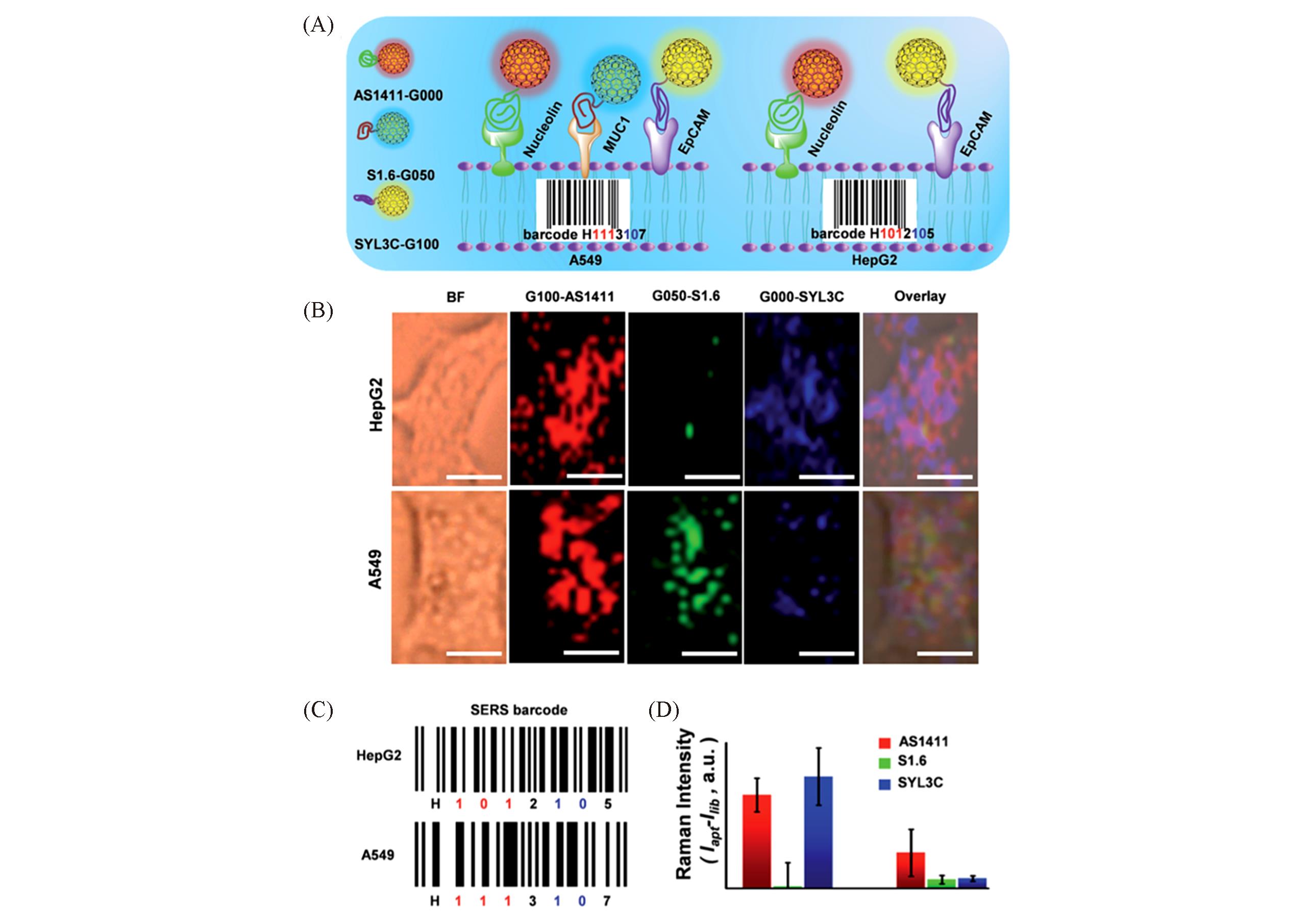
Fig.8 Cancer cell pattern recognition with aptamer?functionalized isotopic GIAN?encoders(A) Schematic illustration of pattern recognition and discrimination of cancer cell lines with multiplexed GIAN encoders; (B) SERS images of cancer cells(scale bar: 10 mm. G100, G050 and G000 conjugated with DSPE-PEG-linked aptamer AS1411, S1.6 and SYL3C, respectively); (C) SERS barcodes of HepG2 and A549 cell lines; (D) statistics of normalized SERS signals shown in (B). Adapted with permission from Ref.[43]. Copyright 2018, Royal Society of Chemistry.
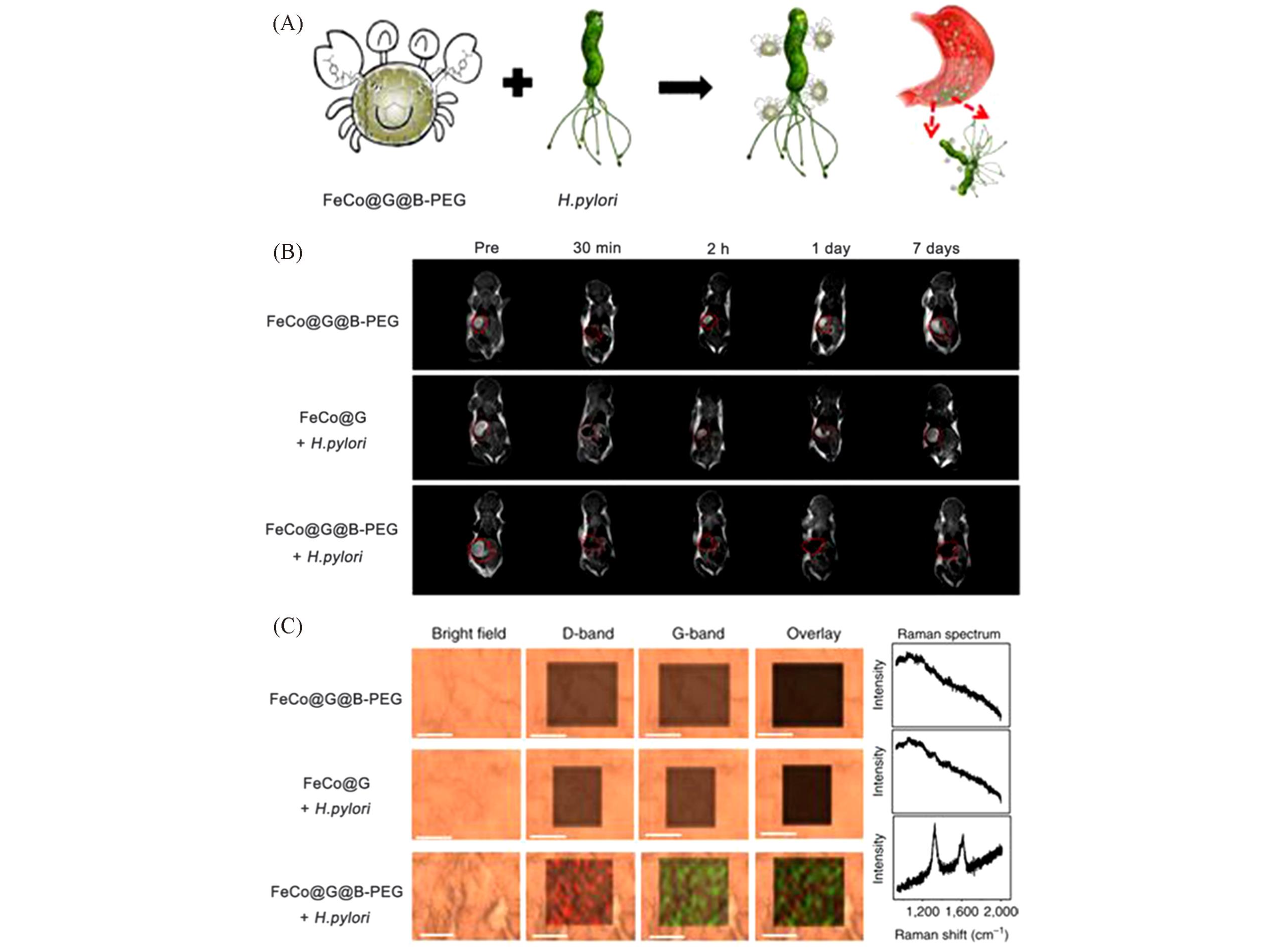
Fig.9 In situ targeted MRI detection of H. pylori with FeCo@G(A) Schematic illustration of H. pylori detection with FeCo@G@B-PEG; (B) T2-weighted images at different times with or without FeCo@G@B-PEG treatments; (C) Raman images and spectra of mice gastric mucosa used in (B)(scale bar: 10 mm; slice thickness: 50 mm). Adapted with permission from Ref.[30]. Copyright 2017, Nature Publishing Group.
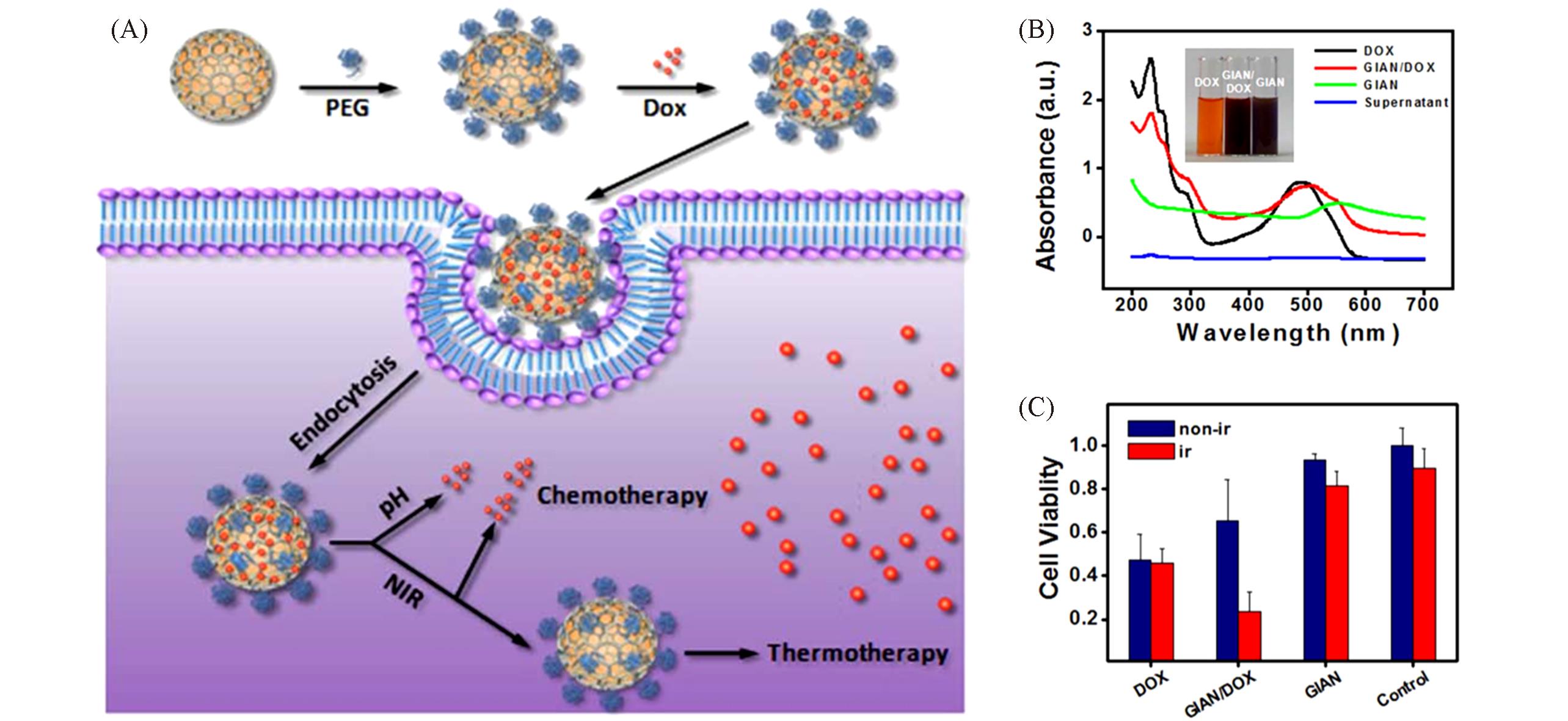
Fig.10 NIR photothermal enhanced chemotherapy of GIAN/DOX complexes(A) NIR photothermal enhanced chemotherapy mechanism of GIAN/DOX; (B) UV-Vis characterization of the DOX-loaded GIANs; (C) cell viability of MCF-7 cells with and without NIR laser irradiation after incubation with free DOX, GIAN, and GIAN/DOX, respectively. Adapted with permission from Ref.[23]. Copyright 2014, Nature Publishing Group.
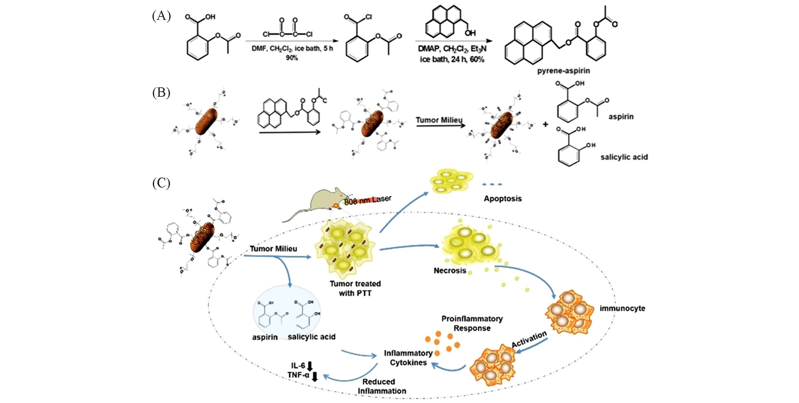
Fig.11 Pyrene?aspirin?loaded AuNR@G for simultaneous photothermal and anti?inflammatory therapy(A) Synthesis of P-aspirin; (B) illustration of surface functionalization and P-aspirin releasing from AuNR@G; (C) illustration of the anti-inflammatory mechanism underlying the inhibition of PTT-associated inflammation by AuNR@G-P-aspirin complexes. Adapted with permission from Ref.[24]. Copyright 2018, Wiley-VCH.
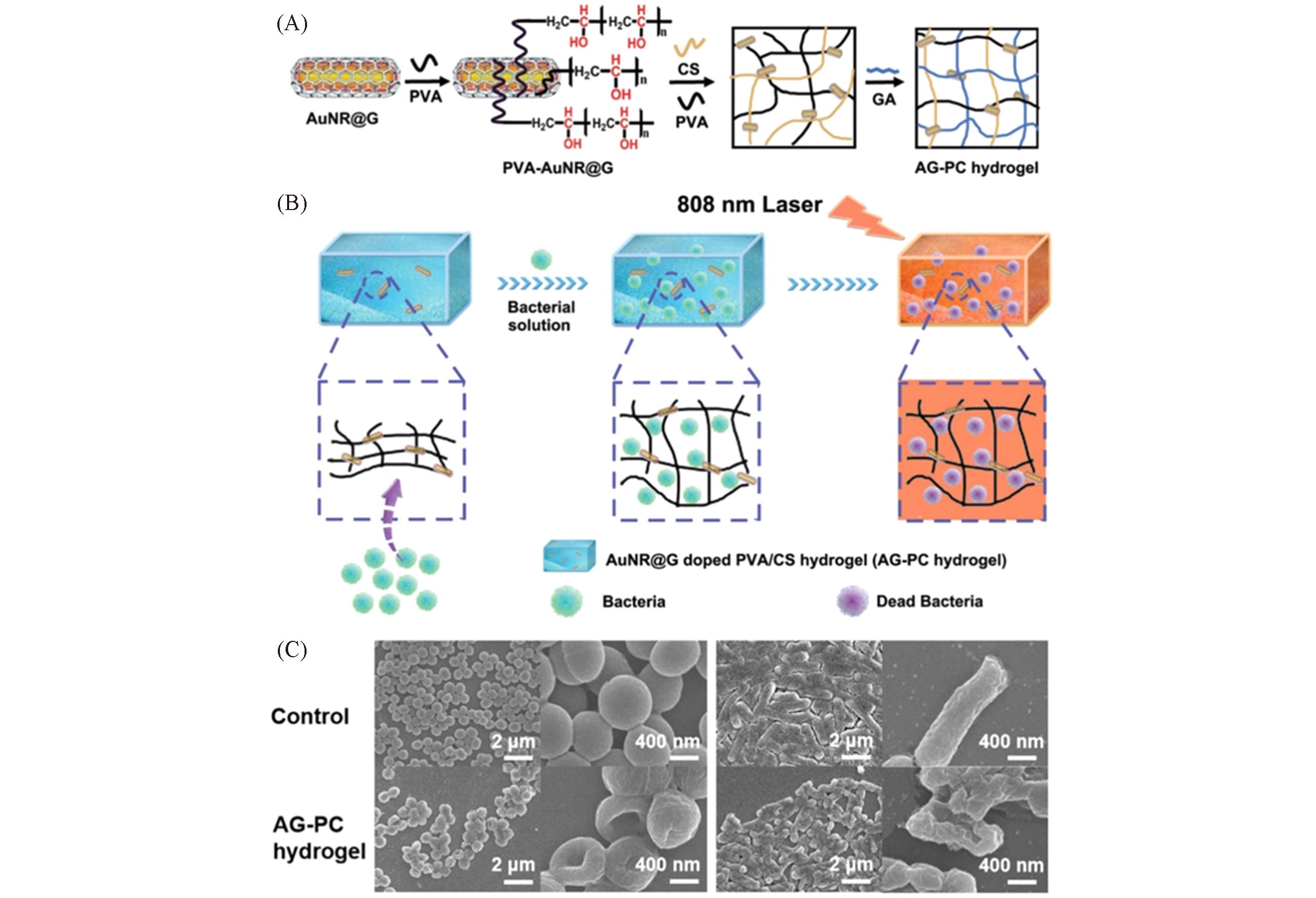
Fig.12 AuNR@G doped poly(vinyl alcohol)/chitosan(AG?PC) hydrogel for photothermal antibacterial applications(A) Preparation of the AG-PC hydrogel; (B) NIR laser-induced antibacterial experiments using the AG-PC hydrogel.Adapted with permission from Ref.[25]. Copyright 2019, Royal Society of Chemistry.
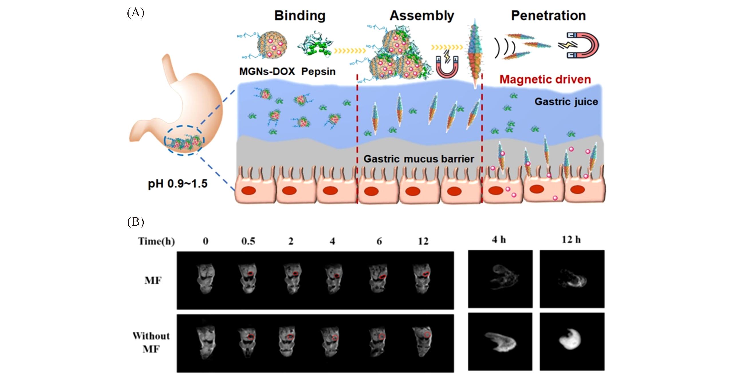
Fig.13 In situ pepsin?assisted needle assembly of MGNs for enhanced gastric retention and mucus penetration(A) Illustration of gastric retention and penetration in vivo; (B) T2-weighted MRI of BALB/c mice(left) and isolated mouse stomach(right) after administration with MGNs at different time with and without application of MF. Adapted with permission from Ref.[31]. Copyright 2021, Elseiver.
| 1 | Du F. L., Wu B. X., Liu J., Xu C. C., Li G. F., Wang X., Chem. J. Chinese. Universities, 2021, 42(1), 1—11(杜芳林, 吴冰昕, 刘娇, 徐聪聪, 李国锋, 王兴. 高等学校化学学报, 2021, 42(1), 1—11) |
| 2 | Wang B. W., Ma R., Wu F., Liu Z. H., Li L. F., Zhang X., Liu D. K., Yang N., Li M. H., Yang D. F., Sun Q., Chem. J. Chinese. Universities, 2020, 41(9), 2099—2106(王博蔚, 马瑞, 吴凡, 刘志辉, 李凌锋, 张骁, 刘定坤, 杨楠, 李美慧, 杨德峰, 孙琪. 高等学校化学学报, 2020, 41(9), 2099—2106) |
| 3 | Cheng Y., Wang K., Qi Y., Liu Z. F., Acta Phys. Chim. Sin., 2022, 38(2), 2006046(程熠, 王坤, 亓月, 刘忠范. 物理化学学报, 2022, 38(2), 2006046) |
| 4 | Zhu A., Qu Q., Shao X., Kong B., Tian Y., Angew. Chem. Int. Ed., 2012, 124, 7297—7301 |
| 5 | Han G., Zhao J., Zhang R., Tian X., Liu Z., Wang A., Liu R., Liu B., Han M. Y., Gao X., Zhang Z., Angew. Chem. Int. Ed., 2019, 58, 7087—7091 |
| 6 | Lee J., Kim J., Kim S., Min D. H., Adv. Drug Delivery Rev., 2016, 105, 275—287 |
| 7 | MoralesNarváez E., Merkoçi A., Adv. Mat., 2019, 31, 1805043 |
| 8 | Shen H., Zhang L., Liu M., Zhang Z., Theranostics, 2012, 2, 283—294 |
| 9 | Ghawanmeh A. A., Ali G. A. M., Algarni H., Sarkar S. M., Chong K. F., Nano Res., 2019, 12, 973—990 |
| 10 | Ding D., Xu Y., Zou Y., Chen L., Chen Z., Tan W., Nanoscale, 2017, 9, 10529—10543 |
| 11 | Liu Z., Li S., Xia X., Zhu Z., Chen L., Chen Z., Small Methods, 2019, 4, 1900440 |
| 12 | Xu Y. T., Chen L., Chen Z., Acta Phys. Chim. Sin., 2017, 33(1), 28—39(徐逸婷, 陈龙, 陈卓. 物理化学学报, 2017, 33(1), 28—39) |
| 13 | Leem J., Wang M. C., Kang P., Nam S., Nano Lett., 2015, 15, 7684—7690 |
| 14 | Tong Y., Chen P., Zhou T., Xu K., Chu W., Wu C., Xie Y., Angew. Chem. Int. Ed., 2017, 56, 7121—7125 |
| 15 | Yin P. T., Shah S., Chhowalla M., Lee K. B., Chem. Rev., 2015, 115, 2483—2531 |
| 16 | Chen Y., Fan Z., Zhang Z., Niu W., Li C., Yang N., Chen B., Zhang H., Chem. Rev., 2018, 118, 6409—6455 |
| 17 | Li S., Xu J., Wang S., Xia X., Chen L., Chen Z., Chin. Chem. Lett., 2019, 30, 1581—1592 |
| 18 | Zhang Y., Zou Y., Liu F., Xu Y., Wang X., Li Y., Liang H., Chen L., Chen Z., Tan W., Anal. Chem., 2016, 88, 10611—10616 |
| 19 | Zou Y., Chen L., Song Z., Ding D., Chen Y., Xu Y., Wang S., Lai X., Zhang Y., Sun Y., Chen Z., Tan W., Nano Res., 2016, 9, 1418—1425 |
| 20 | Zou Y., Zhang Y., Xu Y., Chen Y., Huang S., Lyu Y., Duan H., Chen Z., Tan W., Anal. Chem., 2018, 90, 13687—13694 |
| 21 | Song Z. L., Chen Z., Bian X., Zhou L. Y., Ding D., Liang H., Zou Y. X., Wang S. S., Chen L., Yang C., Zhang X. B., Tan W., J. Am. Chem. Soc., 2014, 136, 13558—13561 |
| 22 | Lai X. F., Zou Y. X., Wang S. S., Zheng M., Hu X., Liang H., Xu Y. T., Wang X. W., Ding D., Chen L., Chen Z., Tan W., Anal. Chem., 2016, 88, 5385—5391 |
| 23 | Bian X., Song Z. L., Qian Y., Gao W., Cheng Z. Q., Chen L., Liang H., Ding D., Nie X. K., Chen Z., Tan W., Sci. Rep., 2015, 4, 6093 |
| 24 | Dong Q., Wang X., Hu X., Xiao L., Zhang L., Song L., Xu M., Zou Y., Chen L., Chen Z., Tan W., Angew. Chem. Int. Ed., 2018, 57, 177—181 |
| 25 | Xu M. L., Guan L. Y., Li S. K., Chen L., Chen Z., Chem. Commun., 2019, 55, 5359—5362 |
| 26 | Song Z. L., Zhao X. H., Liu W. N., Ding D., Bian X., Liang H., Zhang X. B., Chen Z., Tan W., Small, 2013, 9, 951—957 |
| 27 | Han Y., Li P., Xu Y., Li H., Song Z., Nie Z., Chen Z., Yao S., Small, 2015, 11, 877—885 |
| 28 | Zhang L., Zhang J., Zheng Z., Liao Y., Xu Y., Li Z., Li S., Zhang L., Liu Z., Yi H., Chen Z., Tan W., Anal. Chem., 2019, 91, 8762—8766 |
| 29 | Nie X. K., Xu Y. T., Song Z. L., Ding D., Gao F., Liang H., Chen L., Bian X., Chen Z., Tan W., Nanoscale, 2014, 6, 13097—13103 |
| 30 | Li Y., Hu X., Ding D., Zou Y., Xu Y., Wang X., Zhang Y., Chen L., Chen Z., Tan W., Nat. Commun., 2017, 8, 15653 |
| 31 | Cai X., Xu Y., Zhao L., Xu J., Li S., Wen C., Xia X., Dong Q., Hu X., Wang X., Chen L., Chen Z., Tan W., Nano Today, 2021, 36, 101032 |
| 32 | Bystrzejewski M., Cudziło S., Huczko A., Lange H., Soucy G., Cota-Sanchez G., Kaszuwara W., Biomol. Eng., 2007,24, 555—558 |
| 33 | Sadhasivam S., Savitha S., Wu C. J., Lin F. H., Stobiński L., Int. J. Pharm., 2015, 480, 8—14 |
| 34 | Sun X., Li Y., Angew. Chem., 2004, 116, 607—611 |
| 35 | Sun X., Tabakman S. M., Seo W. S., Zhang L., Zhang G., Sherlock S., Bai L., Dai H., Angew. Chem. Int. Ed., 2009, 48, 939—942 |
| 36 | Ding D., Song Z. L., Cheng Z. Q., Liu W. N., Nie X. K., Bian X., Chen Z., Tan W., J. Mater. Chem. A, 2014, 2, 472—477 |
| 37 | Liu Y., Hu Y., Zhang J., J. Phys. Chem. C, 2014, 118, 8993—8998 |
| 38 | Yan K., Fu L., Peng H., Liu Z., Acc. Chem. Res., 2013, 46, 2263—2274 |
| 39 | Yan Z., Peng Z., Tour J. M., Acc. Chem. Res., 2014, 47, 132737 |
| 40 | Li X., Cai W., An J., Kim S., Nah J., Yang D., Piner R., Velamakanni A., Jung I., Tutuc E., Banerjee S. K., Colombo L., Ruoff R. S., Science, 2009, 324, 1312—1314 |
| 41 | Lee H. C., Liu W. W., Chai S. P., Mohamed A. R., Lai C. W., Khe C. S., Voon C. H., Hashim U., Hidayah N. M. S., Procedia Chem., 2016, 19, 916—921 |
| 42 | Liu F., Zhang L. F., Dong Q., Chen Z., Acta Phys.⁃Chim. Sin., 2019, 35(6), 651—656(刘芳, 张鲁凤, 董倩, 陈卓. 物理化学学报, 2019, 35(6), 651—656) |
| 43 | Zou Y., Huang S., Liao Y., Zhu X., Chen Y., Chen L., Liu F., Hu X., Tu H., Zhang L., Liu Z., Chen Z., Tan W., Chem. Sci., 2018, 9, 2842—2849 |
| 44 | Song Z. L., Dai X., Li M., Song Z., Chen Z., Luo X., Chem. Commun., 2018, 54, 8618—8621 |
| 45 | Ding S. Y., Yi J., Li J. F., Ren B., Wu D. Y., Panneerselvam R., Tian Z. Q., Nat. Rev. Mater., 2016, 1, 1—16 |
| 46 | Wang X., Huang S. C., Hu S., Yan S., Ren B., Nat. Rev. Phys., 2020, 2, 253—271 |
| 47 | Bell S. E., Charron G., Cortés E., Kneipp J., de la Chapelle M. L., Langer J., Procházka M., Tran V., Schlücker S., Angew. Chem. Int. Ed., 2020, 59, 5454—5462 |
| 48 | Zheng T., Zhou Y., Feng E., Tian Y., Chin. J. Chem., 2020, 39, 745—756 |
| 49 | Li S., Zhu Z., Cai X., Song M., Wang S., Hao Q., Chen L., Chen Z., Chin. J. Chem., 2021, 39, 1491—1497 |
| 50 | Ma C., Wu J. W., Zhu L., Han X. X., Ruan W. D., Song W., Wang X., Zhao B., Acta Chim. Sinica, 2019, 77, 1024—1030(马超, 武佳炜, 朱琳, 韩晓霞, 阮伟东, 宋薇, 王旭, 赵冰. 化学学报, 2019, 77, 1024—1030) |
| 51 | Liu J., Sun H. L., Yin L., Yuan Y. X., Xu M. M., Yao J. L., Acta Chim. Sinica, 2019, 77, 257—262(刘娇, 孙海龙, 印璐, 袁亚仙, 徐敏敏, 姚建林. 化学学报, 2019, 77, 257—262) |
| 52 | Mosier⁃Boss P. A., Nanomaterials, 2017, 7, 142 |
| 53 | Wu J., Zhang L., Huang F., Ji X., Dai H., Wu W., J. Hazard. Mater., 2020, 387, 121714 |
| 54 | Miškovský P., Jancura D., SanchezCortés S., Kočišová E., Chinsky L., J. Am. Chem. Soc., 1998, 120, 6374—6379 |
| 55 | Marks H., Schechinger M., Garza J., Locke A., Coté G., Nanophotonics, 2017, 6, 681—701 |
| 56 | Cecchini M. P., Turek V. A., Paget J., Kornyshev A. A., Edel J. B., Nat. Mater., 2013, 12, 16571 |
| 57 | Fang Y., Zheng G., Yang J., Tang H., Zhang Y., Kong B., Lv Y., Xu C., M. Asiri A., Zi J., Zhang F., Zhao D., Angew. Chem. Int. Ed., 2014, 53, 5366—5370 |
| 58 | Zhang L., Liu F., Zou Y., Hu X., Huang S., Xu Y., Zhang L., Dong Q., Liu Z., Chen L., Chen Z., Tan W., Anal. Chem., 2018, 90, 11183—11187 |
| 59 | Erol M., Han Y., Stanley S. K., Stafford C. M., Du H., Sukhishvili S., J. Am. Chem. Soc., 2009, 131, 7480—7481 |
| 60 | Cao W., ElsayedAli H. E., Mater. Lett., 2009, 63, 2263—2266 |
| 61 | Tang S., Qi T., Xia D., Xu M., Zhu A., Shen W., Lee H. K., Anal. Chem., 2019, 91, 5888—5895 |
| 62 | Guo Z., Park S., Yoon J., Shin I., Chem. Soc. Rev., 2014, 43, 16—29 |
| 63 | Martinić I., Eliseeva S. V., Nguyen T. N., Pecoraro V. L., Petoud S., J. Am. Chem. Soc., 2017, 139, 8388—8391 |
| 64 | Wang S., Liu Z., Zou Y. X., Lai X. F., Ding D., Chen L., Zhang L., Wu Y., Chen Z., Tan W., Analyst, 2016, 141, 3337—3342 |
| 65 | Willets K. A., Chem. Soc. Rev., 2014, 43, 3854—3864 |
| 66 | Harmsen S., Wall M. A., Huang R., Kircher M. F., Nat. Protoc., 2017, 12, 1400—1414 |
| 67 | Bi Y., Di H., Zeng E., Li Q., Li W., Yang J., Liu D., Anal. Chem., 2020, 92, 9574—9582 |
| 68 | Wei X., Sun Y., Liu C., Li Z., Zou X., Zhang D., Zhang W., Shi J., Huang X., Li Y., Sensor. Actuat. B: Chem., 2021, 329, 129075 |
| 69 | Lu D., Lin X., Chen C., Lu Y., Feng S., Huang Z., You R., Chen J., Wu Y., Anal. Chim. Acta, 2020, 1138, 150—157 |
| 70 | Geethanath S., Vaughan J. T., Imaging, 2019, 49, e65—e77 |
| 71 | Lee N., Yoo D., Ling D., Cho M. H., Hyeon T., Cheon J., Chem. Rev., 2015, 115, 10637—10689 |
| 72 | Fan H., Yan G., Zhao Z., Hu X., Zhang W., Liu H., Fu X., Fu T., Zhang X., Tan W., Angew. Chem. Int. Ed., 2016, 55, 5477—5482 |
| 73 | Zhou Z., Yang L., Gao J., Chen X., Adv. Mater., 2019, 31, 1804567 |
| 74 | Na H. B., Lee J. H., An K., Park Y. I., Park M., Lee I. S., Nam D. H., Kim S. T., Kim S. H., Kim S. W., Lim K. H., Kim K. S., Kim S. O., Hyeon T., Angew. Chem., 2007, 119, 5493—5497 |
| 75 | Zhang Z., Wang J., Chen C., Adv. Mater., 2013, 25, 3869—3880 |
| 76 | Min Y., Li J., Liu F., Yeow E. K. L., Xing B., Angew. Chem. Int. Ed., 2014, 126, 1030—1034 |
| 77 | Park S. M., Aalipour A., Vermesh O., Yu J. H., Gambhir S. S., Nat. Rev. Mater., 2017, 2, 17014 |
| 78 | Huang X., Zhang W., Guan G., Song G., Zou R., Hu J., Acc. Chem. Res., 2017, 50, 2529—2538 |
| 79 | Wang J., Zhang Y., Jin N., Mao C., Yang M., ACS Appl. Mater. Interfaces, 2019, 11, 11136—11143 |
| 80 | Kim F., Song J. H., Yang P. J., Am. Chem. Soc., 2002, 124, 14316—14317 |
| 81 | Miranda O. R., Ahmadi T. S., J. Phys. Chem. B, 2006, 109, 15724 |
| 82 | Melamed J. R., Edelstein R. S., Day E. S., ACS Nano, 2015, 9, 6—11 |
| 83 | Drew D. A., Cao Y., Chan A. T., Nat. Rev. Cancer, 2016, 16, 173—186 |
| 84 | Yue W., Yang C. S., DiPaola R. S., Tan X. L., Cancer Prev. Res., 2014, 7, 388—397 |
| 85 | Harms A., Maisonneuve E., Gerdes K., Science, 2016, 354, 1390—1390 |
| 86 | Bhargava P., Collins J. J., Cell Metab., 2015, 21, 154—155 |
| 87 | Wang X., Wang C., Zhang Q., Cheng Y., Chem. Commun., 2016, 52, 978—981 |
| 88 | Qiu M., Wang D., Liang W., Liu L., Zhang Y., Chen X., Sang D. K., Xing C., Li Z., Dong B., Xing F., Fan D., Bao S., Zhang H., Cao Y., Proc. Natl. Acad. Sci. USA, 2018, 115, 501—506 |
| 89 | Yang X., Ma C., Chen Z., Liu J., Liu F., Xie R., Zhao H., Deng G., Chen A. T., Gong N., Yao L., Zuo P., Zhi K., Wang J., Gao X., Wang J., Fan L., Zhou J., Nano Res., 2019, 12, 2468—2476 |
| 90 | Yun Y., Cho Y. W., Park K., Adv. Drug Delive. Rev., 2013, 65, 822—832 |
| 91 | Xu X., Hou S., Wattanatorn N., Wang F., Yang Q., Zhao C., Yu X., Tseng H. R., Jonas S. J., Weiss P. S., ACS Nano, 2018, 12, 4503—4511 |
| 92 | Yang Z., Deng L., Lan Y., Zhang X., Gao Z., Chu C. W., Cai D., Ren Z., Proc. Natl. Acad. Sci. USA, 2014, 111, 10966—10971 |
| [1] | 刘树威, 晋皓, 尹万忠, 张皓. 用于卵巢癌化疗-光热联合治疗的吉西他滨/聚吡咯复合纳米粒子[J]. 高等学校化学学报, 2022, 43(8): 20220345. |
| [2] | 王雪丽, 宋相伟, 解颜宁, 杜妮阳, 王振新. 部分还原氧化石墨烯的制备、 表征及对人宫颈癌HeLa细胞的体外杀伤作用[J]. 高等学校化学学报, 2022, 43(2): 20210595. |
| [3] | 刘苗, 刘瑞波, 刘巴蒂, 钱鹰. 溶酶体靶向吲哚氟硼二吡咯光敏剂的合成、 双光子荧光成像及光动力治疗[J]. 高等学校化学学报, 2022, 43(10): 20220326. |
| [4] | 黄池宝, 康帅, 潘淇, 吕国岭. 衍生于咔唑的双氰基二苯代乙烯型双光子荧光脂筏探针[J]. 高等学校化学学报, 2021, 42(8): 2443. |
| [5] | 魏敏敏, 袁泽, 闾敏, 马辉, 谢小吉, 黄岭. 稀土掺杂上转换纳米颗粒-金属有机骨架复合材料的研究进展[J]. 高等学校化学学报, 2021, 42(8): 2313. |
| [6] | 葛浩英, 杜健军, 龙飒然, 孙文, 樊江莉, 彭孝军. 功能化金纳米材料在肿瘤诊疗中的研究与应用[J]. 高等学校化学学报, 2021, 42(4): 1202. |
| [7] | 陆峰, 贡祎, 赵婷, 赵宁, 琚雯雯, 范曲立, 黄维. 二元混合表面活性剂用于窄吸收金纳米棒的无种子合成[J]. 高等学校化学学报, 2021, 42(3): 700. |
| [8] | 柯梦婷, 袁江培, 张恒, 方煜. 多孔配位聚合物靶向亚细胞器用于生物成像和诊疗[J]. 高等学校化学学报, 2021, 42(11): 3295. |
| [9] | 谌委菊, 陈诗雅, 薛曹叶, 刘波, 郑晶. 缺氧响应荧光探针的成像及治疗应用[J]. 高等学校化学学报, 2021, 42(11): 3433. |
| [10] | 陈仲辉, 李金秋, 林伟, 俞柳敏, 涂海健, 陈宇, 蔡宗苇, 林振宇. 多色彩可视化半定量检测方法用于COVID-19患者治疗过程中抗体浓度变化的快速监测[J]. 高等学校化学学报, 2021, 42(11): 3509. |
| [11] | 赵卓, 王雪强. 核酸适体偶联药物的生物偶联构建技术与应用[J]. 高等学校化学学报, 2021, 42(11): 3367. |
| [12] | 沈扬, 朱方, 沈湾湾, 范倩倩, 李乙文, 程义云. 植物多酚基元辅助递送siRNA的构效关系研究[J]. 高等学校化学学报, 2020, 41(4): 633. |
| [13] | 邬丰任,刘永佳,陆学民,朱邦尚. 聚多巴胺修饰金纳米花的可控制备及在光热治疗中的应用[J]. 高等学校化学学报, 2020, 41(3): 465. |
| [14] | 武文博,刘斌. 可双光子激发的聚集诱导发光光敏剂及其生物医学应用[J]. 高等学校化学学报, 2020, 41(2): 191. |
| [15] | 袁中文, 贺利贞, 陈填烽. 单原子催化剂的生物医学应用[J]. 高等学校化学学报, 2020, 41(12): 2690. |
| 阅读次数 | ||||||
|
全文 |
|
|||||
|
摘要 |
|
|||||