

高等学校化学学报 ›› 2025, Vol. 46 ›› Issue (5): 20240524.doi: 10.7503/cjcu20240524
罗奎1, 林珈希2, 李建平1,2( )
)
收稿日期:2024-11-29
出版日期:2025-05-10
发布日期:2025-02-14
通讯作者:
李建平
E-mail:likianping@263.net
基金资助:
LUO Kui1, LIN Jiaxi2, LI Jianping1,2()
Received:2024-11-29
Online:2025-05-10
Published:2025-02-14
Contact:
LI Jianping
E-mail:likianping@263.net
Supported by:摘要:
构建了一种快速识别检测乳腺癌PD-L1阳性外泌体的糖基印迹电化学传感器. 以乳腺癌阳性外泌体糖蛋白PD-L1过表达的糖链Galβ1-4GlcNAcβ1-3Galβ1-4GlcNAcβ1-3Gal为模板分子, 3-氨基苯硼酸为功能单体, 采用电聚合法制备了糖基印迹聚合物(GIP); 经洗脱去除模板分子后得到了可特异性识别PD-L1阳性外泌体的印迹膜. 利用铁氰化钾作为探针测量了GIP电极的DPV电流值. 用RIPA裂解外泌体扣除游离蛋白干扰, 记录电流值变化(ΔI). ΔI随着重吸附PD-L1阳性外泌体浓度的增大而逐渐降低, 并与浓度的对数值呈线性正相关, 检测范围为2.36×10²~1.18×10⁷ particles/mL, 检出限为93 particles/mL. 将该方法用于临床样本中乳腺癌PD-L1阳性外泌体的检测, 加标回收率为93.82%~105.32%. 通过糖基化程度差异, 该传感器可用于临床样本中乳腺癌的筛查.
中图分类号:
TrendMD:
罗奎, 林珈希, 李建平. 糖基印迹电化学传感器的制备及乳腺癌PD-L1阳性外泌体的快速检测. 高等学校化学学报, 2025, 46(5): 20240524.
LUO Kui, LIN Jiaxi, LI Jianping. Development of a Glycosyl-imprinted Sensor and Rapid Detection of PD-L1 Positive Exosomes in Breast Cancer. Chem. J. Chinese Universities, 2025, 46(5): 20240524.
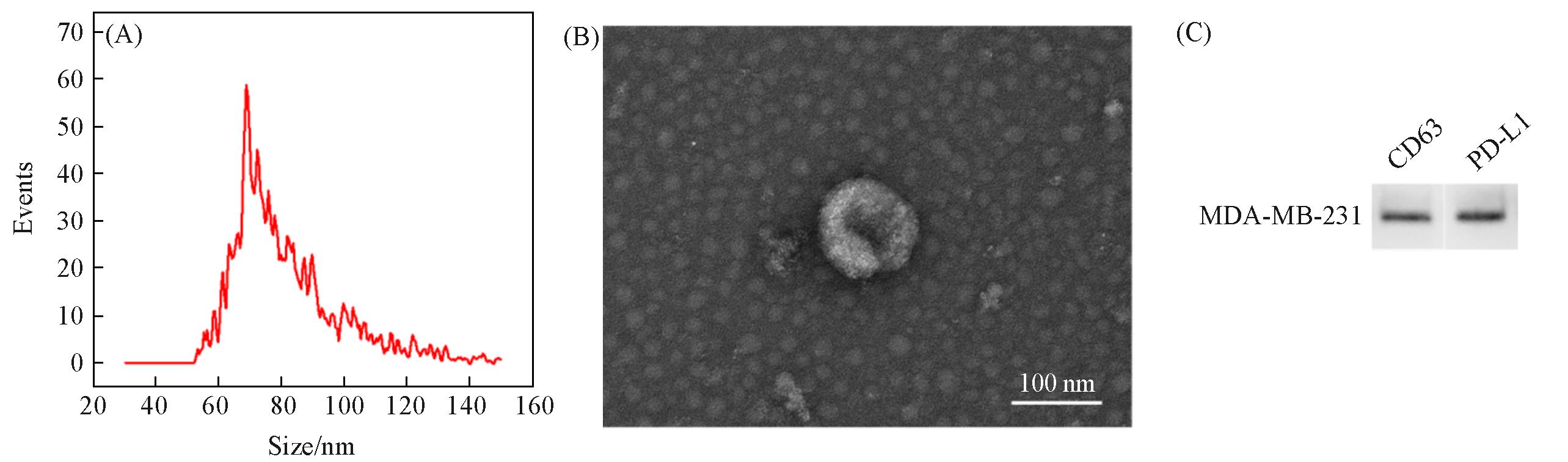
Fig.1 Characterization of size(A), morphology(B) and surface markers(C) of PD⁃L1 positive exosomes
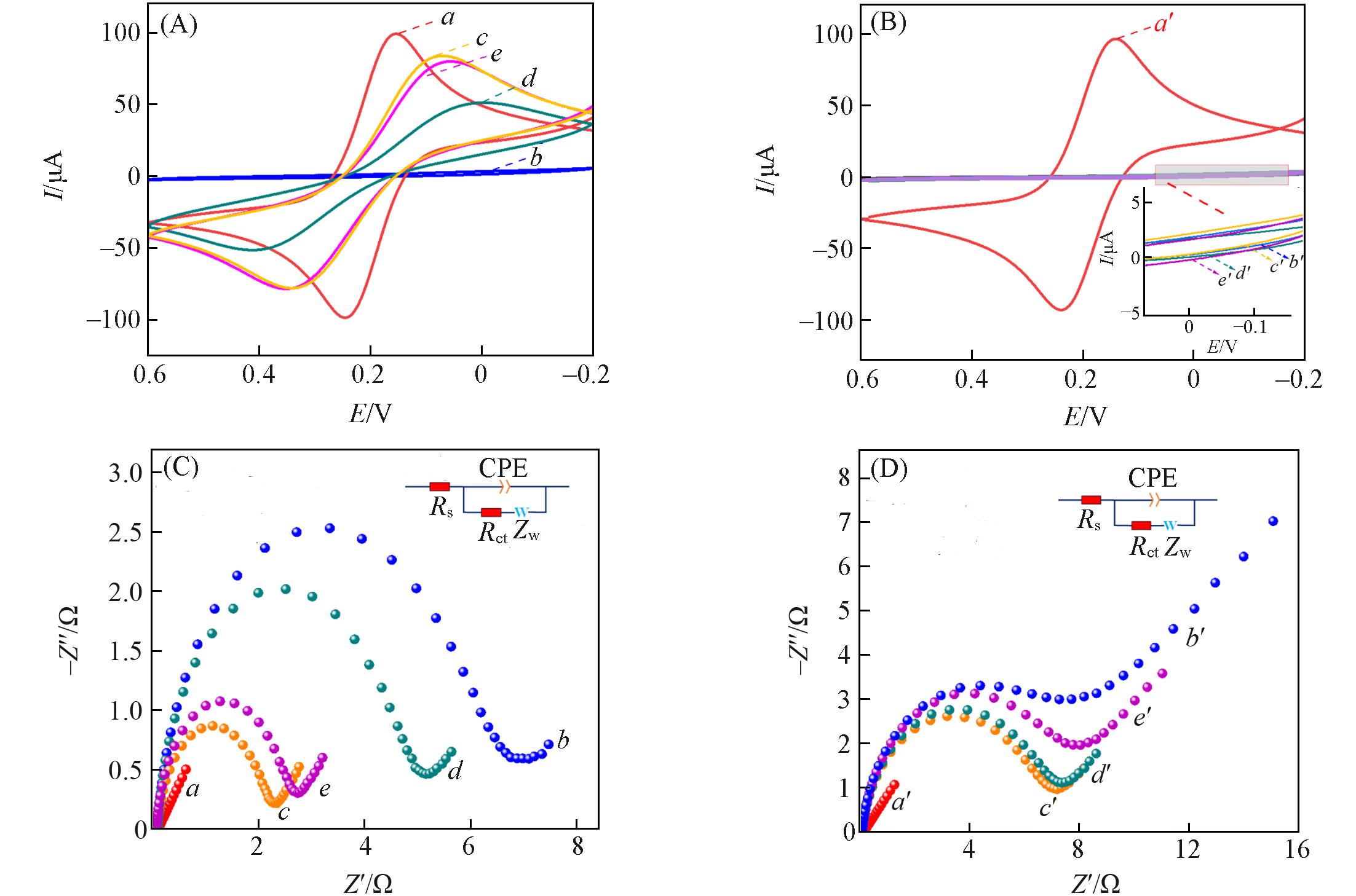
Fig.2 CV responses of GIP(A) and nGIP(B) sensors and EIS characterization of GIP(C) and nGIP(D) sensor under different modification conditions(A, C) a. Bare GCE, b. GIP, c. elution, d. rebound of exosomes, e. lysis of exosomes; (B, D) a′. bare GCE, b′. nGIP, c′. elution, d′. rebound of exosomes, e′. lysis of exosomes.
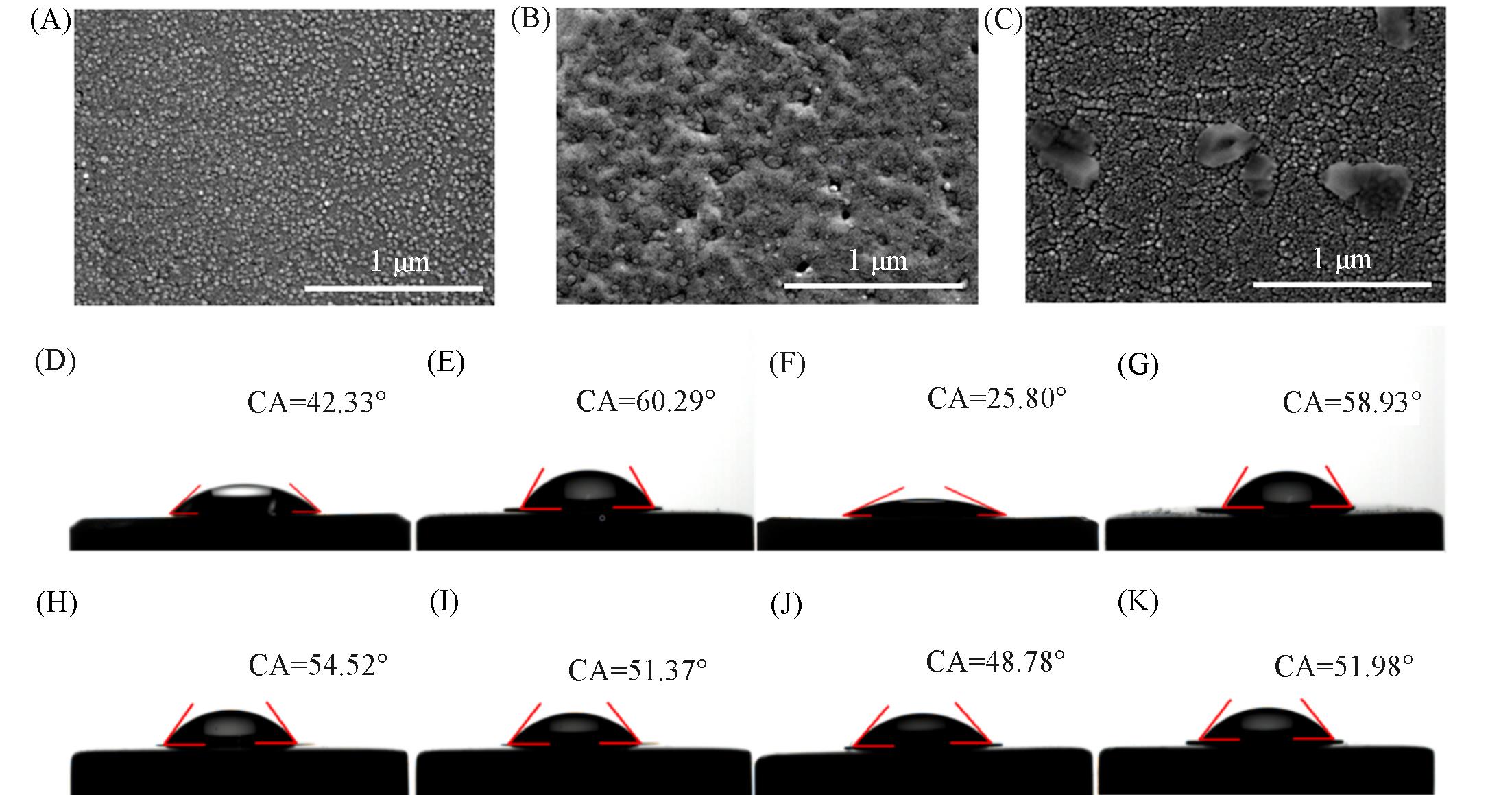
Fig.3 SEM(A—C) and contact angle(D—K) characterizations of GIP membrane under different modification conditions(A, D) GIP membrane; (B, E) GIP membrane after elution; (C) GIP membrane resorbing exosomes; (F) GIP membrane after rebound exosome; (G) GIP membrane after lysis of exosomes; (H) nGIP membrane; (I) nGIP membrane after elution; (J) nGIP membrane after rebound exosome; (K) nGIP membrane after lysis of exosomes.
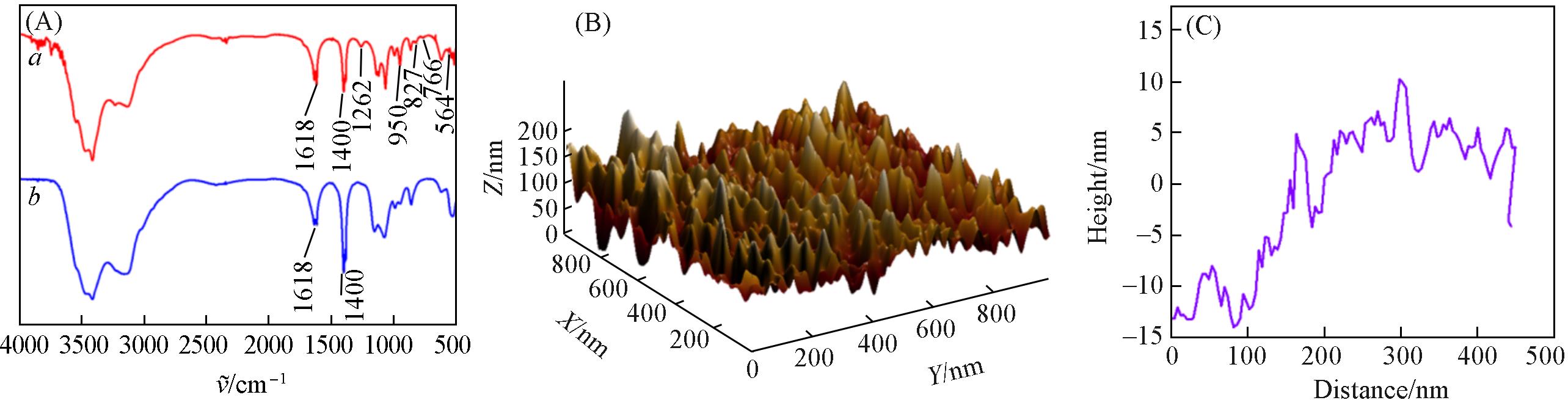
Fig.4 FTIR characterization(A) of GIP membrane before and after elution, the thickness(B) and AFM cross section curve(C) of GIP membrane
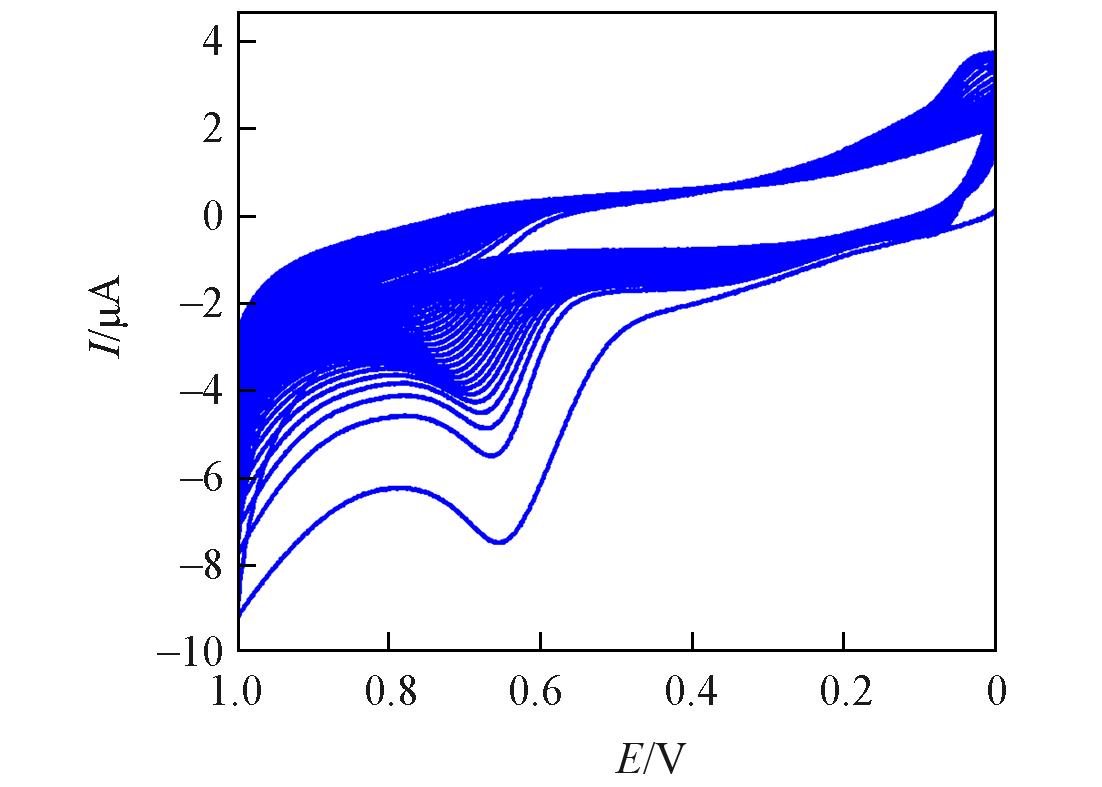
Fig.5 Optimization of the number of electropolymerization cycles
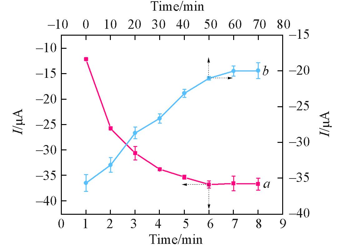
Fig.6 Optimization of elution(a) and rebound(b) time
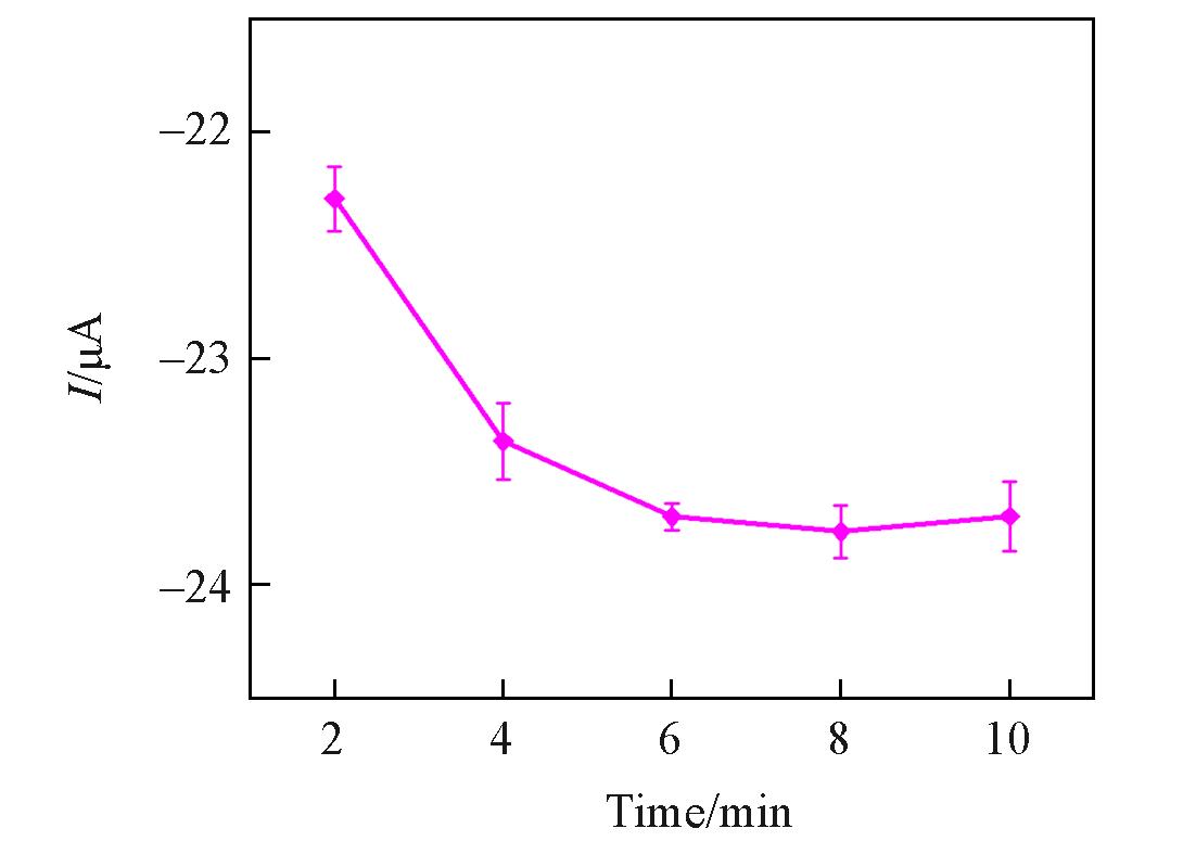
Fig.7 Optimization of time for lysis of exosomes
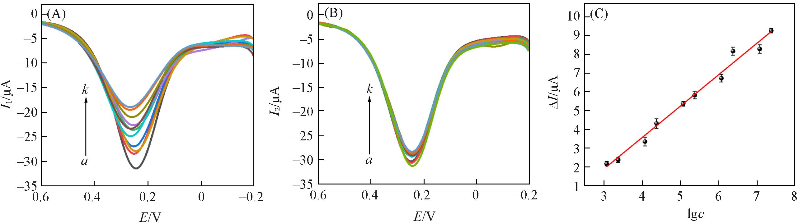
Fig.8 DPV responses of GIP sensor to different concentrations of exosomes(A), DPV responses of GIP sensor after lysing different concentrations of exosomes(B), and calibration curve of GIP sensor for exosomes(C)a—k, c/(particles·mL-1): 0, 2.36×10², 1.18×10³, 2.36×10³, 1.18×10⁴, 2.36×10⁴, 1.18×10⁵, 2.36×10⁵, 1.18×10⁶, 2.36×10⁶, 1.18×10⁷.
| Method | Line range/(particles·mL-1) | LOD/(particles·mL-1) | Ref. |
|---|---|---|---|
| Electrochemical sensor | 1.0×10³—1.0×10¹⁰ | 3.34×10² | [ |
| Surface plasmon resonance | 1.0×10³—1.0×10⁷ | 31.9 | [ |
| Chemiluminescence immunosensor | 4.75×10³—4.75×10⁸ | 7.76×10² | [ |
| Electrochemical biosensor | 1.2×10²—1.2×10⁷ | 38 | [ |
| Fluorescence aptasensor | 5.0×10²—5.0×10⁹ | 13 | [ |
| GIP electrochemical sensor | 2.36×10²—1.18×10⁷ | 93 | This work |
Table 1 Comparison of the GIP sensor with other reported detection methods
| Method | Line range/(particles·mL-1) | LOD/(particles·mL-1) | Ref. |
|---|---|---|---|
| Electrochemical sensor | 1.0×10³—1.0×10¹⁰ | 3.34×10² | [ |
| Surface plasmon resonance | 1.0×10³—1.0×10⁷ | 31.9 | [ |
| Chemiluminescence immunosensor | 4.75×10³—4.75×10⁸ | 7.76×10² | [ |
| Electrochemical biosensor | 1.2×10²—1.2×10⁷ | 38 | [ |
| Fluorescence aptasensor | 5.0×10²—5.0×10⁹ | 13 | [ |
| GIP electrochemical sensor | 2.36×10²—1.18×10⁷ | 93 | This work |
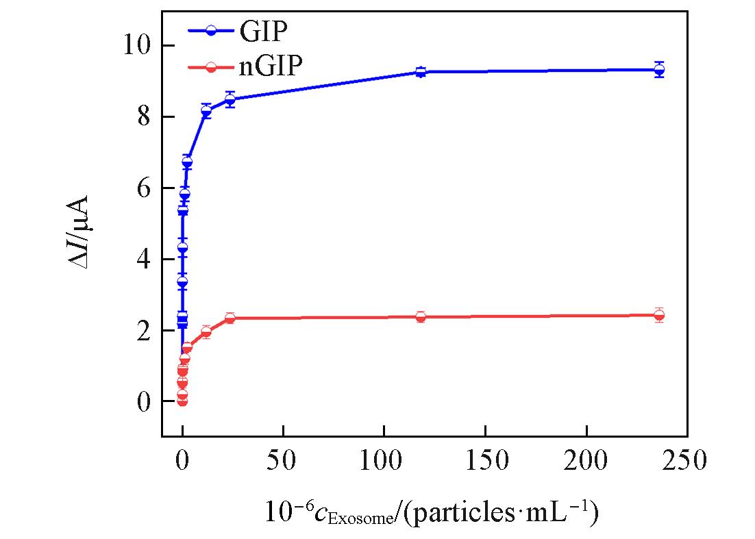
Fig.9 Isothermal adsorption properties of GIP and nGIP sensors for different concentrations of exosomes
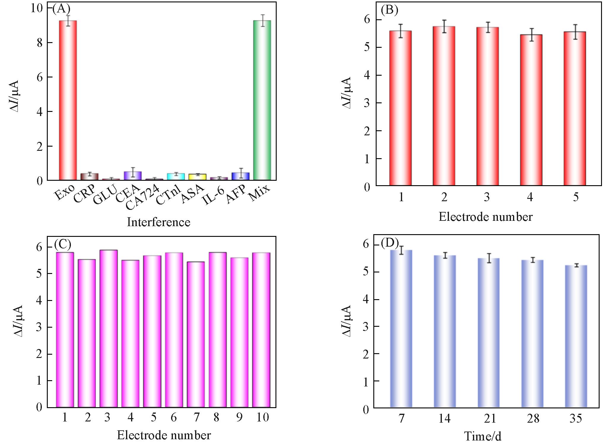
Fig.10 Anti⁃interference(A), inter⁃batch (B) and intra⁃batch(C) reproducibility and stability(D) of the GIP sensor
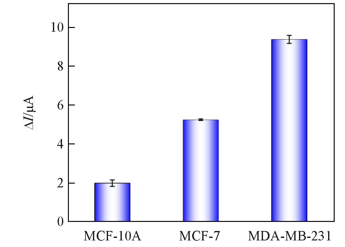
Fig.11 Recognition performance of GIP sensor for exosomes from different sources
| Sample | Found/(particles·mL-1) | RSD(n=5, %) | Added/(particles·mL-1) | Total found/(particles·mL-1) | RSD(n=5, %) | Recovery(%) |
|---|---|---|---|---|---|---|
| Health 1 | 3.35×10⁴ | 2.60 | 1.18×10⁴ | 4.25×10⁴ | 4.29 | 93.82 |
| Health 2 | 6.71×10⁴ | 3.49 | 2.36×10⁴ | 9.03×10⁴ | 2.88 | 99.56 |
| BC 1 | 1.06×10⁶ | 4.23 | 2.36×10⁶ | 3.43×10⁶ | 2.77 | 100.29 |
| BC 2 | 5.35×10⁶ | 3.54 | 2.36×10⁶ | 8.12×10⁶ | 4.43 | 105.32 |
Table 2 Analytical performance of the GIP sensor in clinical samples
| Sample | Found/(particles·mL-1) | RSD(n=5, %) | Added/(particles·mL-1) | Total found/(particles·mL-1) | RSD(n=5, %) | Recovery(%) |
|---|---|---|---|---|---|---|
| Health 1 | 3.35×10⁴ | 2.60 | 1.18×10⁴ | 4.25×10⁴ | 4.29 | 93.82 |
| Health 2 | 6.71×10⁴ | 3.49 | 2.36×10⁴ | 9.03×10⁴ | 2.88 | 99.56 |
| BC 1 | 1.06×10⁶ | 4.23 | 2.36×10⁶ | 3.43×10⁶ | 2.77 | 100.29 |
| BC 2 | 5.35×10⁶ | 3.54 | 2.36×10⁶ | 8.12×10⁶ | 4.43 | 105.32 |
| 1 | Bray F., Laversanne M., Sung H., Ferlay J., Siegel R. L., Soeriomataram I., Jemal A., CA Cancer J. Clin., 2024, 74(3), 229—263 |
| 2 | Xu Y. Y., Gong M. Y., Wang Y., Yang Y., Liu S., Zeng Q. B., Sci. Data, 2023, 10(1), 334 |
| 3 | McDonald E. S., Clark A. S., Tchou J., Zhang P., Freedman G. M. J., Nucl. Med., 2016, 57, 9S—16S |
| 4 | Bevers T. B., Helvie M., Bonaccio E., Calhoun K. E., Daly M. B., Farrar W. B., Garber J. E., Gary R., Greenberg C. C., Greenup R., Hansen N. M., Harris R. E., Heerdt A. S., Helsten T., Hodgkiss L., Hoyt T. L., Huff J. G., Jacobs L., Lehman C. D., Monsees B., Niell B. L., Parker C. C., Pearlman M., Philpotts L., Shepardson L. B., Smith M. L., Stein M., Tumyan L., Williams C., Bergman M. A., Kumar R. J., Natl. Compr. Canc. Netw, 2018, 16(11), 1362—1389 |
| 5 | Jeong S., Park M. J., Song W., Kim H. S., Clin. Biochem., 2020, 78, 43—57 |
| 6 | Yu W., Hurley J., Roberts D., Chakrabortty S. K., Enderle D., Noerholm M., Berakefield X. O., Skog J. K., Ann. Oncol., 2021, 32(4), 466—477 |
| 7 | Jabbari N., Akbariazar E., Feqhhi M., Rahbarghazi R., Rezaie J. J., Cell. Physiol., 2020, 235(10), 6345—6356 |
| 8 | Xu R., Rai A., Chen M., Suwakulsiri W., Greening D. W., Simpson R. J., Nat. Rev. Clin. Oncol., 2018, 15(10), 617—638 |
| 9 | Tang M. K. S., Wong A. S., Cancer Lett., 2015, 367(1), 26—33 |
| 10 | Jenjaroenpun P., Kremenska Y., Nair V. M., Kremenskoy M., Joseph B., Kurochkin I. V., PeerJ, 2013, 1, e201 |
| 11 | Fan W. J., Han P. H., Feng Q. Y., Sun Y. Y., Ren W., Lawson T., Liu C. H., Anal. Chem., 2022, 94(4), 2172—2179 |
| 12 | Yu Z. Q., Yang Y., Fang W. M., Hu P., Liu Y. B., Shi J. L., ACS Nano, 2023, 17(12), 11384-11395 |
| 13 | Li C.W., Lim S. O., Chung E. M., Kim Y. S., Park A. H., Yao J., Cha J. H., Xia W. Y., Chan L. C., Kim T., Chang S. S., Lee H. H., Chou C. K., Liu Y. L., Yeh H. C., Perillo E. P., Dunn A. K., Kuo C. W., Khoo K. H., Hsu J. L., Wu Y., Hsu J. M., Yamaguchi H., Huang T. H., Sahin A. A., Hortobagyi G. N., Yoo S. S., Hung M. C., Cancer Cell, 2018, 33(2), 187—201. e10 |
| 14 | Benicky J., Sanda M., Brnakova Kennedy Z., Grant O. C., Woods R. J., Zwart A., Goldman R. J., Proteome Res., 2020, 20(1), 485—497 |
| 15 | Li C. W., Lim S. O., Xia W. Y., Lee H. H., Chan L. C., Kuo C. W., Khoo K. H., Chang S. S., Cha J. H., Kim T., Hsu J. L., Wu Y., Hsu J. M., Yamaguchi H., Ding Q. Q., Wang Y., Yao J., Lee C. C., Wu H. J., Sahin A. A., Yu D. H., Hortobagyi G. N., Hung M. C., Nat. Commun., 2016, 7(1), 12632 |
| 16 | Zhu L., Sun H. T., Wang S., Huang S. L., Zheng Y., Wang C. Q., Hu B. Y., Qin W., Zou T. T., Fu Y., Shen X. T., Zhu W. W., Geng Y., Lu L., Jia H. L., Qin L. X., Dong Q. Z., J. Hematol. Oncol., 2020, 13, 1—24 |
| 17 | Zhang M. D., Jin K., Gao L., Zhang Z. K., Li F., Zhou F. F., Zhang L., Small Methods, 2018, 2(9), 1800021 |
| 18 | Hu J. J., Mao Z. H., Lu Y. K., Chen Q., Xia J. J., Deng H., Chen H. X., Biosens. Bioelectron., 2023, 235, 115379 |
| 19 | Zhang X. Y., Zhu X. Y., Li Y. F., Hai X., Bi S., Talanta, 2023, 258, 124456 |
| 20 | Zhou J. Q., Lin Q. Y., Huang Z. P., Xiong H. W., Yang B., Chen H., Kong J. L., Anal. Chem., 2022, 94(15), 5723—5728 |
| 21 | Diao X. K., Li X. L., Hou S. P., Li H. J., Qi G. H., Jin Y. D., Anal. Chem., 2023, 95(19), 7552—7559 |
| 22 | Wu L. C., Li X. L., Miao H. H., Xu J. J., Pan G. Q., View, 2022, 3(3), 20200170 |
| 23 | Mazzotta E., Di Giulio T., Malitesta C., Anal. Bioanal. Chem., 2022, 414(18), 5165—5200 |
| 24 | Jiang Z. J., Luo K., Zeng H. H., Li J. P., ACS Sens., 2024, 9(3), 1252—1260 |
| 25 | Li J. P., Ma X. H., Li M. X., Zhang Y., Biosens. Bioelectron., 2018, 99, 438—442 |
| 26 | Li P. F., Liu Z., Chem. Soc. Rev., 2024, 53, 1870—1891 |
| 27 | Cao Y., Wang Y., Yu X. M., Jiang X. H., Li G., Zhao J., Biosens. Bioelectron., 2020, 166, 112452 |
| 28 | Liu H. Z., Zhou Y. Y., Chang W. W., Zhao X. L., Hu X. J., Koh K., Chen H. X., Biosens. Bioelectron., 2024, 262, 116527 |
| 29 | Wang M. L., Shu J. N., Wang Y. S., Zhang W. C., Zheng K. Y., Zhou S. N., Yang D. L., Cui H., ACS Sens., 2024, 9(6), 3444—3454 |
| 30 | Wang M. H., Lin Y. X., Wu S., Deng Y., Zhang Y. Y., Yang J., Li G. X., Sens. Actuators, B, 2022, 362, 131813 |
| 31 | Zhu N. N., Li G. H., Zhou J., Kang K., Ying B. W., Yi Q. Y., Wu Y. J., Mater. Chem., 2021, 9(10), 2483—2493 |
| [1] | 吴梦鸽, 楚占营, 孟波, 叶子弘, 翟睿. 外泌体处理温度对其蛋白质组学分析的影响[J]. 高等学校化学学报, 2025, 46(5): 20250023. |
| [2] | 宁佳雨, 郝鹏飞, 王峰, 叶家全, 崇羽. 功能性多酚-精氨酸自组装纳米药物用于乳腺癌放射增敏[J]. 高等学校化学学报, 2025, 46(1): 20240226. |
| [3] | 马爽, 吕明杨, 张赐童, 刘轶. 化疗-光热治疗-自促进饥饿治疗纳米治疗平台的构筑及对乳腺癌的治疗效果[J]. 高等学校化学学报, 2025, 46(1): 20240467. |
| [4] | 郑德论, 张锐龙. 基于p-n异质结CuO/TiO2复合物高效的载流子分离能力构建超灵敏AFP光电化学分析[J]. 高等学校化学学报, 2024, 45(8): 20240183. |
| [5] | 韦朝鲜, 李南盛, 庞元昊, 张云, 金文英, 袁亚利. 基于低共熔策略合成碳纳米聚合物用于多种生物小分子的同时电化学检测[J]. 高等学校化学学报, 2024, 45(7): 20240103. |
| [6] | 陈晓萍, 王旭潭, 刘宁, 汪庆祥, 倪建聪, 杨伟强, 林振宇. MOFs基微流控电化学芯片对多种重金属离子的实时在线检测[J]. 高等学校化学学报, 2024, 45(2): 20230395. |
| [7] | 李嘉慧, 张剑, 严龙, 丰芸, 章家立, 刘永鑫, 杨绍明. 诺氟沙星印迹电化学传感器的制备及检测性能[J]. 高等学校化学学报, 2024, 45(12): 20240322. |
| [8] | 李世宣, 蒙化, 尹学虎, 易锦飞, 马丽红, 张艳丽, 王红斌, 杨文荣, 庞鹏飞. 基于石墨烯/金纳米粒子/碳化钛复合材料构建双室酶生物燃料电池自供能葡萄糖生物传感器[J]. 高等学校化学学报, 2024, 45(12): 20240301. |
| [9] | 曹宜青, 侯静欣, 刘建业, 李嫣. 体液外泌体代谢组学研究进展和挑战[J]. 高等学校化学学报, 2024, 45(11): 20240324. |
| [10] | 靳莹, 张俊杰, 张毅欣, 袁悦, 韩珍珍. 外泌体分离和蛋白质组学分析的研究进展[J]. 高等学校化学学报, 2024, 45(11): 20240305. |
| [11] | 李玉龙, 谢发婷, 管燕, 刘嘉丽, 张贵群, 姚超, 杨通, 杨云慧, 胡蓉. 基于银离子与DNA相互作用的比率型电化学传感器用于银离子的检测[J]. 高等学校化学学报, 2022, 43(8): 20220202. |
| [12] | 魏闯宇, 陈艳丽, 姜建壮. 基于乙硫基取代的三层酞菁铕二聚体修饰ITO电极构筑电化学多巴胺和尿酸传感器[J]. 高等学校化学学报, 2022, 43(1): 20210582. |
| [13] | 吴杨仪, 陈建平, 艾益静, 汪庆祥, 高飞, 高凤. 2-(2-羟基-3-甲氧基苯基)-C60的合成及在花椰菜花叶病毒启动子DNA传感检测中的应用[J]. 高等学校化学学报, 2021, 42(6): 1754. |
| [14] | 黄玲, 庄梓健, 李翔, 石沐玲, 刘高强. 基于核酸适体的外泌体分子识别研究进展[J]. 高等学校化学学报, 2021, 42(11): 3493. |
| [15] | 付可飞, 连惠婷, 魏晓峰, 孙向英, 刘斌. 环糊精基阻抗型传感器的制备及对L-半胱氨酸的识别[J]. 高等学校化学学报, 2020, 41(4): 706. |
| 阅读次数 | ||||||
|
全文 |
|
|||||
|
摘要 |
|
|||||