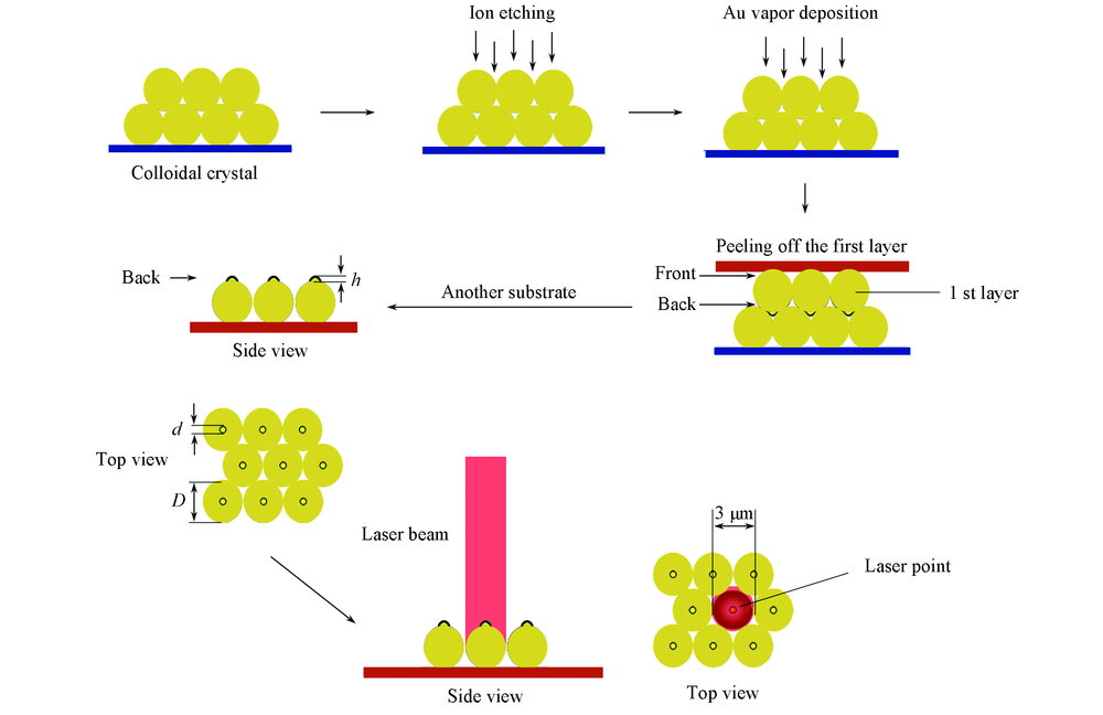
| [1] |
Fleischmann M., Hendra P. J., McQuillan A. J., Chem. Phys. Lett., 1974, 26, 163—166
|
| [2] |
Jeanmaire D. L., van Duyne R. P., J. Electroanal. Chem. Interf. Electrochem., 1977, 84, 1—20
|
| [3] |
Sebastian S., Angew. Chem. Int. Ed., 2014, 53, 4756—4795
|
| [4] |
Cialla D., Marz A., Bohme R., Anal. Bioanal. Chem., 2012, 403(1), 27—54
|
| [5] |
Han X. X., Zhao B., Ozaki Y., Anal. Bioanal. Chem., 2009, 394(8), 1719—1727
|
| [6] |
Yui H., Anal. Bioanal. Chem., 2010, 397(3), 1181—1190
|
| [7] |
Eduardo G. R., Ramon A. A. P., Luca G., Chem. Soc. Rev., 2018, 47, 4909—4923
|
| [8] |
Halvorson R. A., Vikesland P. J., Environ. Sci. Technol., 2010, 44(20), 7749—7755
|
| [9] |
Sebastien B., Sophie L., Chem. Phys. Chem., 2018, 19, 8—18
|
| [10] |
Chou S. Y., Yu C. C., Yen Y. T., Lin K. T., Chen H. L., Su W. F., Anal. Chem., 2015, 87, 6017—6024
|
| [11] |
Wu H. Y., Huang W. L., Michael H. H., Cryst. Growth Des., 2007, 7(4), 831—835
|
| [12] |
Sun Y. G., Xia Y. N., Science, 2002, 298, 2176—2179
|
| [13] |
Kim D. Y., Yu T., Cho E. C., Angew. Chem. Int. Ed., 2011, 50(28), 6328—6331
|
| [14] |
Chen C. L., Furusho H., Mori H., Nanotechnology, 2009, 20(40), 5605—5608
|
| [15] |
Ashkin A., Dziedzic J. M., Bjorkholm J. E., Chu S., Opt. Lett., 1986, 11, 288—290
|
| [16] |
Svoboda K., Block M., Opt. Lett., 1994, 19, 930—932
|
| [17] |
Prikulis J., Svedberg F., Kall M., Enger J., Ramser K., Goksor M., Hanstorp D., Nano Lett., 2004, 4, 115—118
|
| [18] |
Svedberg F., Kall M., Svedberg F., Kall M., Faraday Discuss., 2006, 132, 35—44
|
| [19] |
Zhang G., Wang D.Y., Chem. Asian J., 2009, 4(2), 236—245
|
| [20] |
Ai B., Mohwald H., Wang D. Y., Zhang G., Adv. Mater. Interfaces, 2017, 4, 1600271
|
| [21] |
Zhang G., Wang D. Y., Mohwald H., Nano Lett., 2005, 5(1), 143—146
|
| [22] |
Zhang G., Wang D. Y., Mohwald H., Angew. Chem. Int. Ed., 2005, 44(47), 7767—7770
|
| [23] |
Xu H., Aizpurua J., Kall M., Apell P., Phys. Rev. E, 2000, 62, 4318—4324
|
| [24] |
Liao P. F., Wokaun A., J. Chem. Phys., 1982, 76, 751—752
|
| [25] |
Boyd G. T., Rasing T., Leite J. R. R., Shen Y. R., Phys. Rev. B, 1984, 30, 519—522
|
| [26] |
Chen C. Y., Burstein E., Phys. Rev. Lett., 1980, 45, 1287—1291
|
| [27] |
Inoue M., Ohtaka K., J. Phys. Soc. Jpn., 1983, 52, 3853—3864
|
| [28] |
Dollish F. R., Fateley W. G., Bentley F. F., Characteristic Raman Frequencies of Organic Compounds, John Willey & Sons, New York, 1973
|
| [29] |
Lee I., Han S. W., Kim K., J. Raman Spectrosc., 2001, 32, 947—952
|
| [30] |
Carron K. T., Hurley L. G., J. Phys. Chem. B, 2005, 95, 9979—9984
|
| [31] |
Nie S. M., Lipscomb L. A., Yu N. T., Appl. Spectrosc. Rev., 1991, 26(3), 203—276
|
| [32] |
Majoube M., Henry M., Spectrochim. Acta, 1991, 47A, 1459—1466
|
 )
)