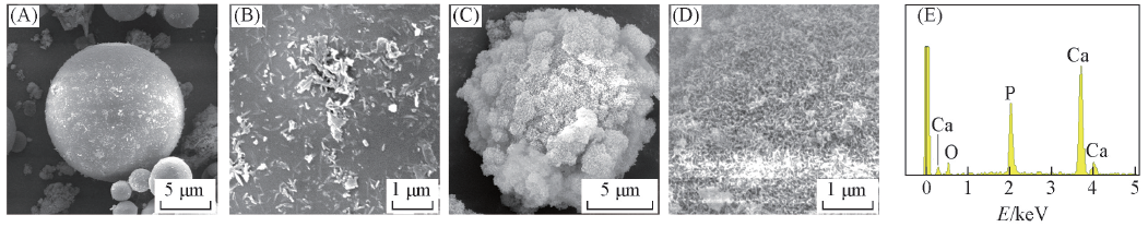
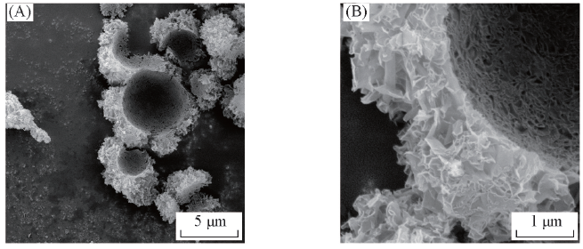
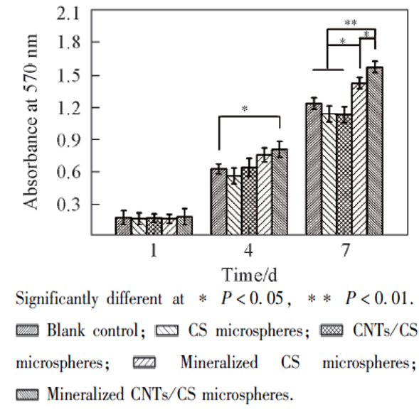
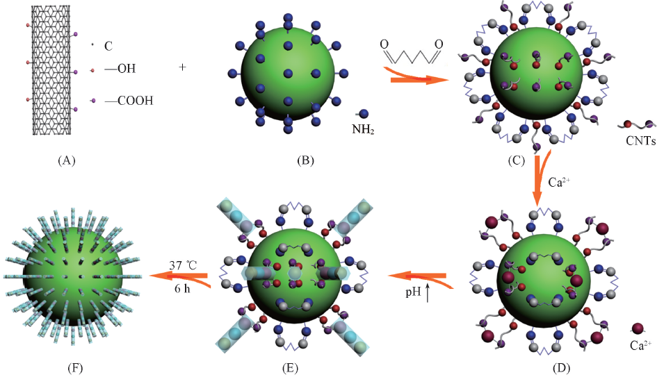
| [1] |
Tian H. Y., Tang Z. H., Zhuang X. L., Chen X. S., Jing X. B., Prog. Polym. Sci., 2012, 37(2), 237—280
|
| [2] |
Xie J., Li X., Jiang C. J., Lee R. J., Zhou Y. L., Teng L. S., Chem. Res. Chinese Universities, 2013, 29(5), 1003—1005
|
| [3] |
Ruckh T. T., Kumar K., Kipper M. J., Popat K. C., Acta Biomater., 2010, 6(8), 2949—2959
|
| [4] |
Anderson J. M., Shive M. S., Adv. Drug Del. Rev., 2012, 64, 72—82
|
| [5] |
Li B., Xu W. F., Liao X. L., J. Inorg. Mater., 2014, 29(10), 1009—1017
|
|
(李波, 徐文峰, 廖晓玲. 无机材料学报,2014, 29(10), 1009—1017)
|
| [6] |
Chen R., Yang J. R., Yang X. Y., Wang Y. G., Li R. S., Wang R. L., Wang Y. S., Chem. J. Chinese Universities, 2012, 33(7), 1586—1590
|
|
(陈柔, 杨金荣, 杨晓英, 王玉玫, 李蓉珊, 王润玲, 王银松. 高等学校化学学报,2012, 33(7), 1586—1590)
|
| [7] |
Zhou Z. H., Zhou J. N., Liu L. H., Yi Q. F., Liu Q. Q., Zeng W. N., Yang Z. M., J. Macromol. Sci. B, 2012, 51(4), 777—785
|
| [8] |
Qian Z., Zhang Z. C., Li H. M., Liu H. R., Hu Z. Q., J. Polym. Sci. Pol. Chem., 2008, 46(1), 228—237
|
| [9] |
Kong M., Chen X. G., Liu C. S., Liu C. G., Meng X. H., Yu L. J., Colloid Surface B Biointerfaces, 2008, 65(2), 197—202
|
| [10] |
Huang D., Zuo Y., Zou Q., Wang Y. Y., Gao S. B., Wang X. Y., Liu H. H., Li Y. B., J. Biomed. Mater. Res. B Appl. Biomater., 2012, 100(1), 51—57
|
| [11] |
Yang C. C., Lin C. C., Liao J. W., Yen S. K., Mater. Sci. Eng. C, 2013, 33(4), 2203—2212
|
| [12] |
Li J., Han Z. J., Wei Y., Niu L. L., Liu Y., Lu G. Y., Huang D., J. Inorg. Mater., 2014, 29(12), 1327—1332
|
|
(李健, 韩志军, 魏延, 牛璐璐, 刘宇, 路国运, 黄棣. 无机材料学报,2014, 29(12), 1327—1332)
|
| [13] |
Eder D., Chem. Rev., 2010, 110(3), 1348—1385
|
| [14] |
Balasubramanian K., Burghard M., Small, 2005, 1(2), 180—192
|
| [15] |
Doane T. L., Burda C., Chem. Soc. Rev., 2012, 41(7), 2885—2911
|
| [16] |
Zhang J., Zou H. L., Qing Q., Yang Y. L., Li Q. W., Liu Z. F., Guo X. Y., Du Z. L., J. Phys. Chem. B, 2003, 107(16), 3712—3718
|
| [17] |
Harper A., Anderson M. R., Sensors, 2010, 10(9), 8248—8274
|
| [18] |
Feng K. J., Yao Y. L., Shen G. L., Yu R. Q., Chem. J. Chinese Universities, 2012, 33(8), 1676—1680
|
|
(封科军, 姚艳玲, 沈国励, 俞汝勤. 高等学校化学学报,2012, 33(8), 1676—1680)
|
| [19] |
Supronowicz P. R., Ajayan P. M., Ullmann K. R., Arulanandam B. P., Metzger D. W., Bizios R., J. Biomed. Mater. Res. A, 2002, 59(3), 499—506
|
| [20] |
Saito T., Matsushige K., Tanaka K., Physica B: Condensed Matter, 2002, 323(1), 280—283
|
| [21] |
Desai K., Kit K., Li J. J., Zivanovic S., Biomacromolecules, 2008, 9(3), 1000—1006
|
| [22] |
Zou Q., Li Y. B., Zhang L., Zuo Y., Li J. F., Li J. D., J. Biomed. Mater. Res. A, 2009, 89(4), 1108—1117
|
| [23] |
Lu X. Y., Wang X. H., Qu S. X., Weng J., J. Inorg. Mater., 2008, 23(2), 332—336
|
|
(卢晓英, 王秀红, 屈树新, 翁杰. 无机材料学报,2008, 23(2), 332—336)
|
| [24] |
Yang J., Chen Z. Q., Hu C. H., J. Third Mil. Med. Univ., 2013, 34(14), 1480—1484
|
|
(杨俊, 陈治清, 胡常红. 第三军医大学学报,2013, 34(14), 1480—1484)
|
 ), 杜晶晶1, 魏延1, 胡超凡1, 叶家业1, 陈维毅1,2(