

高等学校化学学报 ›› 2024, Vol. 45 ›› Issue (11): 20240324.doi: 10.7503/cjcu20240324
曹宜青, 侯静欣, 刘建业, 李嫣( )
)
收稿日期:2024-07-01
出版日期:2024-11-10
发布日期:2024-08-23
通讯作者:
李嫣
E-mail:yanli@fudan.edu.cn
基金资助:
CAO Yiqing, HOU Jingxin, LIU Jianye, LI Yan()
Received:2024-07-01
Online:2024-11-10
Published:2024-08-23
Contact:
LI Yan
E-mail:yanli@fudan.edu.cn
Supported by:摘要:
外泌体作为疾病诊断、 监测和治疗的非侵入性生物标志物, 在体液中广泛存在. 作为细胞间信使, 外泌体携带有丰富的蛋白质、 核酸和代谢物, 目前大多数研究集中在蛋白质和RNA上. 最近, 外泌体代谢组学在疾病检测和疾病病理生理学研究中展示出临床价值和潜在优势, 但仍存在诸多挑战, 特别是外泌体分离和代谢物检测. 本文综合评述了外泌体分离和代谢物检测方面的技术进展和挑战, 并通过案例研究, 展示了不同类型体液的外泌体代谢物作为生物标志物在疾病早期诊断和治疗中的潜力.
中图分类号:
TrendMD:
曹宜青, 侯静欣, 刘建业, 李嫣. 体液外泌体代谢组学研究进展和挑战. 高等学校化学学报, 2024, 45(11): 20240324.
CAO Yiqing, HOU Jingxin, LIU Jianye, LI Yan. Advances and Challenges of Exosome Metabolomics in Body Fluids. Chem. J. Chinese Universities, 2024, 45(11): 20240324.
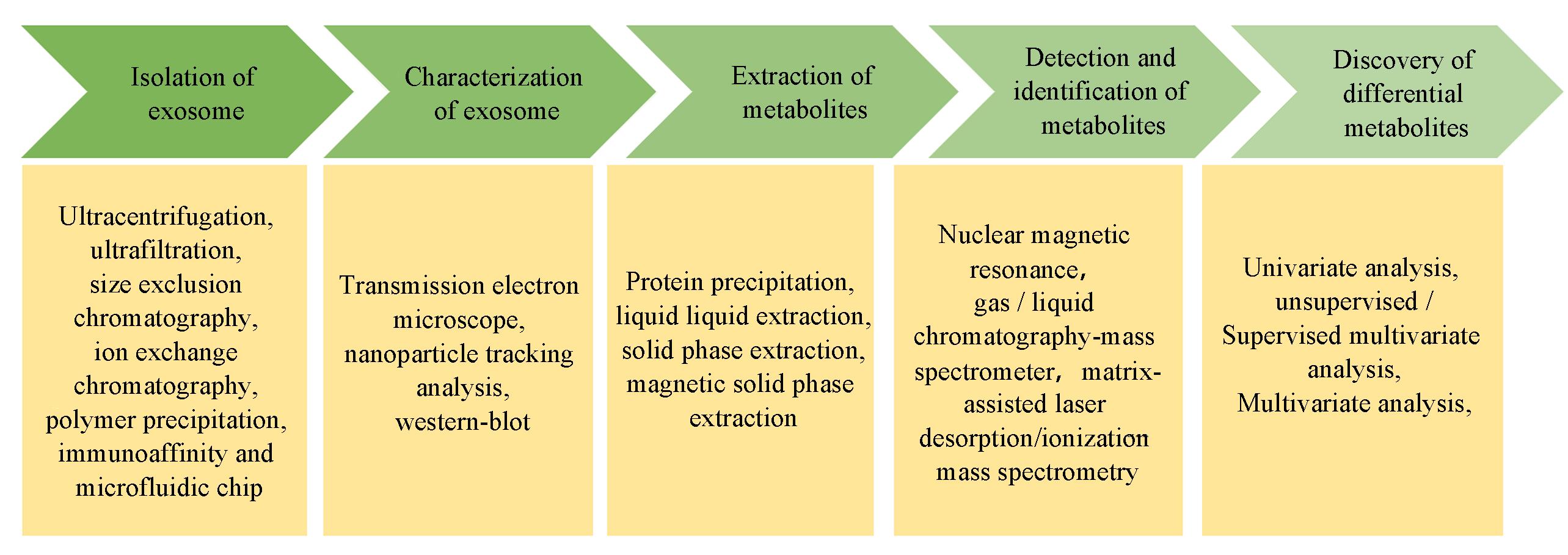
Fig.1 Basic workflow for exosome metabolomics research
| Separation technique | Principle | Advantage | Disadvantage |
|---|---|---|---|
| Ultracentrifugation | Density | Mature method, “gold standard” | Expensive equipment, time⁃consuming(>4 h) |
| Ultrafiltration | Size | Simple operation, suitable for small volumes | Low purity, affects exosome integrity |
| Polymer precipitation | Solubility | Simple, no expensive equipment required | Low purity, affects exosome integrity |
| Size exclusion chromatography | Size | High purity, good reproducibility | High maintenance cost, low concentration, requires additional concentration steps |
| Ion exchange chromatography | Charge | High purity | Requires multiple optimizations, affects exosome integrity |
| Immunoaffinity | Affinity | High purity, high specificity | High cost, not suitable for large⁃scale |
| Microfluidic chip | Fluid dynamics | Automation and high throughput | High equipment cost, technical complexity |
Table 1 Methods for separating exosomes from body fluids
| Separation technique | Principle | Advantage | Disadvantage |
|---|---|---|---|
| Ultracentrifugation | Density | Mature method, “gold standard” | Expensive equipment, time⁃consuming(>4 h) |
| Ultrafiltration | Size | Simple operation, suitable for small volumes | Low purity, affects exosome integrity |
| Polymer precipitation | Solubility | Simple, no expensive equipment required | Low purity, affects exosome integrity |
| Size exclusion chromatography | Size | High purity, good reproducibility | High maintenance cost, low concentration, requires additional concentration steps |
| Ion exchange chromatography | Charge | High purity | Requires multiple optimizations, affects exosome integrity |
| Immunoaffinity | Affinity | High purity, high specificity | High cost, not suitable for large⁃scale |
| Microfluidic chip | Fluid dynamics | Automation and high throughput | High equipment cost, technical complexity |
| Nanomaterial | Mechanism of exosome separation | Target | Source of exosome | Analyte | Analysis platform | Ref. |
|---|---|---|---|---|---|---|
| CD9⁃HPLC⁃IAC | Antibody affinity | CD9 | Serum | Protein | LC⁃MS/MS | [ |
| CoMPC@Au⁃Apt | Aptamer affinity | CD63 | Urine | Metabolite | MALDI⁃MS | [ |
| TiO2 | Ti⁃O and phosphate groups interaction | Lipid bilayer | Serum | Protein | LC⁃MS/MS | [ |
| Fe3O4@PDA@UiO⁃66⁃NH2 | Zr⁃O and phosphate groups interaction | Lipid bilayer | Urine | Phosphoryl⁃peptide | LC⁃MS/MS | [ |
| Fe3O4@SiO2@Eu2O3 | Eu⁃O and phosphate groups interaction | Lipid bilayer | Plasma | Metabolite | LC⁃MS/MS | [ |
| CaTiO3/Al3+/Pr3+/Sm3+ | CaTiO3, Al3+, Pr3+, Sm3+ and phosphate groups interaction | Lipid bilayer | Serum | Protein | MALDI⁃MS | [ |
| Phospholipid⁃MIP | MIP recognition for phosphatidylserine(PS) | Lipid bilayer | Plasma | Protein | LC⁃MS/MS | [ |
| Heparin⁃agarose beads | Heparin and proteoglycans interaction | Lipid bilayer | Plasma | RNA | RT⁃qPCR | [ |
| EXODUS | Size exclusion | — | Plasma | Metabolite | LC⁃MS/MS | [ |
| SNAPs | Size exclusion | — | Urine | Protein | LC⁃MS/MS | [ |
Table 2 Nanomaterials for exosome isolation
| Nanomaterial | Mechanism of exosome separation | Target | Source of exosome | Analyte | Analysis platform | Ref. |
|---|---|---|---|---|---|---|
| CD9⁃HPLC⁃IAC | Antibody affinity | CD9 | Serum | Protein | LC⁃MS/MS | [ |
| CoMPC@Au⁃Apt | Aptamer affinity | CD63 | Urine | Metabolite | MALDI⁃MS | [ |
| TiO2 | Ti⁃O and phosphate groups interaction | Lipid bilayer | Serum | Protein | LC⁃MS/MS | [ |
| Fe3O4@PDA@UiO⁃66⁃NH2 | Zr⁃O and phosphate groups interaction | Lipid bilayer | Urine | Phosphoryl⁃peptide | LC⁃MS/MS | [ |
| Fe3O4@SiO2@Eu2O3 | Eu⁃O and phosphate groups interaction | Lipid bilayer | Plasma | Metabolite | LC⁃MS/MS | [ |
| CaTiO3/Al3+/Pr3+/Sm3+ | CaTiO3, Al3+, Pr3+, Sm3+ and phosphate groups interaction | Lipid bilayer | Serum | Protein | MALDI⁃MS | [ |
| Phospholipid⁃MIP | MIP recognition for phosphatidylserine(PS) | Lipid bilayer | Plasma | Protein | LC⁃MS/MS | [ |
| Heparin⁃agarose beads | Heparin and proteoglycans interaction | Lipid bilayer | Plasma | RNA | RT⁃qPCR | [ |
| EXODUS | Size exclusion | — | Plasma | Metabolite | LC⁃MS/MS | [ |
| SNAPs | Size exclusion | — | Urine | Protein | LC⁃MS/MS | [ |
| Exosome source | Isolation method | Metabolomic profiling | Disease type | Application | Ref. |
|---|---|---|---|---|---|
| Plasma | Differential ultracentrifugation | UHPLC⁃Q⁃TOF⁃MS/MS | Nonalcoholic fatty liver disease(NAFLD) | A distinct change in fatty acids and related pathways in nonalcoholic fatty liver disease patients | [ |
| Plasma | Ultracentrifugation | LC⁃MS | Breast cancer | Targeting succinic acid and L⁃lactic acid in patients with RD after NAC to improve their disease course | [ |
| Plasma | A home⁃constructed device called EXODUS | UPLC⁃MS/MS | Esophageal squamous cell carcinoma | Novel biomarkers for diagnosis and prognosis of ESCC | [ |
| Plasma | Invitrogen total exosome isolation kit | UPLC⁃TOF⁃MS/MS | COVID⁃19 | GM3⁃enriched exosomes may partake in pathological processes related to COVID⁃19 pathogenesis | [ |
| Serum | Ultracentrifugation | LC⁃MS/MS | Castration resistant prostate cancer(CRPC) | Diagnostic TFC, PCa and CRPC by differential metabolites | [ |
| Serum | Size exclusion chromatography | UPLC⁃MS/MS | High⁃altitude cerebral edema(HACE) | Distinguish the HCs and HACE patients | [ |
| Serum | qEV column | UPLC⁃MS/MS | Acute mountain sicknesses | Identified 5 metabolites to distinguish hypoxic preconditioning participants and control subjects | [ |
| Serum | ExoQuick, a fast⁃ acting exosome precipitation solution | UPLC⁃MS/MS | Diabetic nephropathy(DN) and diabetic retinopathy(DR) | 1⁃MH loss may be linked to the pathogenicity of diabetic endothelial dysfunction in DR/DN | [ |
| Serum | qEV column | UPLC⁃MS/MS | Bipolar disorder | 15 Exosomal metabolites to distinguish BD patients and other major psychiatric diseases | [ |
| Urine | MXene@TiO2/Fe3O4 | LDI MS | Bladder cancer | Three biomarkers are indication of treatment in individual patients | [ |
| Urine | HPL⁃SEC | MALDI⁃TOF MS | Systemic lupus erythematosus(SLE) | Screen differential expressions of metabolite signals in the HC and SLE groups | [ |
| Urine | Serial centrifugation | MS | Prostate cancer | Potential prostate cancer biomarkers | [ |
| Urine | Ultracentrifuged | UHPLC⁃MS | Prostate cancer | Distinguish prostate cancer pathogenesis and progression | [ |
| Urine | Ultracentrifuged | 1H NMR | Cardiovascular risk | Three metabolites can be CV risk biomarkers | [ |
| Tissue and urine | Ultrafiltration and ultracentrifugation | LC⁃ESI⁃MS/MS | Prostate cancer | Prove uEVs are potential prostate cancer biomarkers | [ |
| Pleural effusions | Differential ultracentrifugation | LC⁃MS/MS | Tuberculosis and malignancy | Identifying novel biomarkers for diagnosing TPE and MPE | [ |
| Frontal cortex tissues | Ultracentrifuged | nESI⁃ UHRAMS and HCD⁃MS/MS | AD | AD BDEVs have a unique lipid signature | [ |
| Follicular fluid | Exosomes Isolation Reagent and Ultracentrifugation | GC⁃TOFMS | — | Reveal age⁃related changes in ovarian follicular fluid | [ |
| Bile juice | EX⁃03 kit | UPLC⁃Orbitrap⁃MS | Gallbladder cancers(GBCCs) | Activation of PI3K/AKT pathway is found in the gallbladder cancer group | [ |
| Femoral bone tissue | Ultracentrifugation | UPLC⁃MS/MS | Osteonecrosis of the femoral head (ONFH) | Lipid metabolism disorder is an important pathological factor in ONFH | [ |
Table 3 Applications of exosome metabolomics in disease research across different types of body fluids
| Exosome source | Isolation method | Metabolomic profiling | Disease type | Application | Ref. |
|---|---|---|---|---|---|
| Plasma | Differential ultracentrifugation | UHPLC⁃Q⁃TOF⁃MS/MS | Nonalcoholic fatty liver disease(NAFLD) | A distinct change in fatty acids and related pathways in nonalcoholic fatty liver disease patients | [ |
| Plasma | Ultracentrifugation | LC⁃MS | Breast cancer | Targeting succinic acid and L⁃lactic acid in patients with RD after NAC to improve their disease course | [ |
| Plasma | A home⁃constructed device called EXODUS | UPLC⁃MS/MS | Esophageal squamous cell carcinoma | Novel biomarkers for diagnosis and prognosis of ESCC | [ |
| Plasma | Invitrogen total exosome isolation kit | UPLC⁃TOF⁃MS/MS | COVID⁃19 | GM3⁃enriched exosomes may partake in pathological processes related to COVID⁃19 pathogenesis | [ |
| Serum | Ultracentrifugation | LC⁃MS/MS | Castration resistant prostate cancer(CRPC) | Diagnostic TFC, PCa and CRPC by differential metabolites | [ |
| Serum | Size exclusion chromatography | UPLC⁃MS/MS | High⁃altitude cerebral edema(HACE) | Distinguish the HCs and HACE patients | [ |
| Serum | qEV column | UPLC⁃MS/MS | Acute mountain sicknesses | Identified 5 metabolites to distinguish hypoxic preconditioning participants and control subjects | [ |
| Serum | ExoQuick, a fast⁃ acting exosome precipitation solution | UPLC⁃MS/MS | Diabetic nephropathy(DN) and diabetic retinopathy(DR) | 1⁃MH loss may be linked to the pathogenicity of diabetic endothelial dysfunction in DR/DN | [ |
| Serum | qEV column | UPLC⁃MS/MS | Bipolar disorder | 15 Exosomal metabolites to distinguish BD patients and other major psychiatric diseases | [ |
| Urine | MXene@TiO2/Fe3O4 | LDI MS | Bladder cancer | Three biomarkers are indication of treatment in individual patients | [ |
| Urine | HPL⁃SEC | MALDI⁃TOF MS | Systemic lupus erythematosus(SLE) | Screen differential expressions of metabolite signals in the HC and SLE groups | [ |
| Urine | Serial centrifugation | MS | Prostate cancer | Potential prostate cancer biomarkers | [ |
| Urine | Ultracentrifuged | UHPLC⁃MS | Prostate cancer | Distinguish prostate cancer pathogenesis and progression | [ |
| Urine | Ultracentrifuged | 1H NMR | Cardiovascular risk | Three metabolites can be CV risk biomarkers | [ |
| Tissue and urine | Ultrafiltration and ultracentrifugation | LC⁃ESI⁃MS/MS | Prostate cancer | Prove uEVs are potential prostate cancer biomarkers | [ |
| Pleural effusions | Differential ultracentrifugation | LC⁃MS/MS | Tuberculosis and malignancy | Identifying novel biomarkers for diagnosing TPE and MPE | [ |
| Frontal cortex tissues | Ultracentrifuged | nESI⁃ UHRAMS and HCD⁃MS/MS | AD | AD BDEVs have a unique lipid signature | [ |
| Follicular fluid | Exosomes Isolation Reagent and Ultracentrifugation | GC⁃TOFMS | — | Reveal age⁃related changes in ovarian follicular fluid | [ |
| Bile juice | EX⁃03 kit | UPLC⁃Orbitrap⁃MS | Gallbladder cancers(GBCCs) | Activation of PI3K/AKT pathway is found in the gallbladder cancer group | [ |
| Femoral bone tissue | Ultracentrifugation | UPLC⁃MS/MS | Osteonecrosis of the femoral head (ONFH) | Lipid metabolism disorder is an important pathological factor in ONFH | [ |
| 1 | Raposo G., Stahl P. D., Nat. Rev. Mol. Cell Biol., 2019, 20(9), 509—510 |
| 2 | Margolis L., Sadovsky Y., PLoS Biol., 2019, 17(7), e3000363 |
| 3 | Yan H., Li Y. T., Cheng S. B., Zeng Y., Anal. Chem., 2021, 93(11), 4739—4774 |
| 4 | van Niel G., D’Angelo G., Raposo G., Nat. Rev. Mol. Cell Biol., 2018, 19(4), 213—228 |
| 5 | Fordjour F. K., Daaboul G. G., Gould S. J., J. Bilo. Chem., 2022, 298(10), 102394 |
| 6 | Gurung S., Perocheau D., Touramanidou L., Baruteau J., Cell Commun. Signal., 2021, 19(1), 47 |
| 7 | Ibrahim A., Marbán E., Annu. Rev. Physiol., 2016, 78(1), 67—83 |
| 8 | Qin J., Xu Q., Acta Pol. Pharm., 2014, 71(4), 537—543 |
| 9 | Lakhal S., Wood M. J. A., BioEssays, 2011, 33(10), 737—741 |
| 10 | Ratajczak M. Z., Ratajczak J., Leukemia, 2020, 34(12), 3126—3135 |
| 11 | Pathan M., Fonseka P., Chitti S. V., Kang T., Sanwlani R., van Deun J., Hendrix A., Mathivanan S., Nucleic Acids Res., 2019, 47(D1), D516—D519 |
| 12 | Doyle L. M., Wang M. Z., Cells, 2019, 8(7), 727 |
| 13 | Jurj A., Zanoaga O., Braicu C., Sevastre A. S., Irimie A., Cojocneanu R., Pavel I. Z., Gherman C. D., Berindan⁃Neagoe I., Mol. Cancer, 2020, 19, 58 |
| 14 | Harding C., Stahl P., Biochem. Biophys. Res. Commun., 1983, 113(2), 650—658 |
| 15 | Pan B. T., Johnstone R. M., Cell, 1983, 33(3), 967—978 |
| 16 | Johnstone R. M., Adam M., Hammond J. R., Orr L., Turbide C., J. Biol. Chem., 1987, 262(19), 9412—9420 |
| 17 | Li J., Zhang Y., Dong P. Y., Yang G. M., Gurunathan S., Biomed. Pharmacother., 2023, 165, 115087 |
| 18 | Thery C., F1000 Biol. Rep., 2011, 3, 15 |
| 19 | Tkach M., Kowal J., Zucchetti A. E., Enserink L., Jouve M., Lankar D., Saitakis M., Martin⁃Jaular L., Théry C., EMBO J., 2017, 36(20), 3012—3028 |
| 20 | Sobo⁃Vujanovic A., Munich S., Vujanovic N. L., Cell Immunol., 2014, 289(1/2), 119—127 |
| 21 | Guan S. S., Li Q. R., Liu P. P., Xuan X. Y., Du Y., Cent. Eur. J. Immunol., 2014, 39(2), 142—151 |
| 22 | Chernomordik L. V., Melikyan G. B., Chizmadzhev Y. A., Biochim. Biophys. Acta, 1987, 906(3), 309—352 |
| 23 | Jahn R., Südhof T. C., Annu. Rev. Biochem., 1999, 68, 863—911 |
| 24 | Prada I., Meldolesi J., Int. J. Mol. Sci., 2016, 17(8), 1296 |
| 25 | Joshi B. S., de Beer M. A., Giepmans B. N. G., Zuhorn I. S., ACS Nano, 2020, 14(4), 4444—4455 |
| 26 | Tian T., Zhu Y. L., Hu F. H., Wang Y. Y., Huang N. P., Xiao Z. D., J. Cell Physiol., 2013, 228(7), 1487—1495 |
| 27 | Tian T., Wang Y. Y., Wang H. T., Zhu Z. Q., Xiao Z. D., J. Cell Biochem., 2010, 111(2), 488—496 |
| 28 | Derkus B., Emregul K. C., Emregul E., Cell Biol. Int., 2017, 41(5), 466—475 |
| 29 | Boukouris S., Mathivanan S., Proteom. Clin. Appl., 2015, 9(3/4), 358—367 |
| 30 | Alegre E., Zubiri L., Perez⁃Gracia J. L., Gonzalez⁃Cao M., Soria L., Martin⁃Algarra S., Gonzalez A., Clin. Chim. Acta, 2016, 454, 28—32 |
| 31 | Altadill T., Campoy I., Lanau L., Gill K., Rigau M., Gil⁃Moreno A., Reventos J., Byers S., Colas E., Cheema A. K., PLoS One, 2016, 11(3), e0151339 |
| 32 | Vaiselbuh S. R., Cancer Res. Front., 2015, 1, 11—24 |
| 33 | Bhargava P., Anthony D., Mult. Scler., 2020, 26(5), 591—598 |
| 34 | Gallart⁃Ayala H., Teav T., Ivanisevic J., Bioessays, 2020, 42(12), e2000052 |
| 35 | Ma G. C., Wang T. S., Wang J., Feng Y. Q., Biomed. Chromatogr., 2020, 34(3), e4739 |
| 36 | Williams C., Palviainen M., Reichardt N. C., Siljander P. R., Falcón⁃Pérez J. M., Metabolites, 2019, 9(11), 276 |
| 37 | Guo W., Ying P. Y., Ma R. Y., Jing Z. Q., Ma G., Long J., Li G. C., Liu Z., Cytokine Growth Factor Rev., 2023, 73, 69—77 |
| 38 | Royo F., Théry C., Falcón⁃Pérez J. M., Nieuwland R., Witwer K. W., Cells, 2020, 9(9), 1955 |
| 39 | Yang Y. M., Choi S., Chae J., Microfluid. Nanofluid., 2010, 8(4), 477—484 |
| 40 | Lin B. Q., Lei Y. M., Wang J. X., Zhu L., Wu Y. Q., Zhang H. M., Wu L. L., Zhang P., Yang C. Y., Small Methods, 2021, 5(3), e2001131 |
| 41 | Gurunathan S., Kang M. H., Jeyaraj M., Qasim M., Kim J. H., Cells, 2019, 8(4), 307 |
| 42 | Abhange K., Makler A., Wen Y., Ramnauth N., Mao W. J., Asghar W., Wan Y., Bioact. Mater., 2021, 6(11), 3705—3743 |
| 43 | Zhang L., Wang H. Y., Zhao G. F., Li N., Wang X. F., Li Y. M., Jia Y. C., Qiao X. Q., Anal. Chem., 2021, 93(16), 6534—6543 |
| 44 | Yang K. G., Jia M. Q., Cheddah S., Zhang Z. Y., Wang W. W., Li X. Y., Wang Y., Yan C., Bioact. Mater., 2021, 15, 343—354 |
| 45 | Jiao F. L., Gao F. Y., Liu Y. Y., Fan Z. Y., Xiang X. C., Xia C. S., Lv Y. Y., Xie Y. P., Bai H. H., Zhang W. J., Qin W. J., Qian X. H., Talanta, 2021, 223, 121776 |
| 46 | Yoshioka Y., Kosaka N., Konishi Y., Ohta H., Okamoto H., Sonoda H., Nonaka R., Yamamoto H., Ishii H., Mori M., Furuta K., Nakajima T., Hayashi H., Sugisaki H., Higashimoto H., Kato T., Takeshita F., Ochiya T., Nat. Commun., 2014, 5, 3591 |
| 47 | Balaj L., Atai N. A., Chen W. L., Mu D., Tannous B. A., Breakefield X. O., Skog J., Maguire C. A., Sci. Rep., 2015, 5, 10266 |
| 48 | Samsonov R., Shtam T., Burdakov V., Glotov A., Tsyrlina E., Berstein L., Nosov A., Evtushenko V., Filatov M., Malek A., Prostate, 2016, 76(1), 68—79 |
| 49 | Conde⁃Vancells J., Rodriguez⁃Suarez E., Embade N., Gil D., Matthiesen R., Valle M., Elortza F., Lu S. C., Mato J. M., Falcom⁃Perez J. M., J. Proteome. Res., 2008, 7(12), 5157—5166 |
| 50 | Li P., Kaslan M., Lee S. H., Yao J., Gao Z. Q., Theranostics, 2017, 7(3), 789—804 |
| 51 | Zhu J. H., Zhang J., Ji X. H., Tan Z. J., Lubman D. M., J. Proteome. Res., 2021, 20(10), 4901—4911 |
| 52 | Chen H. L., Huang C. W., Wu Y. L., Sun N. R., Deng C. H., ACS Nano, 2022, 16(8), 12952—12963 |
| 53 | Gao F. Y., Jiao F. L., Xia C. S., Zhao Y., Ying W. T., Xie Y. P., Guan X. Y., Tao M., Zhang Y. J., Qin W. J., Qian X. H., Chem. Sci., 2018, 10(6), 1579—1588 |
| 54 | Zhao L. P., Shi J. H., Chang L., Wang Y. H., Liu S., Li Y., Zhang T., Zuo T., Fu B., Wang G. B., Ruan Y. Y., Zhang Y. L., Xu P., ACS Omega, 2021, 6(1), 827—835 |
| 55 | Zhang N., Sun N. R., Deng C. H., Chem. Commun., 2020, 56(90), 13999—14002 |
| 56 | Wu G. Y., Lu F., Zhao J. L., Feng X., Ren Y. J., Hu S. T., Yu. W. J., Dong B., Hu L. H., J. Chromatogr. A, 2024, 1714, 464—543 |
| 57 | Wu G. Y., Geng H. C., Xu R. F., Deng M., Yang C. C., Xun C. F., Wang Y., Cai Q. Y., Chen P., Talanta, 2021, 226, 122186 |
| 58 | Zhou J. T., Cheng X. H., Guo Z. C., Ali M. M., Zhang G. Y., Tao W. A., Hu L. H., Liu Z., Angew. Chem. Int. Ed., 2023, 62(19), e202213938 |
| 59 | Lou D. D., Shi K. Q., Li H. P., Zhu Q. F., Hu L., Luo J. X., Yang R., Liu F., J. Nanobiotechnol., 2022, 20(1), 52 |
| 60 | Li Y. L., Yang K. G., Yuan H. M., Zhang W. J., Sui Z. G., Wang N., Lin H. L., Zhang L. H., Zhang Y. K., Anal. Chem., 2021, 93(50), 16835—16844 |
| 61 | Cai S., Luo B., Jiang P. P., Zhou X. X., Lan F., Yi Q. Y., Wu Y., Nanoscale, 2018, 10(29), 14280—14289 |
| 62 | Chang M. M., Wang Q. Q., Qin W. S., Shi X. Z., Xu G. W., Anal. Chem., 2020, 92(23), 15497—15505 |
| 63 | Agudiez M., Martinez P. J., Martin⁃Lorenzo M., Heredero A., Santiago⁃Hernandez A., Molero D., Garcia⁃Segura J. M., Aldamiz⁃Echevarria G., Alvarez⁃Llamas G., BMC Biol., 2020, 18(1), 192 |
| 64 | Clos⁃Garcia M., Loizaga⁃Iriarte A., Zuñiga⁃Garcia P., Sanchez⁃Mosquera P., Cortazar A.R., Gonzalez E., Torrano V., Alonso C., Perez⁃Cormenzana M., Ugalde⁃Olano A., Lacasa⁃Viscasillas I., Castro A., Royo F., Unda M., Carracedo A., Falcon⁃Perez J. M., J. Extracell. Vesicles, 2018, 7(1), 1470442 |
| 65 | Yang Q. S., Luo J. X., Xu H., Huang L., Zhu X. X., Li H. R., Yang R., Peng B., Sun D., Zhu Q. F., Liu F., J. Nanobiotechnol., 2023, 21(1), 153 |
| 66 | Jiang W. Y., Jin Q. F., Li C. Q., Xun Y. H., Turk. J. Gastroenterol., 2024, 35(2), 125—135 |
| 67 | Liu P. Y., Wang W. X., Wang F., Fan J. Q., Guo J. N., Wu T., Lu D. L., Zhou Q. C., Liu Z. H., Wang Y. L., Shang Z. Q., Chan F. L., Yang W., Li X., Zhao S. C., Zheng Q. Y., Wu D. L., J. Transl. Med., 2023, 21(1), 40 |
| 68 | Joshi S., Garlapati C., Bhattarai S., Su Y. X., Rios⁃Colon L., Deep G., Torres M. A., Aneja R., Int. J. Mol. Sci., 2022, 23(10), 5324 |
| 69 | Tang Q., Fan F. C., Chen L., Chen Y. W., Yuan L., Wang L. L., Xu H., Zhang Y., Cheng Y., Sci. Rep., 2024, 14(1), 11585 |
| 70 | Fan F. C., Du Y., Chen L., Chen Y. W., Zhong Z. F., Li P., Cheng Y., Wang L., Jiang W., Oxid. Med. Cell Longev., 2023, 2023, 5509913 |
| 71 | Yang J., Liu D. W., Liu Z. S., Front. Endocrinol., 2022, 13, 830466 |
| 72 | Loras A., Trassierra M., Sanjuan⁃Herráez D., Martinez⁃Bisbal M. C., Castell J. V., Quintas G., Ruiz⁃Cerda J. L., Sci. Rep., 2018, 8, 9172 |
| 73 | Lima A. R., Bastos M. D. L., Carvalho M., de Pinho P. G., Transl. Oncol., 2016, 9(4), 357—370 |
| 74 | Al⁃Daffaie F. M., Al⁃Mudhafar S. F., Alhomsi A., Tarazi H., Almehdi A. M., El⁃Huneidi W., Abu⁃Gharbieh E., Bustanji Y., Alqudah M. A. Y., Abuhelwa A. Y., Guella A., Alzoubi K. H., Semreen M. H., Int. J. Mol. Sci., 2024, 25(10), 5071 |
| 75 | Posada⁃Ayala M., Zubiri I., Martin⁃Lorenzo M., Sanz⁃Maroto A., Molero D., Gonzales⁃Calero L., Fernandez⁃Fernandez B., de la Cuesta F., Laborde C. M., Barderas M. G., Ortiz A., Vivanco F., Alvarez⁃Loama G., Kidney Int., 2014, 85(1), 103—111 |
| 76 | Skotland T., Ekroos K., Kauhanen D., Simolin H., Seierstad T., Berge V., Sandvig K., Llorente A., Eur. J. Cancer, 2017, 70, 122—132 |
| 77 | Shi C. F., Liu D. D., He A. Q., Wu X. M., Shen X. J., Zhu X. T., Xue Y., Yang J. W., Zhou Y., Chin. J. Pathophysiol., 2023, 39(7), 1244—1252 |
| 石彩凤, 刘丹丹, 何爱琴, 吴小梅, 沈新佳, 朱雪婷, 薛颖, 杨俊伟, 周阳. 中国病理生理杂志, 2023, 39(7), 1244—1252 | |
| 78 | Rohit A., Stapleton F., Brown S. H. J., Mitchell T. W., Willcox M. D. P., Optom. Vis. Sci., 2014, 91(12), 1391—1395 |
| 79 | Botello⁃Marabotto M., Martínez⁃Bisbal M. C., Piazo⁃Durán M. D., Martinez⁃Manez R., Talanta, 2024, 273, 125826 |
| 80 | Yazdani M., Elgstøen K. B. P, Rootwelt H., Shahdadfar A., Utheim O. A., Utheim T. P., Int. J. Mol. Sci., 2019, 20(15), 3755 |
| 81 | Khanna R. K., Catanese S., Emond P., Corcia P., Blasco H., Pisella P. J., Surv. Ophthalmol., 2022, 67(4), 1229—1243 |
| 82 | Lam C. W., Law C. Y., J. Proteome Res., 2014, 13(9), 4040—4046 |
| 83 | Li N., Mao W. M., Gao Y., Wang D., Song Z. B., Chen Z. J., J. Pharm. Biomed. Anal., 2021, 202, 114147 |
| 84 | Luo P., Mao K. M., Xu J. J., Wu F., Wang X., Wang S. F., Zhou M., Duan L. M., Tan Q., Ma G. Z., Yang G. H., Du R. H., Huang H., Huang Q., Li Y. M., Guo M. F., Jin Y., J. Extracell. Vesicles, 2020, 9(1), 1790158 |
| 85 | van der Velpen V., Teav T., Gallart⁃Ayala H., Mehl F., Konz I., Clark C., Oikonomidi A., Peyratout G., Henry H., Delorenzi M., Ivanisevic J., Popp J., Alzheimers Res. Ther., 2019, 11(1), 93 |
| 86 | Su H. Q., Rustam Y. H., Masters C. L., Makalic E., McLean C. A., Hill A. F., Barnham K. J., Reid G. E., Vella L. J., J. Extracell. Vesicles, 2021, 10(7), e12089 |
| 87 | Zhu Q. F., Huang L., Yang Q. S., Ao A., Yang R., Krzesniak J., Lou D. D., Hu L., Dai X. D., Guo F., Liu F., Nanoscale, 2021, 13(39), 16457—16464 |
| 88 | Song J. W., Lam S. M., Fan X., Cao W. J., Wang S. Y., Tian H., Chua G. H., Zhang C., Meng F. P., Xu Z., Fu J. L., Huang L., Xia P., Yang T., Zhang S. H., Li B. W., Jiang T. J., Wang R. X., Wang Z. H., Shi M., Zhan J. Y., Wang F. S., Shui G. H., Cell Metab., 2020, 32(2), 188—202.e5 |
| 89 | Du Y., Dong J. H., Chen L., Liu H., Zheng G. E., Chen G. Y., Cheng Y., Oxid. Med. Cell Longev., 2022, 2022, 5717445 |
| 90 | Chen H. L., Qi Y., Yang C. Y., Tai Q. F., Zhang M., Shen X. Z., Deng C. H., Guo J. M., Jiang S., Sun N. R., ACS Nano, 2023, 17(23), 23924—23935 |
| 91 | Yan S. H., Huang Z. Z., Chen X. F., Chen H. L., Yang X., Gao M. X., Zhang X. M., Anal. Bioanal. Chem., 2023, 415(26), 6411—6420 |
| 92 | Ding T., He W. X., Yan H., Wei Z., Zeng X. F., Hao X. K., Clin. Chim. Acta, 2024, 556, 117845 |
| 93 | Gu Y. Q., Zhang X. Y., Wang R. X., Wei Y. Y., Peng H. Wang K., LI H., Ji Y. Z., Eur. J. Med. Res., 2024, 29(1), 4 |
| 94 | Kong M. Y., Hong D. H., Paudel S., Yoon N. E., Jung B. H., Kim M., Kim T. H., Jeong J., Choi D., Lee H., Biochem. Biophys. Res. Commun., 2024, 705, 149724 |
| 95 | Guo M. K., Zhang J., Metabolomics, 2023, 19(4), 34 |
| [1] | 侯泽金, 李荣其, 李健, 冯怡宁, 靳茜茜, 孙俊红, 曹洁. 基于GC-MS和机器学习的深静脉血栓形成预测[J]. 高等学校化学学报, 2024, 45(9): 20240199. |
| [2] | 靳莹, 张俊杰, 张毅欣, 袁悦, 韩珍珍. 外泌体分离和蛋白质组学分析的研究进展[J]. 高等学校化学学报, 2024, 45(11): 20240305. |
| [3] | 霍志远, 周金萍, 马秀敏, 周严, 黄琳. 基于质谱的单细胞多组学分析技术研究进展[J]. 高等学校化学学报, 2024, 45(11): 20240389. |
| [4] | 段一雄, 杨柏, 李云峰. 纤维素纳米晶的空间受限自组装: 从胶体液晶到功能材料[J]. 高等学校化学学报, 2023, 44(2): 20220474. |
| [5] | 席京, 陈娜, 杨雁冰, 袁荃. 长余辉纳米材料的控制合成及在疾病诊断中的应用[J]. 高等学校化学学报, 2021, 42(11): 3247. |
| [6] | 黄玲, 庄梓健, 李翔, 石沐玲, 刘高强. 基于核酸适体的外泌体分子识别研究进展[J]. 高等学校化学学报, 2021, 42(11): 3493. |
| [7] | 张怡萌, 张慧欣, 刘洋. 外泌体生物分析及其临床应用研究进展[J]. 高等学校化学学报, 2020, 41(11): 2306. |
| [8] | 张楠茜, 吕经纬, 金平, 李晶峰, 边学峰, 张辉, 孙佳明. N-苄基十六碳酰胺促小鼠Leydig细胞增殖和分泌睾酮的 1H NMR代谢组学研究[J]. 高等学校化学学报, 2019, 40(9): 1832. |
| [9] | 李雯雯, 朱爱如, 龙怡静, 王春燕, 韩源平, 段忆翔. 高脂与维生素D缺乏饮食诱导的2型糖尿病小鼠血清和肝脏代谢组学研究[J]. 高等学校化学学报, 2018, 39(11): 2395. |
| [10] | 谢琰, 陈佳, 徐斌, 闫珑, 唐吉军, 谢剑炜. 膦酰化肟对乙酰胆碱酯酶的抑制作用及中毒酶重活化特性研究--- 基于效应标志物质谱定量分析技术[J]. 高等学校化学学报, 2017, 38(5): 758. |
| [11] | 黄玉, 谷彩云, 吴翰钟, 夏晓爽, 李新. 基于超高效液相色谱-四极杆飞行时间质谱的缺血性脑卒中的代谢组学研究[J]. 高等学校化学学报, 2017, 38(10): 1742. |
| [12] | 王曦烨, 单晓彤, 王伊林, 李丹, 赵明, 许良. 丹参多酚酸盐改善扩张性心肌病心肌功能的作用机制[J]. 高等学校化学学报, 2016, 37(5): 844. |
| [13] | 李鹏辉, 邓伶莉, 罗娇, 李巍, 宁晶, 丁健桦, 邬小萍. 多批次肝衰竭患者呼出气体的电喷雾萃取电离质谱检测及代谢组学数据分析[J]. 高等学校化学学报, 2016, 37(4): 626. |
| [14] | 杨永霞, 王琳琳, 郑凌云, 王淑美, 黄榕波, 张磊, 黄耀庭. 基于核磁共振氢谱代谢组学研究黄连解毒汤对胰岛素抵抗大鼠棕色脂肪组织代谢组的影响[J]. 高等学校化学学报, 2014, 35(9): 1883. |
| [15] | 张永建, 刘正堂, 钱一梦, 栗志广, 臧渡洋. 含盐胶体液滴的蒸发图案形成机理[J]. 高等学校化学学报, 2014, 35(6): 1258. |
| 阅读次数 | ||||||
|
全文 |
|
|||||
|
摘要 |
|
|||||