

高等学校化学学报 ›› 2020, Vol. 41 ›› Issue (2): 191.doi: 10.7503/cjcu20190614
• 庆祝《高等学校化学学报》复刊40周年专栏 • 上一篇 下一篇
武文博,刘斌( )
)
收稿日期:2019-11-26
出版日期:2020-02-10
发布日期:2019-12-31
通讯作者:
刘斌
E-mail:chewwe@nus.edu.sg
基金资助:
WU Wenbo,LIU Bin()
Received:2019-11-26
Online:2020-02-10
Published:2019-12-31
Contact:
Bin LIU
E-mail:chewwe@nus.edu.sg
Supported by:摘要:
综合评述了双光子激发的聚集诱导发光(AIE)光敏剂的研究进展, 重点讨论了双光子激发的AIE光敏剂的设计思路及在生物医学中的应用, 并对其发展前景进行了展望.
中图分类号:
TrendMD:
武文博,刘斌. 可双光子激发的聚集诱导发光光敏剂及其生物医学应用. 高等学校化学学报, 2020, 41(2): 191.
WU Wenbo,LIU Bin. Two-photon Excitable Photosensitizers with Aggregation-induced Emission and Their Biomedical Applications †. Chem. J. Chinese Universities, 2020, 41(2): 191.
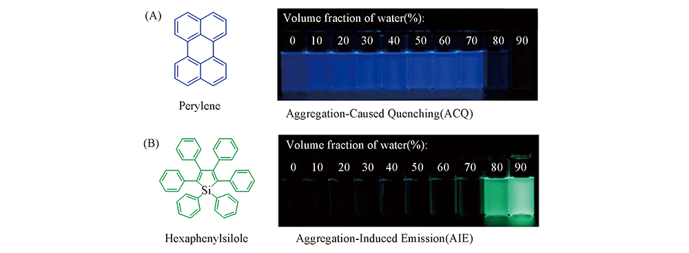
Fig.1 Fluorescent photos of solutions or suspensions of perylene(ACQ fluorophore, A) and hexaphenylsilole(AIE fluorophore, B) in THF/water mixtures with different volume fractions of water under UV lamp Reproduced with permission from Ref.[17], Copyright 2015, American Chemical Society.
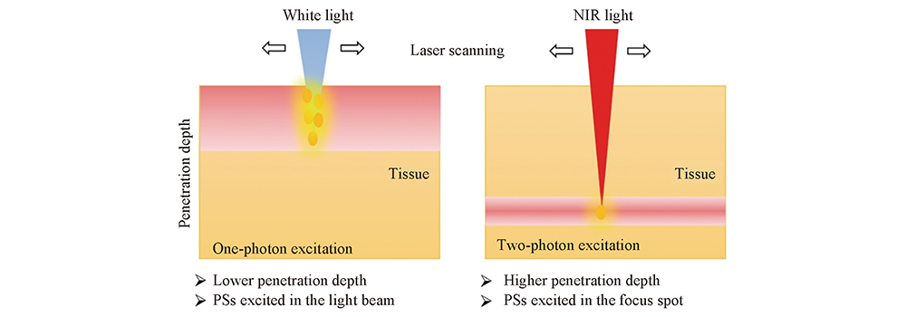
Fig.2 Difference between one-photon excited and two-photon excited photodynamic therapy Reproduced with permission from Ref.[35], Copyright 2018, Royal Society of Chemistry.
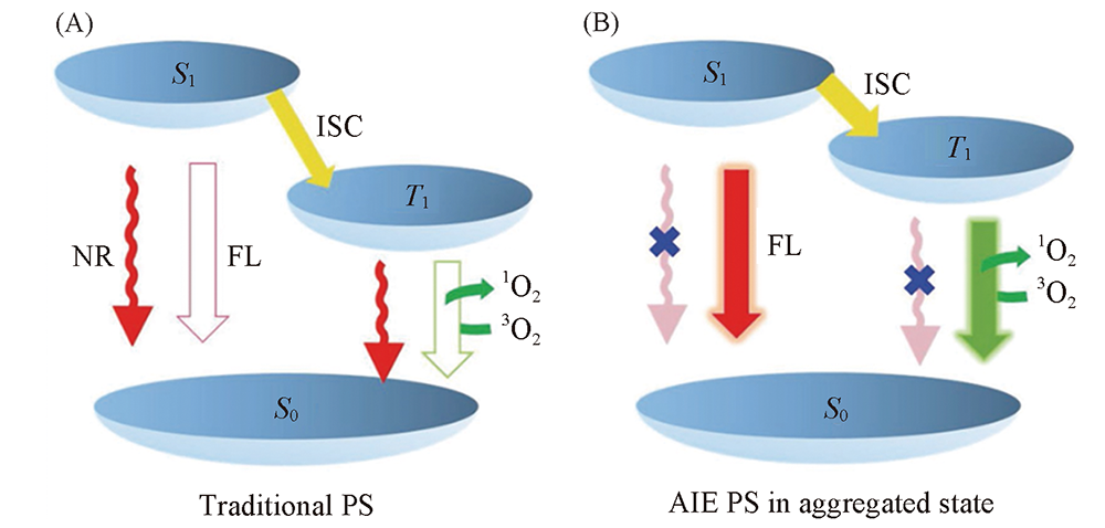
Fig.3 Simplified Jablonski diagram depicting the electron transitions of different types of PSs upon light excitation (A) Traditional PS; (B) AIE PS in aggregated state. ISC from S1 to T1, and energy transfer from T1 to 3O2, generating cytotoxic 1O2. ISC: intersystem crossing, NR: nonradiative decay, FL: fluorescence, 1O2: singlet oxygen, 3O2: normal oxygen. Reproduced with permission from Ref.[20], Copyright 2016, Wiley-VCH.
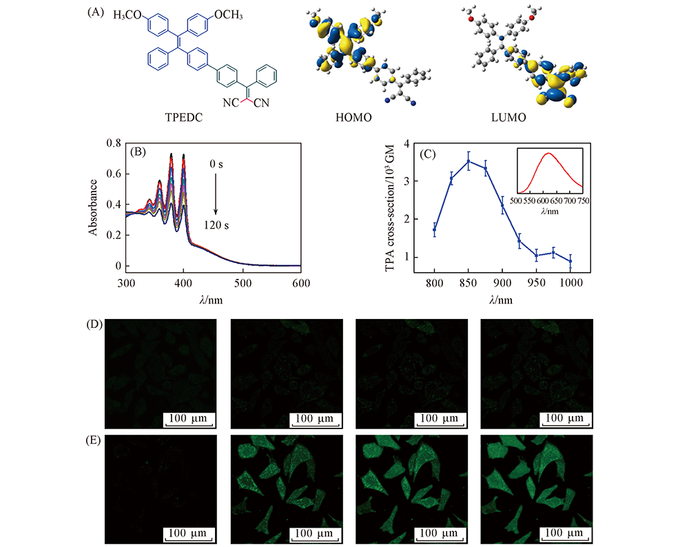
Fig.4 Design and properties of TPEDC (A) Chemical structure, HOMO and LUMO distributions of TPEDC; (B) UV-Vis spectra of ABDA in the presence of TPEDC NPs under light irradiation(60 mW/cm2, 400—700 nm) in water; (C) two-photon absorption cross section of TPEDC NPs at different wavelengths, the inset shows the two-photon-induced fluorescence spectrum; (D, E) detection of intracellular ROS generation using DCF-DA in HeLa cells incubated with(E) and without(D, control) TPEDC NPs followed by different two-photon scans, λex=488 nm; λem=505—525 nm. Fig.(A) was reproduced with permission from Ref.[45], Copyright 2017, Wiley-VCH; Figures(B—E) were reproduced with permission from Ref.[38], Copyright 2017, Wiley-VCH.
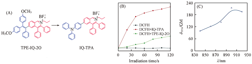
Fig.5 Design and properties of IQ-TPA (A) Chemical structures of TPE-IQ-2O and IQ-TPA; (B) change in fluorescence intensity at 525 nm of TPE-IQ-2O/IQ-TPA and DCFH in PBS upon white light irradiation for different times; (C) two-photon absorption spectrum of IQ-TPA. Reproduced with permission from Ref.[35], Copyright 2018, Royal Society of Chemistry.
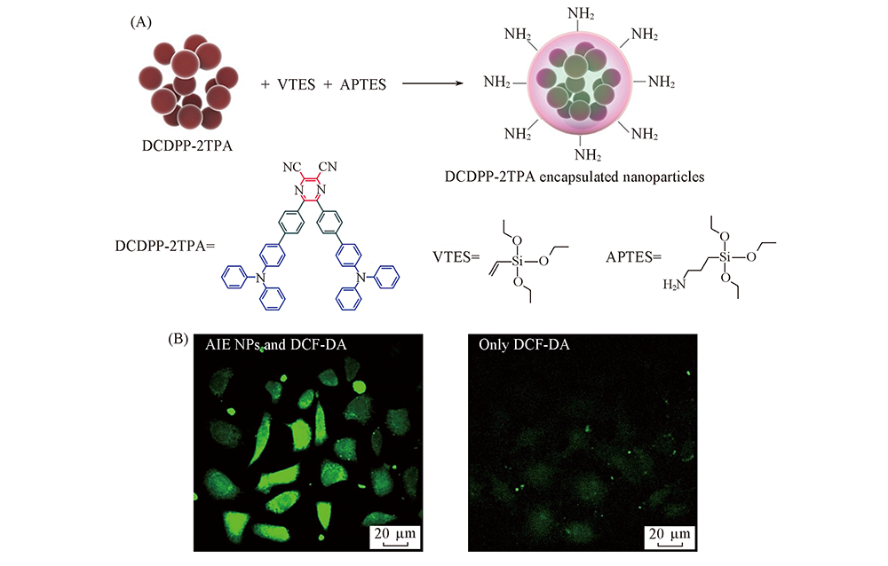
Fig.6 Design and properties of DCDPP nanoparticles (A) Preparation of DCDPP-2TPA-encapsulated silica nanoparticles; (B) two-photon fluorescence imaging of HeLa cells after irradiation for 5 min with a 1040 nm fs laser pretreated with DCDPP-2TPA-encapsulated silica NPs(0.018 mg/mL) and DCF-DA(25 μmol/L), or only DCF-DA(25 μmol/L). Reproduced with permission from Ref.[52], Copyright 2018, Wiley-VCH.
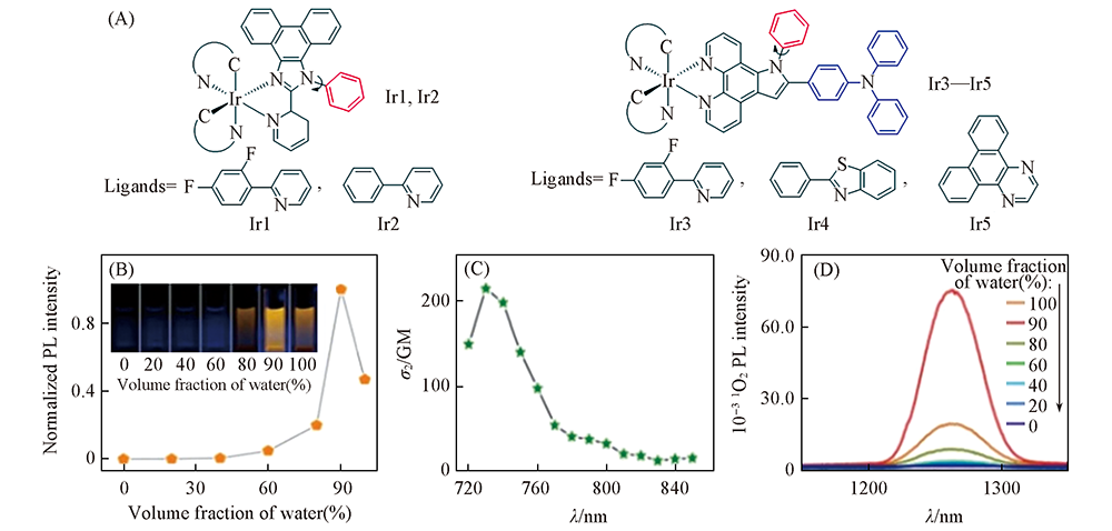
Fig.7 Design and properties of Ir1—Ir5 (A) Chemical structures of Ir1—Ir5; (B) Trajectory of Ir3 emission intensity versus water fraction and visual observation of PL; (C) TPA cross-sections of Ir3; (D) 1O2 emission spectra in the presence of Ir3 and irradiation(405 nm laser) in varying fractions of water-DMSO mixture. Reproduced with permission from Ref.[38], Copyright 2018, Royal Society of Chemistry.
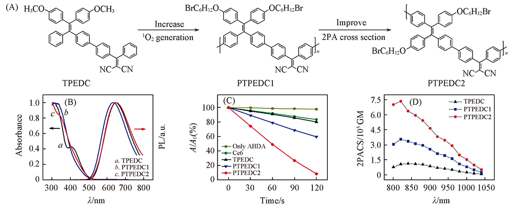
Fig.8 Design and properties of PTPEDC2 (A) Chemical structures of TPEDC, PTPEDC1 and PTPEDC2; (B) normalized absorption and photoluminescence(PL) spectra of AIE PS NPs in aqueous media; (C) normalized degradation percentages of ABDA in the presence of PS NPs in aqueous media upon white light irradiation(400—700 nm, 50 mW/cm2); [AIE PS NPs]=10 μmol/L based on AIE PS; [ABDA]=50 μmol/L; (D) two-photon absorption cross-section spectra of AIE PS NPs in aqueous solution. Reproduced with permission from Ref.[60], Copyright 2019, American Chemical Society.
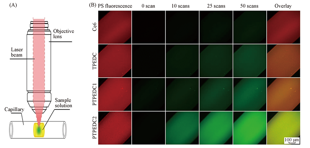
Fig.9 Schematic illustration for the in vitro ROS detection of AIE PS NPs in aqueous media under two-photon excitation(A) and in vitro real-time detection of ROS generation in aqueous solution of PS NPs under two-photon excitation after different scans(B) The image in the last column is the overlay image between the 1th and 5th columns. λex=820 nm, λem: 635—675 nm(red, from PS NPs) and 510—535 nm(green, from DCFH), scanning laser: 820 nm, 6 mW, 5.33 s per scan. Reproduced with permission from Ref.[60], Copyright 2019, American Chemical Society.
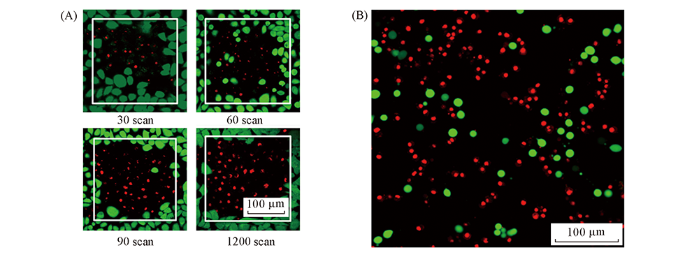
Fig.10 PDT results of TPEDC by two-photon(A) and one-photon(B) excited PDT (A) Live/dead staining of TPEDC NPs(10 μg/mL) treated HeLa cells after different two-photon scans. The live cells were stained by calcein(green), while dead cells were stained by propidium iodide(red). The scanned areas(243 μm×243 μm) were shown by white squares[38]. (B) Live/dead staining of TPEDC NPs(5 μg/mL) treated MDA-MB-231 breast cancer cells after 5 min light irradiation(60 mW/cm2, 400—700 nm). The live cells were stained by fluorescein diacetate(green), while dead cells were stained by propidium iodide(red)[61]. Reproduced with permission from Ref.[38,61], Copyright 2017, Royal Society of Chemistry.
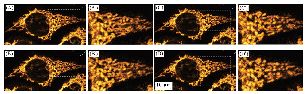
Fig.11 Monitoring the mitochondrial change during two-photon PDT by the fluorescence of IQ-TPA Fluorescence images of HeLa cells were incubated with 1 mmol/L of IQ-TPA for 30 min and then followed by two-photon scans: (A, A') 1 scan, (B, B') 33 scans, (C, C') 66 scans, (D, D') 100 scans. The two-photon excitation condition was at 900 nm(fs Ti: sapphire laser, 5 mW) with a scan area of 60 mm×60 mm and a scan speed of 1.02 s per scan. Reproduced with permission from Ref.[35], Copyright 2018, Royal Society of Chemistry.
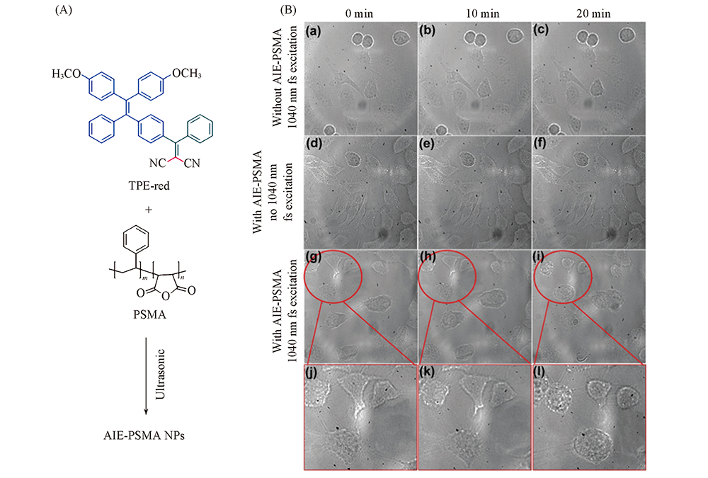
Fig.12 Chemical structures of TPE-red and PSMA as well as the preparation of AIE-PSMA NPs(A) and transmission images of HeLa cells with different treatment(B) Reproduced with permission from Ref.[65], Copyright 2017, Royal Society of Chemistry.
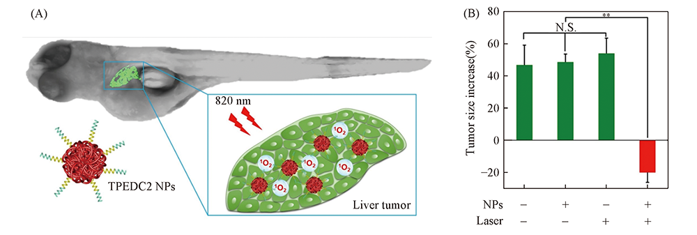
Fig.13 Two-photon excited PDT results of in PTPEDC2 NPs in zebrafish liver tumor model (A) Schematic illustration of in vivo two-photon excited PDT of PTPEDC2 NPs in zebrafish liver tumor model; (B) the relative increase(in percent) in zebrafish tumor size after different treatments. N.S.: data are not significantly different; double asterisks indicate p<0.01, and n=6. Laser: 820 nm fs laser. Reproduced with permission from Ref.[60], Copyright 2019, American Chemical Society.
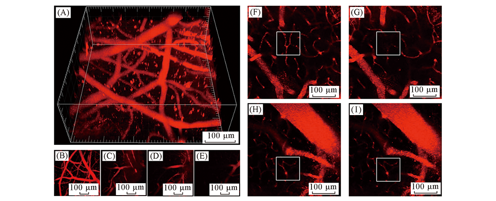
Fig.14 Two-photon excited brain-blood-vessel closure results of TPEDC (A, B): 3D reconstruct(A) and Z-projection(B) of two-photon images of brain blood vessels. (C—E): Two-photon images of brain blood vessels at different vertical depths: 70 μm(C), 140 μm(D), and 200 μm(E). (F, G): Pre-irradiation(F) and post-irradiation(G) images of the brain blood vessels of mouse treated with TPEDC NPs(8 mg/kg based on TPEDC) and two-photon excitation. (H, I): Pre-irradiation(H) and post-irradiation(I) images of the brain blood vessels of a mouse treated with Luminicell NPs and two-photon excitation. The scanned areas are highlighted by white squares. Two-photon excitation condition: 800 nm, 30 mW; λex: 800 nm; λem: 590—630 nm. Reproduced with permission from Ref.[38], Copyright 2017, Wiley-VCH.
| [1] | The website of World Health Organization. |
| [2] | Ye X., Fan W. J., Wang H., Wang J. J., Gu S. Z., Feng W. J., Zhuang Y. P., Liu B. D., Li X. G., Li Y. L., Yang P., Yang X., Yang W. W., Chen J. H., Zhang R., Lin Z. Y., Meng Z. Q., Hu K. W., Liu C., Peng Z. M., Han Y., Jin Y., Lei G. Y., Zhai B., Huang G. H., Chin. J. Lung Cancer, 2017,20, 433— 445 |
| ( 叶欣, 范卫君, 王徽, 王俊杰, 古善智, 冯威健, 庄一平, 刘宝东, 李晓光, 李玉亮, 杨坡, 杨霞, 杨武威, 陈俊辉, 张嵘, 林征宇, 孟志强, 胡凯文, 柳晨, 彭忠民, 韩玥, 靳勇, 雷光焰, 翟博, 黄广慧 . 中国肺癌杂志, 2017,20, 433— 445) | |
| [3] |
Lucky S. S., Soo K. C., Zhang Y., Chem. Rev., 2015,115, 1990— 2042
doi: 10.1021/cr5004198 URL |
| [4] |
Fan W., Huang P., Chen X., Chem. Soc. Rev., 2016,45, 6488— 6519
doi: 10.1039/C6CS00616G URL |
| [5] | Yang B., Chen Y., Shi J., Chem. Rev., 2019,119, 4881— 4985 |
| [6] | Shi L., Yang W. C., Zeng S. Y., Mo T. T., Zhang Z., Cao M. L., Liu H., Chem. J. Chinese Universities, 2016,37(6), 1059— 1068 |
| ( 史蕾, 杨文聪, 曾淑莹, 莫婷婷, 张召, 曹曼丽, 刘海洋 . 高等学校化学学报, 2016,37(6), 1059— 1068) | |
| [7] | Li J., Pu K., Chem. Soc. Rev., 2019,48, 38— 71 |
| [8] | Ethirajan M., Chen Y., Joshi P., Pandey R .K., Chem. Soc. Rev., 2011,40, 340— 362 |
| [9] |
Yang K., Wen J., Chao S., Liu J., Yang K., Pei Y., Pei Z., Chem. Commun., 2018,54, 5911— 5914
doi: 10.1039/C8CC02739K URL |
| [10] |
Zhao J., Xu K., Yang W., Wang Z., Zhong F., Chem. Soc. Rev., 2015,44, 8904— 58939
doi: 10.1039/C5CS00364D URL |
| [11] |
Barth B. M., Altınoglu E. I., Shanmugavelandy S. S., Kaiser J. M., Crespo-Gonzalez D., DiVittore N. A., McGovern C., Goff T. M., Keasey N. R., Adair J. H., Thomas J., Loughran P., Claxton D. F., Kester M., ACS Nano 2011,5, 5325— 5337
doi: 10.1021/nn2005766 URL |
| [12] |
Prier C. K., Rankic D. A., MacMillan D. W., Chem. Rev., 2013,113, 5322— 5363
doi: 10.1021/cr300503r URL |
| [13] |
Singh S., Aggarwal A., Bhupathiraju N. V. S. D. K., Arianna G., Tiwari K., Drain C. M., Chem. Rev., 2015,115, 10261— 10306
doi: 10.1021/acs.chemrev.5b00244 URL |
| [14] |
Yuan Y., Feng G., Qin W., Tang B. Z., Liu B., Chem. Commun., 2014,50, 8757— 8760
doi: 10.1039/C4CC02767A URL |
| [15] |
Hu F., Huang Y., Zhang G., Zhao R., Yang H., Zhang D., Anal. Chem., 2014,86, 7987— 7995
doi: 10.1021/ac502103t URL |
| [16] | Luo J., Xie Z., Lam J. W. Y., Cheng L., Chen H., Qiu C., Kwok H. S., Zhan X., Liu Y., Zhu D., Tang B. Z., Chem. Commun., 2001,37, 1740— 1741 |
| [17] |
Mei J., Leung N. L. C., Kowk R. T. K., Lam J. W. Y., Tang B. Z., Chem. Rev., 2015,115, 11718— 11940
doi: 10.1021/acs.chemrev.5b00263 URL |
| [18] |
Mei J., Hong Y., Lam J. W. Y., Qin A., Tang Y., Tang B. Z., Adv. Mater., 2014,26, 5429— 5479
doi: 10.1002/adma.201401356 URL |
| [19] |
Feng G., Kwok R. T. K., Tang B. Z., Liu B., Appl. Phys. Rev., 2017,4, 021307
doi: 10.1063/1.4984020 URL |
| [20] |
Hu F., Xu S., Liu B., Adv. Mater., 2018,30, 1801350
doi: 10.1002/adma.v30.45 URL |
| [21] |
Wang D., Lee M. M. S., Xu W., Kowk R. T. K., Lam J. W. Y., Tang B. Z., Theranostics, 2018,8, 4925— 4956
doi: 10.7150/thno.27787 URL |
| [22] |
Zhang R., Duan Y., Liu B ., Nanoscale, 2019,11, 19241— 19250
doi: 10.1039/C9NR06012J URL |
| [23] |
Mei J., Huang Y., Tian H., ACS Appl. Mater. Interfaces 2018,10, 12217— 12261
doi: 10.1021/acsami.7b14343 URL |
| [24] |
Gu X., Kowk R. T. K., Lam J. W. Y., Tang B. Z., Biomaterials, 2017,146, 115— 135
doi: 10.1016/j.biomaterials.2017.09.004 URL |
| [25] |
Gao M., Tang B. Z., Coordin. Chem. Rev., 2020,402, 213076
doi: 10.1016/j.ccr.2019.213076 URL |
| [26] |
Liu Z., Zou H., Zhao Z., Zhang P., Shan G. G., Kowk R. T. K., Lam J. W. Y., Zheng L., Tang B. Z., ACS Nano, 2019,13, 11283— 11293
doi: 10.1021/acsnano.9b04430 URL |
| [27] |
Yang Y., Wang L., Cao H., Li Q., Li Y., Han M., Wang H., Li J., Nano Lett, 2019,19, 1821— 1826
doi: 10.1021/acs.nanolett.8b04875 URL |
| [28] |
Wu W., Mao D., Xu S., Panahandeh-Fard M., Duan Y., Hu F., Kong D., Liu B., Adv. Funct. Mater., 2019,29, 1901791
doi: 10.1002/adfm.v29.42 URL |
| [29] |
Kim H. M., Cho B. R., Chem. Rev., 2015,115, 5014— 5055
doi: 10.1021/cr5004425 URL |
| [30] |
Olesiak-Banska J., Waszkielewicz M., Obstarczyk P., Samoc M., Chem. Soc. Rev., 2019,48, 4087— 4117
doi: 10.1039/C8CS00849C URL |
| [31] |
Bolze F., Jenni S., Sour A., Heitz V., Chem. Commun., 2017,53, 12857— 12877
doi: 10.1039/C7CC06133A URL |
| [32] |
Sun Z., Zhang L. P., Wu F., Zhao Y., Adv. Funct. Mater., 2017,27, 1704079
doi: 10.1002/adfm.v27.48 URL |
| [33] |
Brown S ., Nat. Photonics, 2008,2, 394— 395
doi: 10.1038/nphoton.2008.112 URL |
| [34] |
Shen Y., Shuhendler A. J., Ye D., Xu J. J., Chen H. Y., Chem. Soc. Rev., 2016,45, 6725— 6741
doi: 10.1039/C6CS00442C URL |
| [35] |
Jiang M., Kowk R. T. K., Li X., Gui C., Lam J. W. Y., Qu J., Tang B. Z., J. Mater. Chem. B, 2018,6, 2557— 2565
doi: 10.1039/C7TB02609A URL |
| [36] |
Helmchen F., Denk W ., Nat. Methods, 2005,2, 932— 940
doi: 10.1038/nmeth818 URL |
| [37] |
Collins H. A., Khurana M., Moriyama E. H., Mariampillai A., Dahlstedt E., Balaz M., Kuimova M. K., Drobizhev M., Yang V. X. D., Phillips D., Rebane A., Wilson B. C., Anderson H. L., Nat. Photonics, 2008,2, 420— 424
doi: 10.1038/nphoton.2008.100 URL |
| [38] |
Gu B., Wu W., Xu G., Feng G., Yin F., Chong P. H. J., Qu J., Yong K. T., Liu B., Adv. Mater., 2017,29, 1701076
doi: 10.1002/adma.v29.28 URL |
| [39] |
Liu J., Jin C., Yuan B., Liu X., Chen Y., Jia L., Chao H., Chem. Commun., 2017,53, 2052— 2055
doi: 10.1039/C6CC10015E URL |
| [40] |
Vatansever F de Melo W. C. M. A., Vecchio D., Sadasivam M., Gupta A., Chandran. R., Karimi M., Parizotto N. A., Yin R., Tegos G. P., Hamblin M. R., ., FEMS Microbiol. Rev., 2013,37, 955— 989
doi: 10.1111/1574-6976.12026 URL |
| [41] |
Kasha M., Radiat. Res., 1963,20, 55— 70
doi: 10.2307/3571331 URL |
| [42] |
An Z., Zheng C., Tao Y., Chen R., Shi H., Chen T., Wang Z., Li H., Deng R., Liu X., Huang W., Nat. Mater., 2015,14, 685— 690
doi: 10.1038/nmat4259 URL |
| [43] |
Chen Y., Lam J. W. Y., Kowk R. T. K., Liu B., Tang B. Z., Mater. Horiz., 2019,6, 428— 433
doi: 10.1039/C8MH01331D URL |
| [44] |
Xu S., Yuan Y., Cai X., Zhang C. J., Hu F., Liang J., Zhang G., Zhang D., Liu B., Chem. Sci., 2015,6, 5824— 5830
doi: 10.1039/C5SC01733E URL |
| [45] |
Wu W., Mao D., Hu F., Xu S., Chen C., Zhang C. J., Cheng X., Yuan Y., Ding D., Kong D., Adv. Mater., 2017,29, 1700548
doi: 10.1002/adma.v29.33 URL |
| [46] |
Kaiser W., Garrett C .G. B., Phys. Rev. Lett., 1961,7, 229— 231
doi: 10.1103/PhysRevLett.7.229 URL |
| [47] |
He G. S., Tan L., Zheng Q., Prasad P. N., Chem. Rev., 2008,108, 1245— 1330
doi: 10.1021/cr050054x URL |
| [48] |
Wang Y., Wu W., Liu J., Manghnani P. N., Hu F., Ma D., Teh C., Wang B., Liu B., ACS Nano, 2019,13, 6879— 6890
doi: 10.1021/acsnano.9b01665 URL |
| [49] |
Zhuang W., Yang L., Ma B., Kong Q., Li G., Wang Y., Tang B. Z., ACS Appl. Mater. Interfaces 2019,11, 20715— 20724
doi: 10.1021/acsami.9b04813 URL |
| [50] |
Qin W., Zhang P., Li H., Lam J. W. Y., Cai Y., Kowk R. T. K., Qian J., Zhang W., Tang B. Z., Chem. Sci., 2018,9, 2705— 2710
doi: 10.1039/C7SC04820C URL |
| [51] | Kato S., Matsumoto T Ishi-i T., Thiemann T., Shigeiwa M., Gorohmaru H., Maeda S., Yamashita Y., Mataka S., ., Chem. Commun., 2004,40, 2342— 2343 |
| [52] |
Chen M., Xie W., Li D., Zebibula A., Wang Y., Qian J., Qin A., Tang B. Z., Chem. Eur. J., 2018,24, 16603— 16608
doi: 10.1002/chem.v24.62 URL |
| [53] |
Qiu K., Ouyang M., Liu Y., Huang H., Liu C., Chen Y., Ji L., Chao H ., J. Mater. Chem. B, 2017,5, 5488— 5498
doi: 10.1039/C7TB00731K URL |
| [54] |
Qiu K., Huang H., Liu B., Liu Y., Zhang P., Chen Y., Ji L., Chao H , J. Mater. Chem. B, 2015,3, 6690— 6697
doi: 10.1039/C5TB01091H URL |
| [55] |
Yu B., Ouyang C., Qiu K., Zhao J., Ji L., Chao H., Chem. Eur. J., 2015,21, 3691— 3700
doi: 10.1002/chem.v21.9 URL |
| [56] |
Wu W., Mao D., Xu S., Kenry Hu F., Li X., Kong D., Liu B., Chem., 2018,4, 1937— 1951
doi: 10.1016/j.chempr.2018.06.003 URL |
| [57] |
Wu W ., Chem., 2018,4, 1762— 1764
doi: 10.1016/j.chempr.2018.07.017 URL |
| [58] |
Liu S., Zhang H., Li Y., Liu J., Du L., Chen M., Kowk R. T. K., Lam J. W. Y., Phillips D. L., Tang B. Z., Angew. Chem. Int. Ed., 2018,57, 15189— 15193
doi: 10.1002/anie.201810326 URL |
| [59] |
Wu W., Tang R., Li Q., Li Z., Chem. Soc. Rev., 2015,44, 3997— 4022
doi: 10.1039/C4CS00224E URL |
| [60] |
Wang S., Wu W., Manghnani P. N., Xu S., Wang Y, Goh C. C., Ng L. G., Liu B., ACS Nano, 2019,13, 3095— 3105
doi: 10.1021/acsnano.8b08398 URL |
| [61] |
Wu W., Mao D., Xu S., Ji S., Hu F., Ding D., Kong D., Liu B., Mater. Horiz., 2017,4, 1110— 1114
doi: 10.1039/C7MH00469A URL |
| [62] |
Schweitzer C., Schmid R., Chem. Rev., 2003,103, 1685— 1757
doi: 10.1021/cr010371d URL |
| [63] |
Modica-Napolitano J. S., Aprille J. R., Adv. Drug Delivery Rev., 2001,49, 63— 70
doi: 10.1016/S0169-409X(01)00125-9 URL |
| [64] |
Kenry, Duan Y., Liu B., Adv. Mater., 2018,30, 1802394
doi: 10.1002/adma.v30.47 URL |
| [65] |
Alifu N., Dong X., Li D., Sun X., Zebibula A., Zhang D., Zhang G., Qian J., Mater. Chem. Front., 2017,1, 1746— 1753
doi: 10.1039/C7QM00092H URL |
| [66] |
Henderson B. W., Dougherty T. J., Photochem. Photobiol., 1992,55, 145— 157
doi: 10.1111/php.1992.55.issue-1 URL |
| [67] |
SamkoeK. S., Clancy A. A., Karotki A., Wilson B. C., Cramb D. T., J. Biomed. Opt., 2007,12, 034025.
doi: 10.1117/1.2750663 URL |
| [1] | 刘苗, 刘瑞波, 刘巴蒂, 钱鹰. 溶酶体靶向吲哚氟硼二吡咯光敏剂的合成、 双光子荧光成像及光动力治疗[J]. 高等学校化学学报, 2022, 43(10): 20220326. |
| [2] | 赵宇辉, 李明乐, 龙飒然, 樊江莉, 彭孝军. 极性敏感的BDP分子溶剂化效应的光谱性质[J]. 高等学校化学学报, 2020, 41(9): 2018. |
| [3] | 杜宪超, 郝红霞, 秦安军, 唐本忠. 聚集诱导发光分子/适配体/外切酶Ⅰ体系对可卡因的检测[J]. 高等学校化学学报, 2020, 41(3): 411. |
| [4] | 张雨, 荆江博, 邵玥明, 殷鑫, 徐斌, 温晓玉. 基于聚集诱导发光的纳米粒子用于肝癌细胞靶向成像[J]. 高等学校化学学报, 2019, 40(11): 2382. |
| [5] | 魏良晨, 胡伟康, 周世雄, 束俊, 周会东, 胡旭成, 姜毅, 童碧海, 张千峰. 含亚磷酸酯和联吡啶羧酸酯配体的铱配合物及其聚集诱导发光增强和电致发光性能[J]. 高等学校化学学报, 2018, 39(7): 1371. |
| [6] | 凌瑶, 刘雪静, 郝海景, 郝晓辉, 白利斌, 武永刚. 具有聚集诱导发光效应的水溶性糖基荧光聚合物的制备及性能[J]. 高等学校化学学报, 2018, 39(6): 1319. |
| [7] | 马冬冬, 林萍萍, 陈莉莉, 王瑜华, 贺丹丹, 陈婉玲, 张甜甜, 陈奎治, 彭亦如. 新型聚乙二醇-聚L-赖氨酸负载四(对磺酸基 偶氮苯基-4-氨基磺酰基)铝(Ⅲ)氯酞菁的纳米光敏剂的合成及离体光动力活性[J]. 高等学校化学学报, 2012, 33(07): 1456. |
| [8] | 池振国, 何克强, 李海银, 张锡奇, 许炳佳, 刘四委, 张艺, 许家瑞. 二咔唑四苯乙烯多功能发光化合物的合成与性能[J]. 高等学校化学学报, 2012, 33(04): 725. |
| [9] | 钱鹰, 闾新明, 周志强, 崔一平. 三苯胺-噁二唑超支化共轭聚合物的多光子泵浦绿色荧光[J]. 高等学校化学学报, 2011, 32(10): 2441. |
| [10] | 申进波, 佟斌, 石建兵, 孙书, 冯霄, 支俊格, 董宇平. 含磷酰杂菲侧基的苯乙烯衍生物聚集诱导发光特性及其在过渡金属离子检测中的应用[J]. 高等学校化学学报, 2010, 31(8): 1656. |
| [11] | 钱鹰, 胡凯明, 周志强, 吕昌贵, 崔一平. 新型树枝分子的宽带多光子荧光发射[J]. 高等学校化学学报, 2010, 31(11): 2268. |
| [12] | 陈锦灿,陈宏炜,李永东,王俊东,陈耐生,黄金陵,黄明东 . 新型单取代两亲性酞菁锌的制备及其光动力活性研究[J]. 高等学校化学学报, 2008, 29(11): 2131. |
| [13] | 钱鹰,黄维,路志锋 ,朱晓勤,孟康,吕昌贵 ,崔一平 . 多枝[1,3,4]-噁二唑衍生物的双光子吸收和光功率限幅特性[J]. 高等学校化学学报, 2007, 28(12): 2369. |
| [14] | 刘海洋; 郭平叶; 江柏荣; 应晓; 廖世军; 麦乃歧; 张启光 . Corrole光敏剂在光动力治疗中的重原子效应[J]. 高等学校化学学报, 2006, 27(7): 1363. |
| [15] | 黄剑东, 刘尔生, 杨素苓, 薛金萍, 陈耐生, 黄金陵. 抗癌光敏剂ZnPcSP在溶液中的存在状态及其对活性的影响[J]. 高等学校化学学报, 2002, 23(12): 2287. |
| 阅读次数 | ||||||
|
全文 |
|
|||||
|
摘要 |
|
|||||