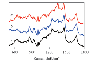
| [1] |
Parkin D. M., Bray F., Ferlay J., Pisani P., Ca. Cancer J. Clin., 2005, 55(2), 74—108
|
| [2] |
Reich O., Lahousen M., Pickel H., Tamussino K., Winter R., Obstet. Gynecol., 2002, 99(2), 193—196
|
| [3] |
Kondepati V. R., Heise H. M., Backhaus J., Anal. Bioanal. Chem., 2008, 390(1), 125—139
|
| [4] |
Krishna C. M., Sockalingum G. D., Vidyasagar M. S., Manfait M., Fernandes D. J., Vadhiraja B. M., Maheedhar K., J. Cancer Res. Ther., 2008, 4(1), 26—36
|
| [5] |
Nemeth T.S., Biopolymer Research Trends, Nova Science Publishers, New York, 2007, 189—209
|
| [6] |
DaCosta R. S., Wilson B. C., Marcon N. E., Scientific World J., 2007, 7, 2046—2071
|
| [7] |
Bigio I. J., Brown S. G., Cancer Biol. Ther., 2004, 3(3), 259—267
|
| [8] |
Quan R. H., Shen A. G., Liao C. X., Wang H., Hu J. M., Chem. J. Chinese Universities,2007, 28(9), 1645—1650
|
|
(权日浩, 沈爱国, 廖长秀, 汪晖, 胡继明. 高等学校化学学报, 2007, 28(9), 1645—1650)
|
| [9] |
Hu C. X., Wang J. X., Zheng C., Xu S. P., Zhang H. P., Liang Y. C., Bi L. R., Fan Z. M., Han B., Xu W. Q., Med. Phys., 2013, 40(6), 063501—063507
|
| [10] |
Jacobs V. R., Paepke S., Schaaf H., Weber B. C., Kiechle-Bahat M., Clin. Breast Cancer,2007, 7(8), 619—623
|
| [11] |
Hu C. X., Zheng C., Zhang H. P., Bi L. R., Xu S. P., Fan Z. M., Han B., Xu W. Q., Chem. J. Chinese Universities,2013, 34(12), 2721—2727
|
|
(胡成旭, 郑超, 张海鹏, 毕丽荣, 徐抒平, 范志民, 韩冰, 徐蔚青. 高等学校化学学报, 2013, 34(12), 2721—2727)
|
| [12] |
Krishna C. M., Kurien J., Mathew S., Rao L., Maheedhar K., Kumar K. K., Chowdary M. V. P., Expert Rev. Mol. Diagn., 2008, 8(2), 149—166
|
| [13] |
Chowdary M. V., Kumar K. K., Kurien J., Mathew S., Krishna C. M., Biopolymers,2006, 83(5), 556—569
|
| [14] |
Yu C., Gestl E., Eckert K., Allara D., Irudayaraj J., Cancer Detect. Prev., 2006, 30(6), 515—522
|
| [15] |
Haka A. S., Shafer-Peltier K. E., Fitzumaurice M., Crowe J., Dasari R. R., Feld M. S. Proc. Natl. Acad. Sci. USA,2005, 102, 12371—12376
|
| [16] |
Shafer-Peltier K. E., Haka A. S., Fitzmaurice M., Crowe J., Myles J., Dasari R. R., Feld M. S., J. Raman Spectrosc., 2002, 33(7), 552—563
|
| [17] |
Marcelo M., Leandro R., Emília Ângelo Loschiavo A., Ana Maria do Espírito S., Edson Aparecido Pereira dos S., Renata Andrade B., Airton Abrahão M., Theor. Chem. Acc., 2009, 125(3—6), 329—334
|
| [18] |
Deng Y. B., Yang T. Q., Spectrosc. Spect. Anal., 2007, 27(7), 1312—315(邓一兵, 杨体强. 光谱学与光谱分析, 2007, 27(7), 1312—1315)
|
| [19] |
Usuda J., Chiu S. M., Azizuddin K., Xue L. Y., Lam M., Nieminen A. L., Oleinick N. L., Photochem Photobiol., 2002, 76(2), 217—223
|
| [20] |
Abramczyk H., Placek I., Brozek-Pluska B., Kurczewski K., Morawiecc Z., Tazbir M., Spectrosc. Int. J., 2008, 22(2/3), 113—121
|
| [21] |
Shen S. J., Liu B. Y., Li Q., Ma X., Song Z. J., Spectrosc. Spect. Anal., 2000, 20(1), 28—30
|
|
(沈世杰, 刘炳玉, 李清, 马溪, 宋占军. 光谱学与光谱分析, 2000, 20(1), 28—30)
|
 ), 韩冰1(