

Chem. J. Chinese Universities ›› 2020, Vol. 41 ›› Issue (2): 191.doi: 10.7503/cjcu20190614
Previous Articles Next Articles
WU Wenbo,LIU Bin( )
)
Received:2019-11-26
Online:2020-02-10
Published:2019-12-31
Contact:
Bin LIU
E-mail:chewwe@nus.edu.sg
Supported by:CLC Number:
TrendMD:
WU Wenbo,LIU Bin. Two-photon Excitable Photosensitizers with Aggregation-induced Emission and Their Biomedical Applications †[J]. Chem. J. Chinese Universities, 2020, 41(2): 191.
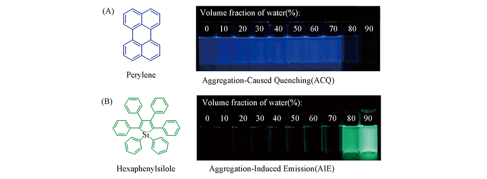
Fig.1 Fluorescent photos of solutions or suspensions of perylene(ACQ fluorophore, A) and hexaphenylsilole(AIE fluorophore, B) in THF/water mixtures with different volume fractions of water under UV lamp Reproduced with permission from Ref.[17], Copyright 2015, American Chemical Society.
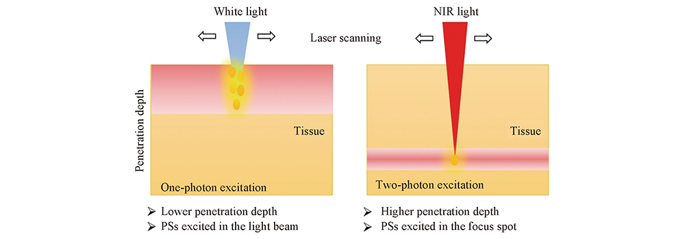
Fig.2 Difference between one-photon excited and two-photon excited photodynamic therapy Reproduced with permission from Ref.[35], Copyright 2018, Royal Society of Chemistry.
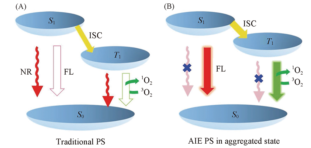
Fig.3 Simplified Jablonski diagram depicting the electron transitions of different types of PSs upon light excitation (A) Traditional PS; (B) AIE PS in aggregated state. ISC from S1 to T1, and energy transfer from T1 to 3O2, generating cytotoxic 1O2. ISC: intersystem crossing, NR: nonradiative decay, FL: fluorescence, 1O2: singlet oxygen, 3O2: normal oxygen. Reproduced with permission from Ref.[20], Copyright 2016, Wiley-VCH.
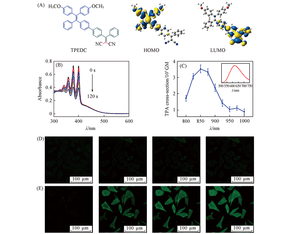
Fig.4 Design and properties of TPEDC (A) Chemical structure, HOMO and LUMO distributions of TPEDC; (B) UV-Vis spectra of ABDA in the presence of TPEDC NPs under light irradiation(60 mW/cm2, 400—700 nm) in water; (C) two-photon absorption cross section of TPEDC NPs at different wavelengths, the inset shows the two-photon-induced fluorescence spectrum; (D, E) detection of intracellular ROS generation using DCF-DA in HeLa cells incubated with(E) and without(D, control) TPEDC NPs followed by different two-photon scans, λex=488 nm; λem=505—525 nm. Fig.(A) was reproduced with permission from Ref.[45], Copyright 2017, Wiley-VCH; Figures(B—E) were reproduced with permission from Ref.[38], Copyright 2017, Wiley-VCH.
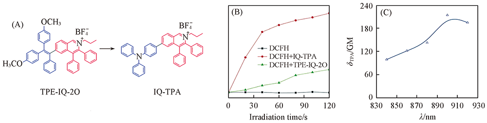
Fig.5 Design and properties of IQ-TPA (A) Chemical structures of TPE-IQ-2O and IQ-TPA; (B) change in fluorescence intensity at 525 nm of TPE-IQ-2O/IQ-TPA and DCFH in PBS upon white light irradiation for different times; (C) two-photon absorption spectrum of IQ-TPA. Reproduced with permission from Ref.[35], Copyright 2018, Royal Society of Chemistry.
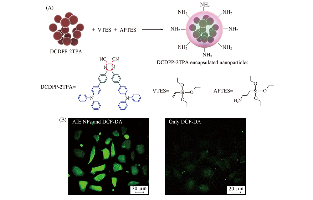
Fig.6 Design and properties of DCDPP nanoparticles (A) Preparation of DCDPP-2TPA-encapsulated silica nanoparticles; (B) two-photon fluorescence imaging of HeLa cells after irradiation for 5 min with a 1040 nm fs laser pretreated with DCDPP-2TPA-encapsulated silica NPs(0.018 mg/mL) and DCF-DA(25 μmol/L), or only DCF-DA(25 μmol/L). Reproduced with permission from Ref.[52], Copyright 2018, Wiley-VCH.
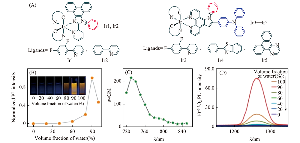
Fig.7 Design and properties of Ir1—Ir5 (A) Chemical structures of Ir1—Ir5; (B) Trajectory of Ir3 emission intensity versus water fraction and visual observation of PL; (C) TPA cross-sections of Ir3; (D) 1O2 emission spectra in the presence of Ir3 and irradiation(405 nm laser) in varying fractions of water-DMSO mixture. Reproduced with permission from Ref.[38], Copyright 2018, Royal Society of Chemistry.
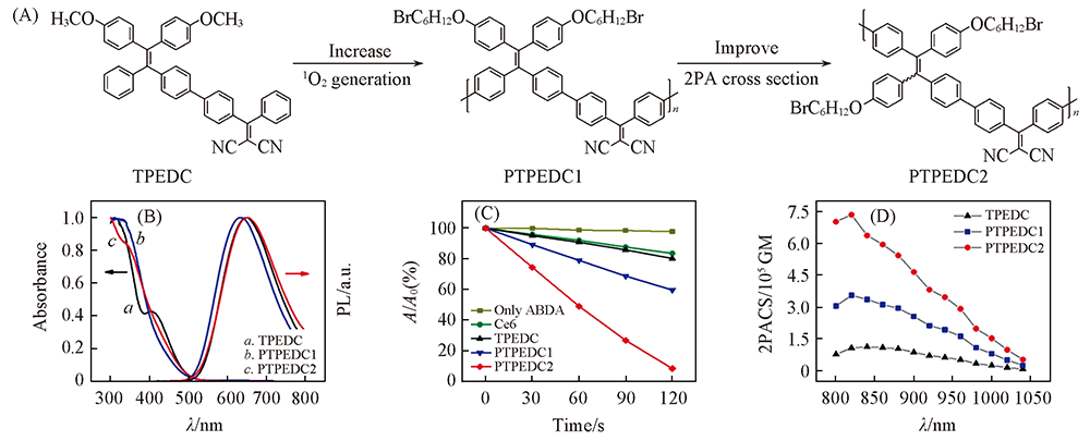
Fig.8 Design and properties of PTPEDC2 (A) Chemical structures of TPEDC, PTPEDC1 and PTPEDC2; (B) normalized absorption and photoluminescence(PL) spectra of AIE PS NPs in aqueous media; (C) normalized degradation percentages of ABDA in the presence of PS NPs in aqueous media upon white light irradiation(400—700 nm, 50 mW/cm2); [AIE PS NPs]=10 μmol/L based on AIE PS; [ABDA]=50 μmol/L; (D) two-photon absorption cross-section spectra of AIE PS NPs in aqueous solution. Reproduced with permission from Ref.[60], Copyright 2019, American Chemical Society.
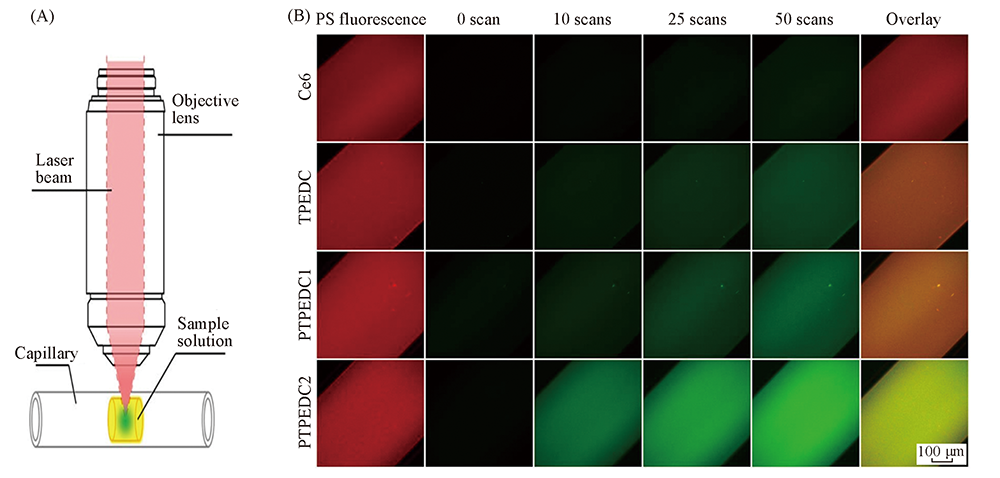
Fig.9 Schematic illustration for the in vitro ROS detection of AIE PS NPs in aqueous media under two-photon excitation(A) and in vitro real-time detection of ROS generation in aqueous solution of PS NPs under two-photon excitation after different scans(B) The image in the last column is the overlay image between the 1th and 5th columns. λex=820 nm, λem: 635—675 nm(red, from PS NPs) and 510—535 nm(green, from DCFH), scanning laser: 820 nm, 6 mW, 5.33 s per scan. Reproduced with permission from Ref.[60], Copyright 2019, American Chemical Society.
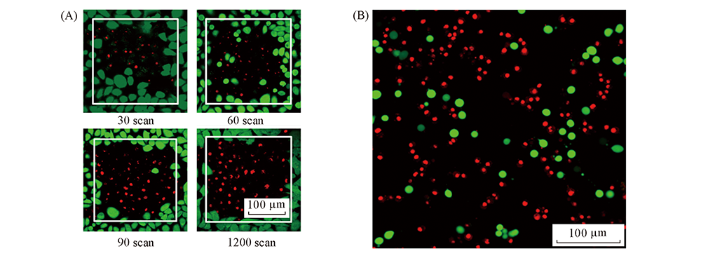
Fig.10 PDT results of TPEDC by two-photon(A) and one-photon(B) excited PDT (A) Live/dead staining of TPEDC NPs(10 μg/mL) treated HeLa cells after different two-photon scans. The live cells were stained by calcein(green), while dead cells were stained by propidium iodide(red). The scanned areas(243 μm×243 μm) were shown by white squares[38]. (B) Live/dead staining of TPEDC NPs(5 μg/mL) treated MDA-MB-231 breast cancer cells after 5 min light irradiation(60 mW/cm2, 400—700 nm). The live cells were stained by fluorescein diacetate(green), while dead cells were stained by propidium iodide(red)[61]. Reproduced with permission from Ref.[38,61], Copyright 2017, Royal Society of Chemistry.
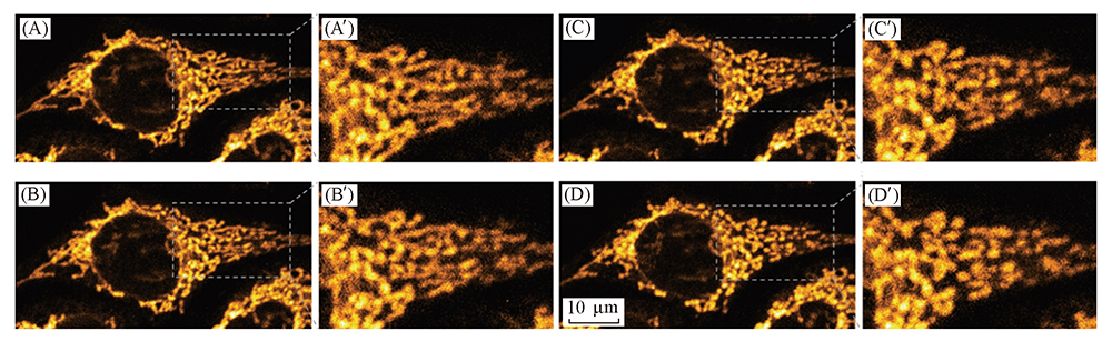
Fig.11 Monitoring the mitochondrial change during two-photon PDT by the fluorescence of IQ-TPA Fluorescence images of HeLa cells were incubated with 1 mmol/L of IQ-TPA for 30 min and then followed by two-photon scans: (A, A') 1 scan, (B, B') 33 scans, (C, C') 66 scans, (D, D') 100 scans. The two-photon excitation condition was at 900 nm(fs Ti: sapphire laser, 5 mW) with a scan area of 60 mm×60 mm and a scan speed of 1.02 s per scan. Reproduced with permission from Ref.[35], Copyright 2018, Royal Society of Chemistry.
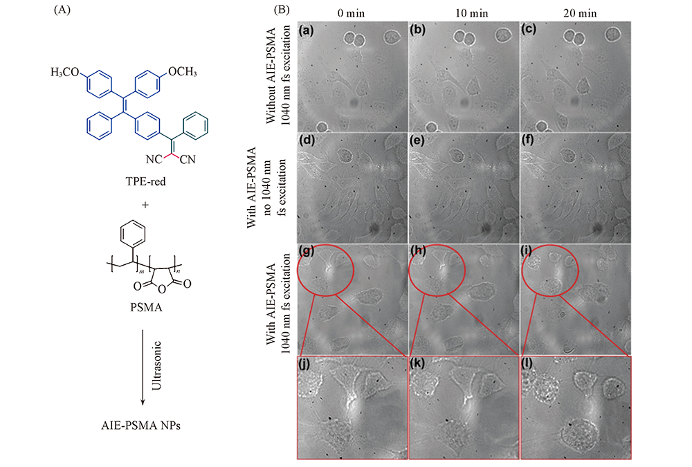
Fig.12 Chemical structures of TPE-red and PSMA as well as the preparation of AIE-PSMA NPs(A) and transmission images of HeLa cells with different treatment(B) Reproduced with permission from Ref.[65], Copyright 2017, Royal Society of Chemistry.
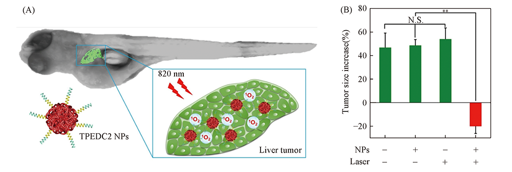
Fig.13 Two-photon excited PDT results of in PTPEDC2 NPs in zebrafish liver tumor model (A) Schematic illustration of in vivo two-photon excited PDT of PTPEDC2 NPs in zebrafish liver tumor model; (B) the relative increase(in percent) in zebrafish tumor size after different treatments. N.S.: data are not significantly different; double asterisks indicate p<0.01, and n=6. Laser: 820 nm fs laser. Reproduced with permission from Ref.[60], Copyright 2019, American Chemical Society.
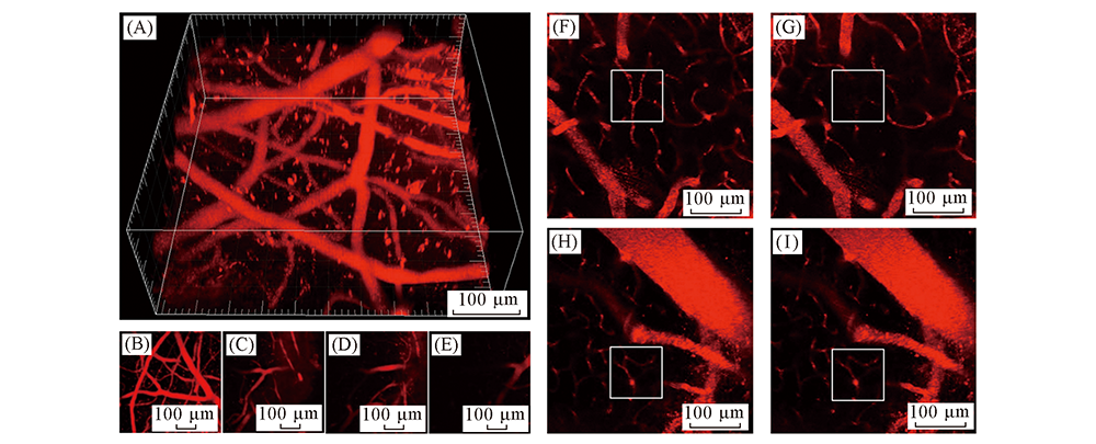
Fig.14 Two-photon excited brain-blood-vessel closure results of TPEDC (A, B): 3D reconstruct(A) and Z-projection(B) of two-photon images of brain blood vessels. (C—E): Two-photon images of brain blood vessels at different vertical depths: 70 μm(C), 140 μm(D), and 200 μm(E). (F, G): Pre-irradiation(F) and post-irradiation(G) images of the brain blood vessels of mouse treated with TPEDC NPs(8 mg/kg based on TPEDC) and two-photon excitation. (H, I): Pre-irradiation(H) and post-irradiation(I) images of the brain blood vessels of a mouse treated with Luminicell NPs and two-photon excitation. The scanned areas are highlighted by white squares. Two-photon excitation condition: 800 nm, 30 mW; λex: 800 nm; λem: 590—630 nm. Reproduced with permission from Ref.[38], Copyright 2017, Wiley-VCH.
| [1] | The website of World Health Organization. |
| [2] | Ye X., Fan W. J., Wang H., Wang J. J., Gu S. Z., Feng W. J., Zhuang Y. P., Liu B. D., Li X. G., Li Y. L., Yang P., Yang X., Yang W. W., Chen J. H., Zhang R., Lin Z. Y., Meng Z. Q., Hu K. W., Liu C., Peng Z. M., Han Y., Jin Y., Lei G. Y., Zhai B., Huang G. H., Chin. J. Lung Cancer, 2017,20, 433— 445 |
| ( 叶欣, 范卫君, 王徽, 王俊杰, 古善智, 冯威健, 庄一平, 刘宝东, 李晓光, 李玉亮, 杨坡, 杨霞, 杨武威, 陈俊辉, 张嵘, 林征宇, 孟志强, 胡凯文, 柳晨, 彭忠民, 韩玥, 靳勇, 雷光焰, 翟博, 黄广慧 . 中国肺癌杂志, 2017,20, 433— 445) | |
| [3] |
Lucky S. S., Soo K. C., Zhang Y., Chem. Rev., 2015,115, 1990— 2042
doi: 10.1021/cr5004198 URL |
| [4] |
Fan W., Huang P., Chen X., Chem. Soc. Rev., 2016,45, 6488— 6519
doi: 10.1039/C6CS00616G URL |
| [5] | Yang B., Chen Y., Shi J., Chem. Rev., 2019,119, 4881— 4985 |
| [6] | Shi L., Yang W. C., Zeng S. Y., Mo T. T., Zhang Z., Cao M. L., Liu H., Chem. J. Chinese Universities, 2016,37(6), 1059— 1068 |
| ( 史蕾, 杨文聪, 曾淑莹, 莫婷婷, 张召, 曹曼丽, 刘海洋 . 高等学校化学学报, 2016,37(6), 1059— 1068) | |
| [7] | Li J., Pu K., Chem. Soc. Rev., 2019,48, 38— 71 |
| [8] | Ethirajan M., Chen Y., Joshi P., Pandey R .K., Chem. Soc. Rev., 2011,40, 340— 362 |
| [9] |
Yang K., Wen J., Chao S., Liu J., Yang K., Pei Y., Pei Z., Chem. Commun., 2018,54, 5911— 5914
doi: 10.1039/C8CC02739K URL |
| [10] |
Zhao J., Xu K., Yang W., Wang Z., Zhong F., Chem. Soc. Rev., 2015,44, 8904— 58939
doi: 10.1039/C5CS00364D URL |
| [11] |
Barth B. M., Altınoglu E. I., Shanmugavelandy S. S., Kaiser J. M., Crespo-Gonzalez D., DiVittore N. A., McGovern C., Goff T. M., Keasey N. R., Adair J. H., Thomas J., Loughran P., Claxton D. F., Kester M., ACS Nano 2011,5, 5325— 5337
doi: 10.1021/nn2005766 URL |
| [12] |
Prier C. K., Rankic D. A., MacMillan D. W., Chem. Rev., 2013,113, 5322— 5363
doi: 10.1021/cr300503r URL |
| [13] |
Singh S., Aggarwal A., Bhupathiraju N. V. S. D. K., Arianna G., Tiwari K., Drain C. M., Chem. Rev., 2015,115, 10261— 10306
doi: 10.1021/acs.chemrev.5b00244 URL |
| [14] |
Yuan Y., Feng G., Qin W., Tang B. Z., Liu B., Chem. Commun., 2014,50, 8757— 8760
doi: 10.1039/C4CC02767A URL |
| [15] |
Hu F., Huang Y., Zhang G., Zhao R., Yang H., Zhang D., Anal. Chem., 2014,86, 7987— 7995
doi: 10.1021/ac502103t URL |
| [16] | Luo J., Xie Z., Lam J. W. Y., Cheng L., Chen H., Qiu C., Kwok H. S., Zhan X., Liu Y., Zhu D., Tang B. Z., Chem. Commun., 2001,37, 1740— 1741 |
| [17] |
Mei J., Leung N. L. C., Kowk R. T. K., Lam J. W. Y., Tang B. Z., Chem. Rev., 2015,115, 11718— 11940
doi: 10.1021/acs.chemrev.5b00263 URL |
| [18] |
Mei J., Hong Y., Lam J. W. Y., Qin A., Tang Y., Tang B. Z., Adv. Mater., 2014,26, 5429— 5479
doi: 10.1002/adma.201401356 URL |
| [19] |
Feng G., Kwok R. T. K., Tang B. Z., Liu B., Appl. Phys. Rev., 2017,4, 021307
doi: 10.1063/1.4984020 URL |
| [20] |
Hu F., Xu S., Liu B., Adv. Mater., 2018,30, 1801350
doi: 10.1002/adma.v30.45 URL |
| [21] |
Wang D., Lee M. M. S., Xu W., Kowk R. T. K., Lam J. W. Y., Tang B. Z., Theranostics, 2018,8, 4925— 4956
doi: 10.7150/thno.27787 URL |
| [22] |
Zhang R., Duan Y., Liu B ., Nanoscale, 2019,11, 19241— 19250
doi: 10.1039/C9NR06012J URL |
| [23] |
Mei J., Huang Y., Tian H., ACS Appl. Mater. Interfaces 2018,10, 12217— 12261
doi: 10.1021/acsami.7b14343 URL |
| [24] |
Gu X., Kowk R. T. K., Lam J. W. Y., Tang B. Z., Biomaterials, 2017,146, 115— 135
doi: 10.1016/j.biomaterials.2017.09.004 URL |
| [25] |
Gao M., Tang B. Z., Coordin. Chem. Rev., 2020,402, 213076
doi: 10.1016/j.ccr.2019.213076 URL |
| [26] |
Liu Z., Zou H., Zhao Z., Zhang P., Shan G. G., Kowk R. T. K., Lam J. W. Y., Zheng L., Tang B. Z., ACS Nano, 2019,13, 11283— 11293
doi: 10.1021/acsnano.9b04430 URL |
| [27] |
Yang Y., Wang L., Cao H., Li Q., Li Y., Han M., Wang H., Li J., Nano Lett, 2019,19, 1821— 1826
doi: 10.1021/acs.nanolett.8b04875 URL |
| [28] |
Wu W., Mao D., Xu S., Panahandeh-Fard M., Duan Y., Hu F., Kong D., Liu B., Adv. Funct. Mater., 2019,29, 1901791
doi: 10.1002/adfm.v29.42 URL |
| [29] |
Kim H. M., Cho B. R., Chem. Rev., 2015,115, 5014— 5055
doi: 10.1021/cr5004425 URL |
| [30] |
Olesiak-Banska J., Waszkielewicz M., Obstarczyk P., Samoc M., Chem. Soc. Rev., 2019,48, 4087— 4117
doi: 10.1039/C8CS00849C URL |
| [31] |
Bolze F., Jenni S., Sour A., Heitz V., Chem. Commun., 2017,53, 12857— 12877
doi: 10.1039/C7CC06133A URL |
| [32] |
Sun Z., Zhang L. P., Wu F., Zhao Y., Adv. Funct. Mater., 2017,27, 1704079
doi: 10.1002/adfm.v27.48 URL |
| [33] |
Brown S ., Nat. Photonics, 2008,2, 394— 395
doi: 10.1038/nphoton.2008.112 URL |
| [34] |
Shen Y., Shuhendler A. J., Ye D., Xu J. J., Chen H. Y., Chem. Soc. Rev., 2016,45, 6725— 6741
doi: 10.1039/C6CS00442C URL |
| [35] |
Jiang M., Kowk R. T. K., Li X., Gui C., Lam J. W. Y., Qu J., Tang B. Z., J. Mater. Chem. B, 2018,6, 2557— 2565
doi: 10.1039/C7TB02609A URL |
| [36] |
Helmchen F., Denk W ., Nat. Methods, 2005,2, 932— 940
doi: 10.1038/nmeth818 URL |
| [37] |
Collins H. A., Khurana M., Moriyama E. H., Mariampillai A., Dahlstedt E., Balaz M., Kuimova M. K., Drobizhev M., Yang V. X. D., Phillips D., Rebane A., Wilson B. C., Anderson H. L., Nat. Photonics, 2008,2, 420— 424
doi: 10.1038/nphoton.2008.100 URL |
| [38] |
Gu B., Wu W., Xu G., Feng G., Yin F., Chong P. H. J., Qu J., Yong K. T., Liu B., Adv. Mater., 2017,29, 1701076
doi: 10.1002/adma.v29.28 URL |
| [39] |
Liu J., Jin C., Yuan B., Liu X., Chen Y., Jia L., Chao H., Chem. Commun., 2017,53, 2052— 2055
doi: 10.1039/C6CC10015E URL |
| [40] |
Vatansever F de Melo W. C. M. A., Vecchio D., Sadasivam M., Gupta A., Chandran. R., Karimi M., Parizotto N. A., Yin R., Tegos G. P., Hamblin M. R., ., FEMS Microbiol. Rev., 2013,37, 955— 989
doi: 10.1111/1574-6976.12026 URL |
| [41] |
Kasha M., Radiat. Res., 1963,20, 55— 70
doi: 10.2307/3571331 URL |
| [42] |
An Z., Zheng C., Tao Y., Chen R., Shi H., Chen T., Wang Z., Li H., Deng R., Liu X., Huang W., Nat. Mater., 2015,14, 685— 690
doi: 10.1038/nmat4259 URL |
| [43] |
Chen Y., Lam J. W. Y., Kowk R. T. K., Liu B., Tang B. Z., Mater. Horiz., 2019,6, 428— 433
doi: 10.1039/C8MH01331D URL |
| [44] |
Xu S., Yuan Y., Cai X., Zhang C. J., Hu F., Liang J., Zhang G., Zhang D., Liu B., Chem. Sci., 2015,6, 5824— 5830
doi: 10.1039/C5SC01733E URL |
| [45] |
Wu W., Mao D., Hu F., Xu S., Chen C., Zhang C. J., Cheng X., Yuan Y., Ding D., Kong D., Adv. Mater., 2017,29, 1700548
doi: 10.1002/adma.v29.33 URL |
| [46] |
Kaiser W., Garrett C .G. B., Phys. Rev. Lett., 1961,7, 229— 231
doi: 10.1103/PhysRevLett.7.229 URL |
| [47] |
He G. S., Tan L., Zheng Q., Prasad P. N., Chem. Rev., 2008,108, 1245— 1330
doi: 10.1021/cr050054x URL |
| [48] |
Wang Y., Wu W., Liu J., Manghnani P. N., Hu F., Ma D., Teh C., Wang B., Liu B., ACS Nano, 2019,13, 6879— 6890
doi: 10.1021/acsnano.9b01665 URL |
| [49] |
Zhuang W., Yang L., Ma B., Kong Q., Li G., Wang Y., Tang B. Z., ACS Appl. Mater. Interfaces 2019,11, 20715— 20724
doi: 10.1021/acsami.9b04813 URL |
| [50] |
Qin W., Zhang P., Li H., Lam J. W. Y., Cai Y., Kowk R. T. K., Qian J., Zhang W., Tang B. Z., Chem. Sci., 2018,9, 2705— 2710
doi: 10.1039/C7SC04820C URL |
| [51] | Kato S., Matsumoto T Ishi-i T., Thiemann T., Shigeiwa M., Gorohmaru H., Maeda S., Yamashita Y., Mataka S., ., Chem. Commun., 2004,40, 2342— 2343 |
| [52] |
Chen M., Xie W., Li D., Zebibula A., Wang Y., Qian J., Qin A., Tang B. Z., Chem. Eur. J., 2018,24, 16603— 16608
doi: 10.1002/chem.v24.62 URL |
| [53] |
Qiu K., Ouyang M., Liu Y., Huang H., Liu C., Chen Y., Ji L., Chao H ., J. Mater. Chem. B, 2017,5, 5488— 5498
doi: 10.1039/C7TB00731K URL |
| [54] |
Qiu K., Huang H., Liu B., Liu Y., Zhang P., Chen Y., Ji L., Chao H , J. Mater. Chem. B, 2015,3, 6690— 6697
doi: 10.1039/C5TB01091H URL |
| [55] |
Yu B., Ouyang C., Qiu K., Zhao J., Ji L., Chao H., Chem. Eur. J., 2015,21, 3691— 3700
doi: 10.1002/chem.v21.9 URL |
| [56] |
Wu W., Mao D., Xu S., Kenry Hu F., Li X., Kong D., Liu B., Chem., 2018,4, 1937— 1951
doi: 10.1016/j.chempr.2018.06.003 URL |
| [57] |
Wu W ., Chem., 2018,4, 1762— 1764
doi: 10.1016/j.chempr.2018.07.017 URL |
| [58] |
Liu S., Zhang H., Li Y., Liu J., Du L., Chen M., Kowk R. T. K., Lam J. W. Y., Phillips D. L., Tang B. Z., Angew. Chem. Int. Ed., 2018,57, 15189— 15193
doi: 10.1002/anie.201810326 URL |
| [59] |
Wu W., Tang R., Li Q., Li Z., Chem. Soc. Rev., 2015,44, 3997— 4022
doi: 10.1039/C4CS00224E URL |
| [60] |
Wang S., Wu W., Manghnani P. N., Xu S., Wang Y, Goh C. C., Ng L. G., Liu B., ACS Nano, 2019,13, 3095— 3105
doi: 10.1021/acsnano.8b08398 URL |
| [61] |
Wu W., Mao D., Xu S., Ji S., Hu F., Ding D., Kong D., Liu B., Mater. Horiz., 2017,4, 1110— 1114
doi: 10.1039/C7MH00469A URL |
| [62] |
Schweitzer C., Schmid R., Chem. Rev., 2003,103, 1685— 1757
doi: 10.1021/cr010371d URL |
| [63] |
Modica-Napolitano J. S., Aprille J. R., Adv. Drug Delivery Rev., 2001,49, 63— 70
doi: 10.1016/S0169-409X(01)00125-9 URL |
| [64] |
Kenry, Duan Y., Liu B., Adv. Mater., 2018,30, 1802394
doi: 10.1002/adma.v30.47 URL |
| [65] |
Alifu N., Dong X., Li D., Sun X., Zebibula A., Zhang D., Zhang G., Qian J., Mater. Chem. Front., 2017,1, 1746— 1753
doi: 10.1039/C7QM00092H URL |
| [66] |
Henderson B. W., Dougherty T. J., Photochem. Photobiol., 1992,55, 145— 157
doi: 10.1111/php.1992.55.issue-1 URL |
| [67] |
SamkoeK. S., Clancy A. A., Karotki A., Wilson B. C., Cramb D. T., J. Biomed. Opt., 2007,12, 034025.
doi: 10.1117/1.2750663 URL |
| [1] | WU Zexin, ZHU Yuanjie, WANG Hongzhong, WANG Junan, HE Ying. Methyl-modified Carbazole/Diphenyl Sulfone-based AIE-TADF Blue Emitter and Its OLEDs [J]. Chem. J. Chinese Universities, 2022, 43(11): 20220371. |
| [2] | LIU Miao, LIU Ruibo, LIU Badi, QIAN Ying. Synthesis, Two-photon Fluorescence Imaging and Photodynamic Therapy of Lysosome-targeted Indole-BODIPY Photosensitizer [J]. Chem. J. Chinese Universities, 2022, 43(10): 20220326. |
| [3] | LIU Wei, YAO Wei, ZHOU Mingming, YOU Qi, NIE Yong, JIANG Xuchuan. Synthesis, Aggregation-induced Emission and Piezofluorochromic Properties of 9,10-Bis(N-phenylindole-3-vinyl)anthracene [J]. Chem. J. Chinese Universities, 2021, 42(8): 2668. |
| [4] | ZHAO Yuhui, LI Mingle, LONG Saran, FAN Jiangli, PENG Xiaojun. Spectroscopic Characterization of Solvation Effect for a Polarity-Sensitive BDP [J]. Chem. J. Chinese Universities, 2020, 41(9): 2018. |
| [5] | DU Xianchao, HAO Hongxia, QIN Anjun, TANG Benzhong. Detection of Cocaine Based on the System of AIEgen, Aptamer and Exonuclease Ⅰ † [J]. Chem. J. Chinese Universities, 2020, 41(3): 411. |
| [6] | SHAO Wei, LEE Jiyoung, LI Fangyuan, LING Daishun. Organic Small Molecule Nanoparticles for Phototheranostics [J]. Chem. J. Chinese Universities, 2020, 41(11): 2356. |
| [7] | ZHANG Yu, JING Jiangbo, SHAO Yueming, YIN Xin, XU Bin, WEN Xiaoyu. Specific Hepatocellular Carcinoma Imaging Based on Aggregation-Induced Emission Nanoparticles † [J]. Chem. J. Chinese Universities, 2019, 40(11): 2382. |
| [8] | LING Yao, LIU Xuejing, HAO Haijing, HAO Xiaohui, BAI Libin, WU Yonggang. Synthesis and Properties of Water-soluble Glycosyl Fluorescent Polymer with Aggregation-induced Emission Effect† [J]. Chem. J. Chinese Universities, 2018, 39(6): 1319. |
| [9] | WANG Xueli,WANG Zhenxin. Preparation of a Targeted Tumor Nanocomposites for Combined Photodynamic-photothermal Therapy Based on Partially Reduced Graphene Oxide† [J]. Chem. J. Chinese Universities, 2018, 39(10): 2185. |
| [10] | JIANG Tingting, WANG Yuanfang, LIU Peilong, TIAN Yixia, LI Hao, HU Yongguo. Vancomycin Derivative Modified Silica-coated Silver Nanoplate for Surface-enhanced Raman Scattering Imaging and Antimicrobial Photodynamic Therapy of Vancomycin Resistant Bacterial Strains† [J]. Chem. J. Chinese Universities, 2017, 38(5): 846. |
| [11] | YANG Hua, QI Qi, WANG Bin-Bin, QIAN Ying. Aggregation-induced Emission Enhance and Electrical Storage Properties of 1,2,4,5-Tetra{4-[N,N-di(4-iodophenyl)amino]styryl}benzene [J]. Chem. J. Chinese Universities, 2013, 34(8): 1880. |
| [12] | MA Dong-Dong, LIN Ping-Ping, CHEN Li-Li, WANG Yu-Hua, HE Dan-Dan, CHEN Wan-Ling, ZHANG Tian-Tian, CHEN Kui-Zhi, PENG Yi-Ru. Synthesis of Poly(ethylene glycol)-poly(L-lysine) Diblock Copolymer Incorporating Tetra-(p-sulfoazophenyl-4-aminosulfonyl)phthalocyanine Chloride Aluminum(Ⅲ) Polyion Nanoparticles and Its in vitro Photodynamic Therapy Efficacy [J]. Chem. J. Chinese Universities, 2012, 33(07): 1456. |
| [13] | CHI Zhen-Guo, HE Ke-Qiang, LI Hai-Yin, ZHANG Xi-Qi, XU Bing-Jia, LIU Si-Wei, ZHANG Yi, XU Jia-Rui. Synthesis and Properties of a Dicarbazolyl Tetraphenylethylene Multi-functional Luminophor [J]. Chem. J. Chinese Universities, 2012, 33(04): 725. |
| [14] | SHEN Jin-Bo, TONG Bin, SHI Jian-Bing, SUN Shu, FENG Xiao, ZHI Jun-Ge, .... Aggregation-Induced Emission Enhancement Properties of Phosphaphenanthrene-containing Styrene Derivant and Its Application of Detection to Transition Metal Ions [J]. Chem. J. Chinese Universities, 2010, 31(8): 1656. |
| [15] | ZHENG Jing-Jing, WANG Lei*, LIU Yang, TAO Xu-Tang*. Aggregation Induced Luminescence in the Λ-Shaped iso-Phoronederivative Nanoparticles [J]. Chem. J. Chinese Universities, 2010, 31(5): 855. |
| Viewed | ||||||
|
Full text |
|
|||||
|
Abstract |
|
|||||