

高等学校化学学报 ›› 2021, Vol. 42 ›› Issue (1): 201.doi: 10.7503/cjcu20200415
所属专题: 分子筛功能材料 2021年,42卷,第1期
凌旸1, 章冠群1( ), 马延航1,2()
收稿日期:2020-07-01
出版日期:2021-01-10
发布日期:2021-01-12
通讯作者:
章冠群
E-mail:zhanggq1@shanghaitech.edu.cn;mayh2@shanghaitech.edu.cn
作者简介:马延航, 男, 博士, 助理教授, 主要从事电子显微学研究. E-mail: 基金资助:
LING Yang1, ZHANG Guanqun1(), MA Yanhang1,2()
Received:2020-07-01
Online:2021-01-10
Published:2021-01-12
Contact:
ZHANG Guanqun
E-mail:zhanggq1@shanghaitech.edu.cn;mayh2@shanghaitech.edu.cn
Supported by:摘要:
透射电子显微镜是解析沸石分子筛新结构、 分析结构缺陷和研究活性位点等的有力工具. 应用于分子筛研究的透射电子显微术总体上可以分为图像法和衍射法, 包括透射电子显微镜和扫描透射电子显微图像、 选区电子衍射和三维电子衍射, 通常结合其中的几种方法进行分析. 近年来, 随着电子显微镜硬件性能的不断提升, 特别是球差矫正器的广泛应用及各种适用于分子筛等电子束敏感材料的探测器和图像处理技术的不断革新, 在原子尺度观察分子筛的结构已成为可能. 此外, 利用原位电子显微镜技术研究分子筛的生长和催化反应机理也在逐步展开. 本文按电子显微镜方法分类, 综述了近些年基于电子显微镜的分子筛研究, 包括新结构解析、 手性确认和金属负载等的最新进展.
中图分类号:
TrendMD:
凌旸, 章冠群, 马延航. 基于透射电子显微镜的沸石分子筛结构研究进展. 高等学校化学学报, 2021, 42(1): 201.
LING Yang, ZHANG Guanqun, MA Yanhang. Progress of Zeolite Structural Analysis Based on Transmission Electron Microscopy. Chem. J. Chinese Universities, 2021, 42(1): 201.
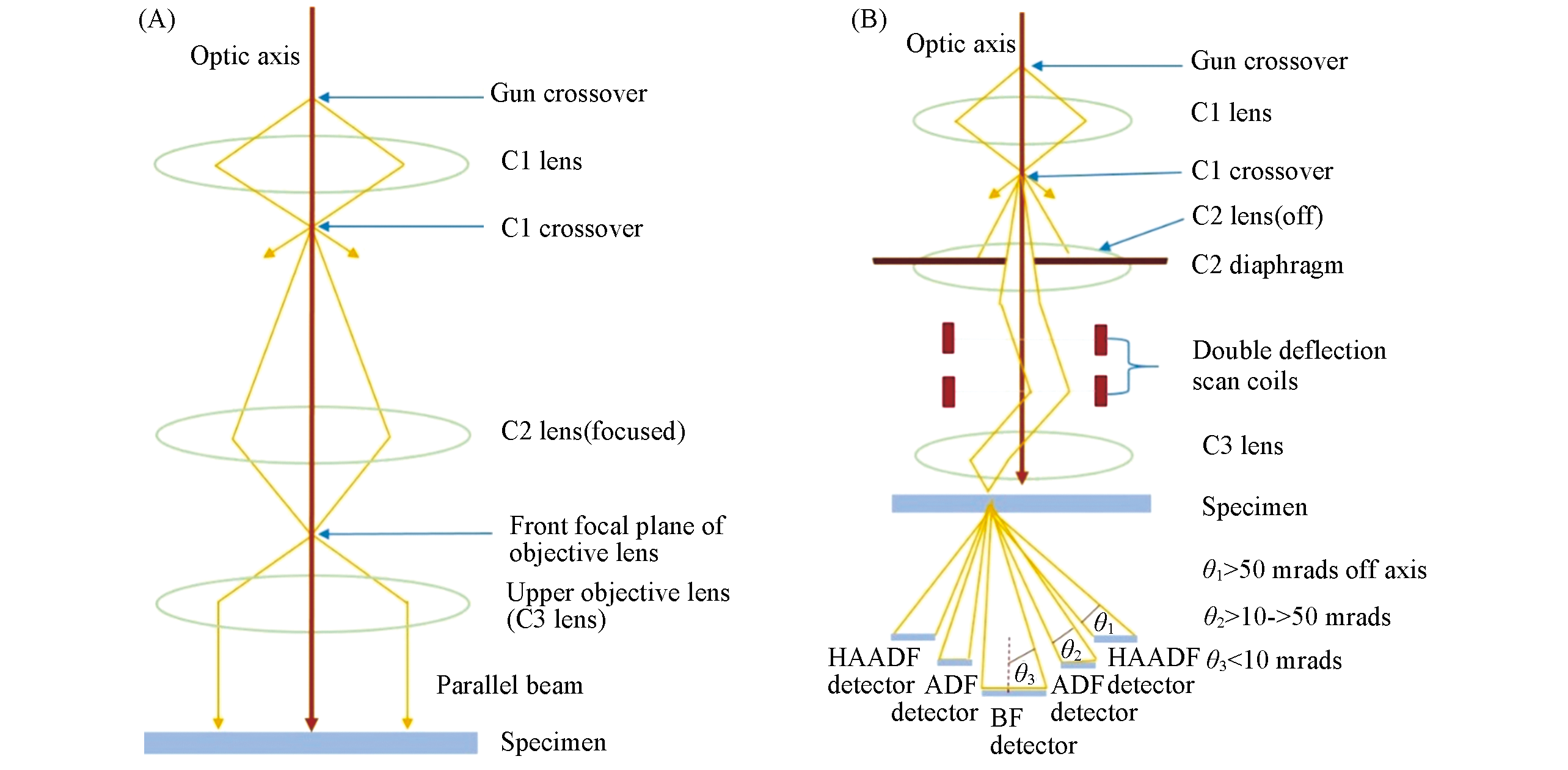
Fig.1 Comparison between TEM(A) and STEM(B)
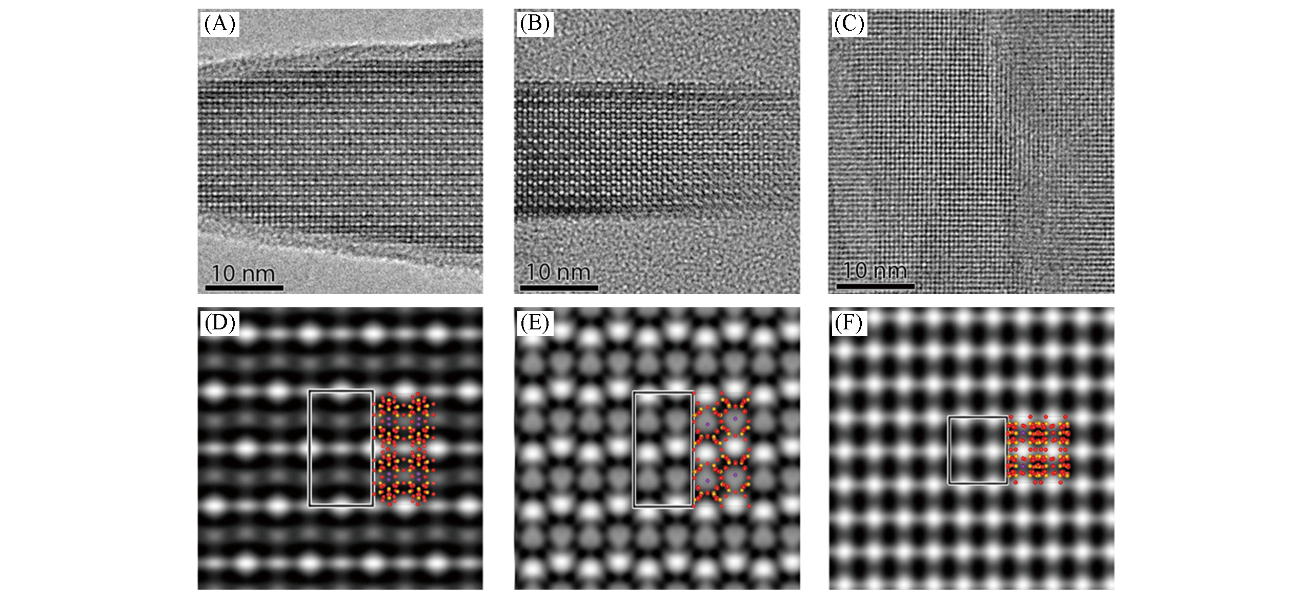
Fig.2 Reconstructed structure projection images(A―C) and corresponding symmetry?imposed lattice?averaged potential maps by Qfocus(D―F) along [001](A, D), [100](B, E) and[010](C, F), respectively[25]Copyright 2017, American Chemical Society.
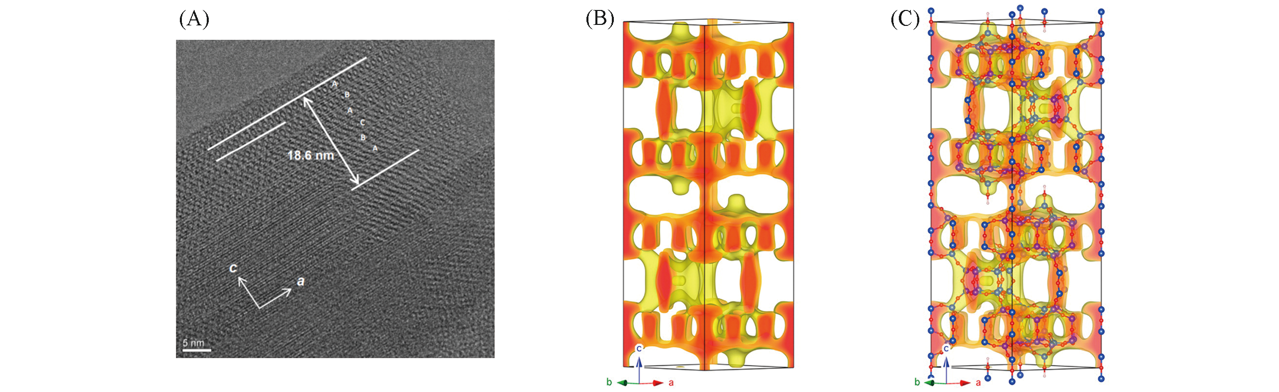
Fig.3 HRTEM image of ECNU?5(A), 3D potential map reconstructed from the HRTEM image(B) and overlap of 3D potential map and 3D structure model by shifting the MWW layers with 1/3 unit cell in ab?plane(C)[28]Copyright 2015, American Chemical Society.
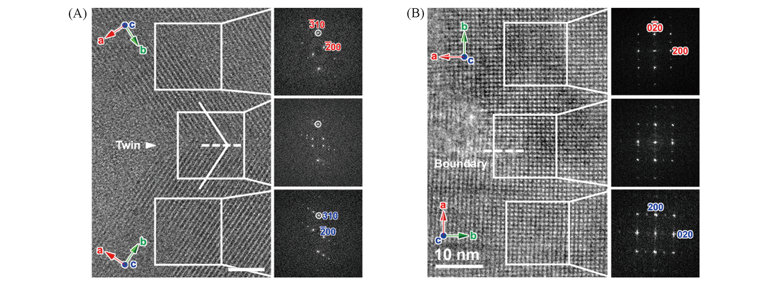
Fig.4 HRTEM images taken along [001] direction of intergrown MTW(A) and MFI(B), respectively[34]Corresponding Fourier diffractograms of HRTEM using selected areas in white rectangles are also given.Copyright 2018, American Chemical Society.
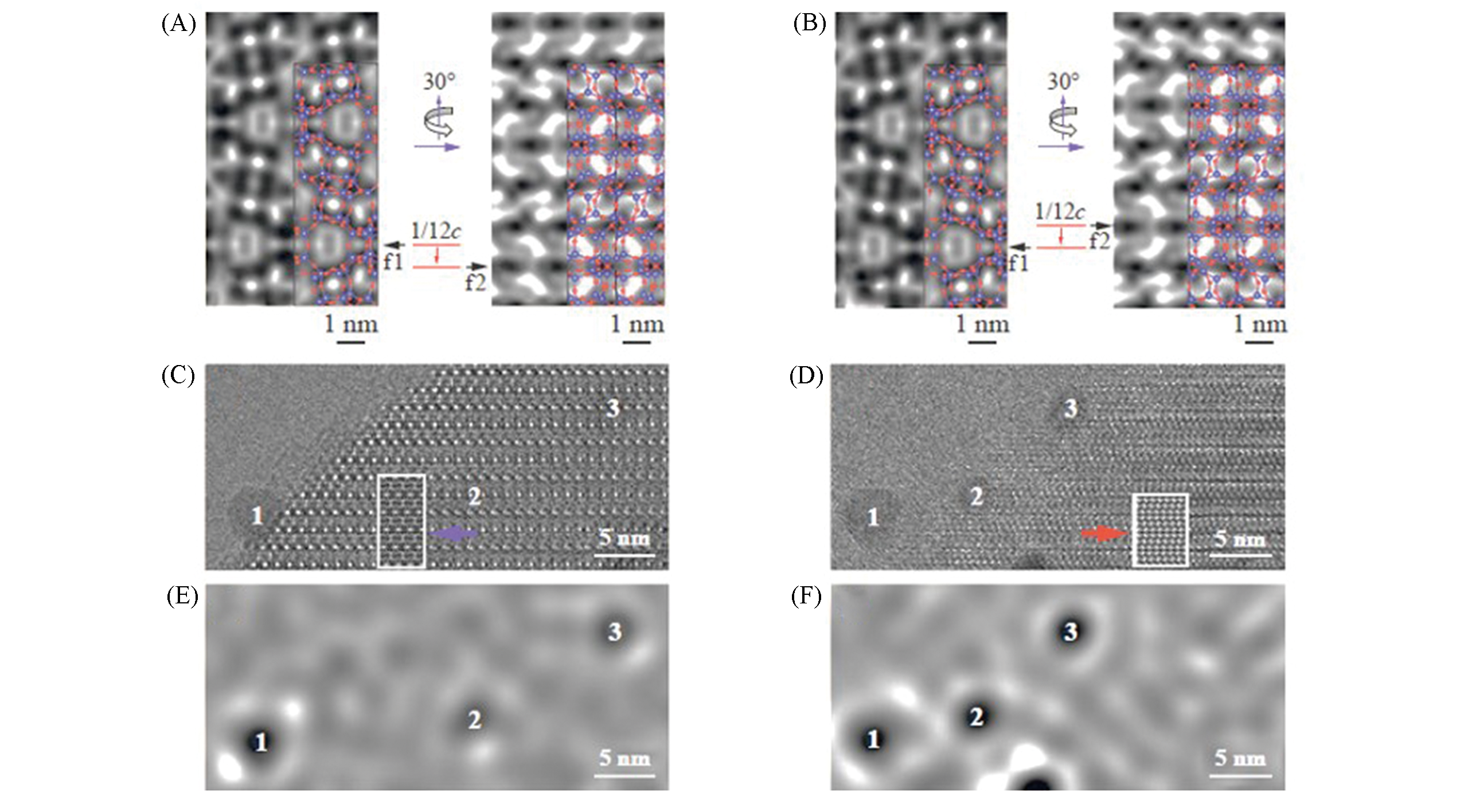
Fig.5 Handedness determination of a chiral zeolite by HRTEM[8](A, B) Simulated HRTEM images of right?handed(A) and left?handed(B) STW structures; (C, D) experimental HRTEM images of a chiral zeolite with gold nanoparticles as markers taken along [21ˉ1ˉ0](C) and [11ˉ00](D); (E, F) processed images by Fourier filtering of images(C) and (D), respectively.Copyright 2019, Springer Nature.
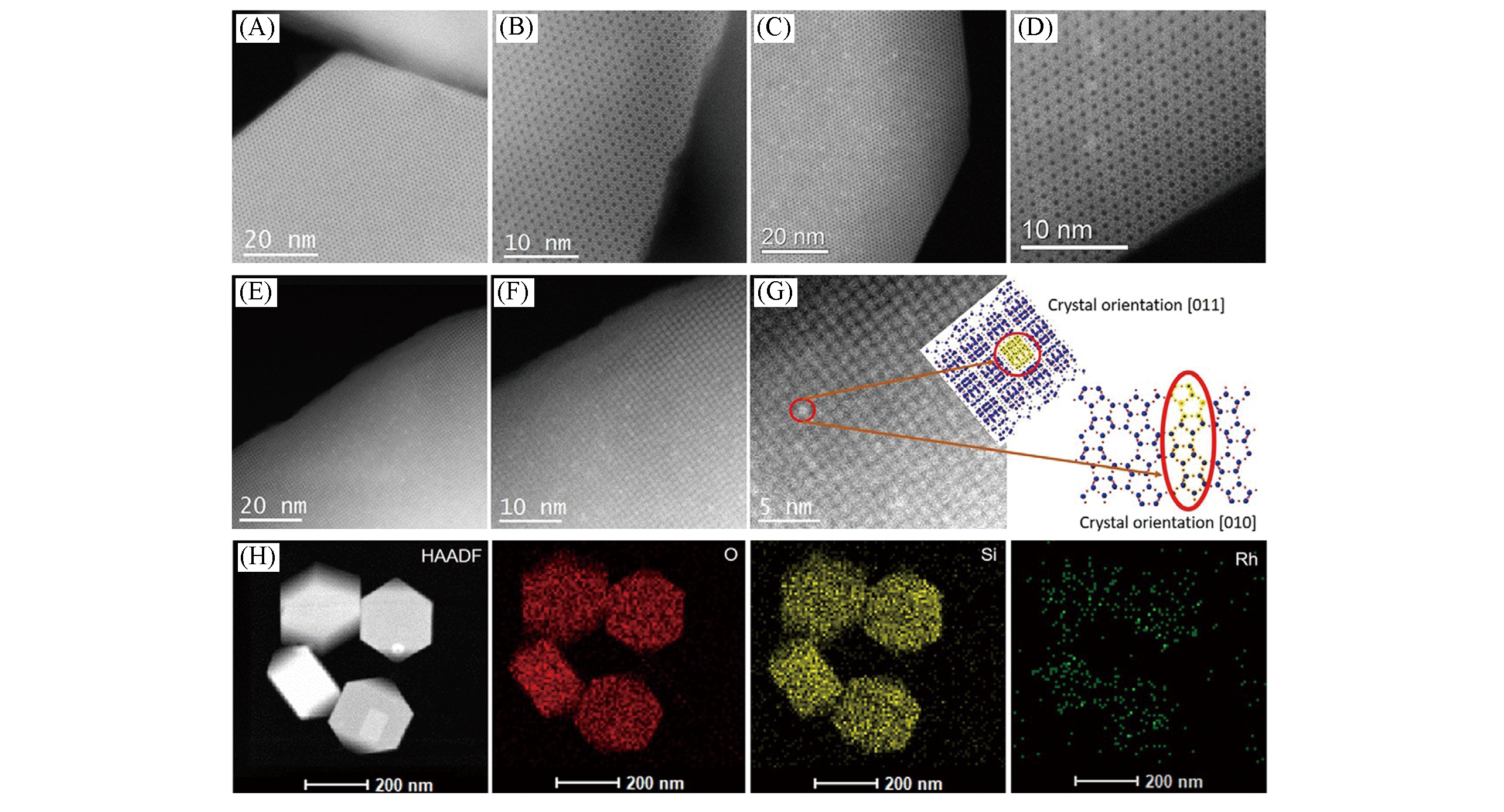
Fig.6 Cs?corrected STEM images of Rh@S?1[45](A―G) Cs?corrected STEM images of Rh@S?1?H(A, B) and Rh@S?1?C(C, D) viewed along b?axis orientation, and Rh@S?1?H viewed along [011] orientation as well as the schematic models along the same projection(E―G); (H) HAADF?STEM image of Rh@S?1?H and the corresponding EDX mapping images for O, Si, and Rh elements.Copyright 2019, Wiley?VCH.
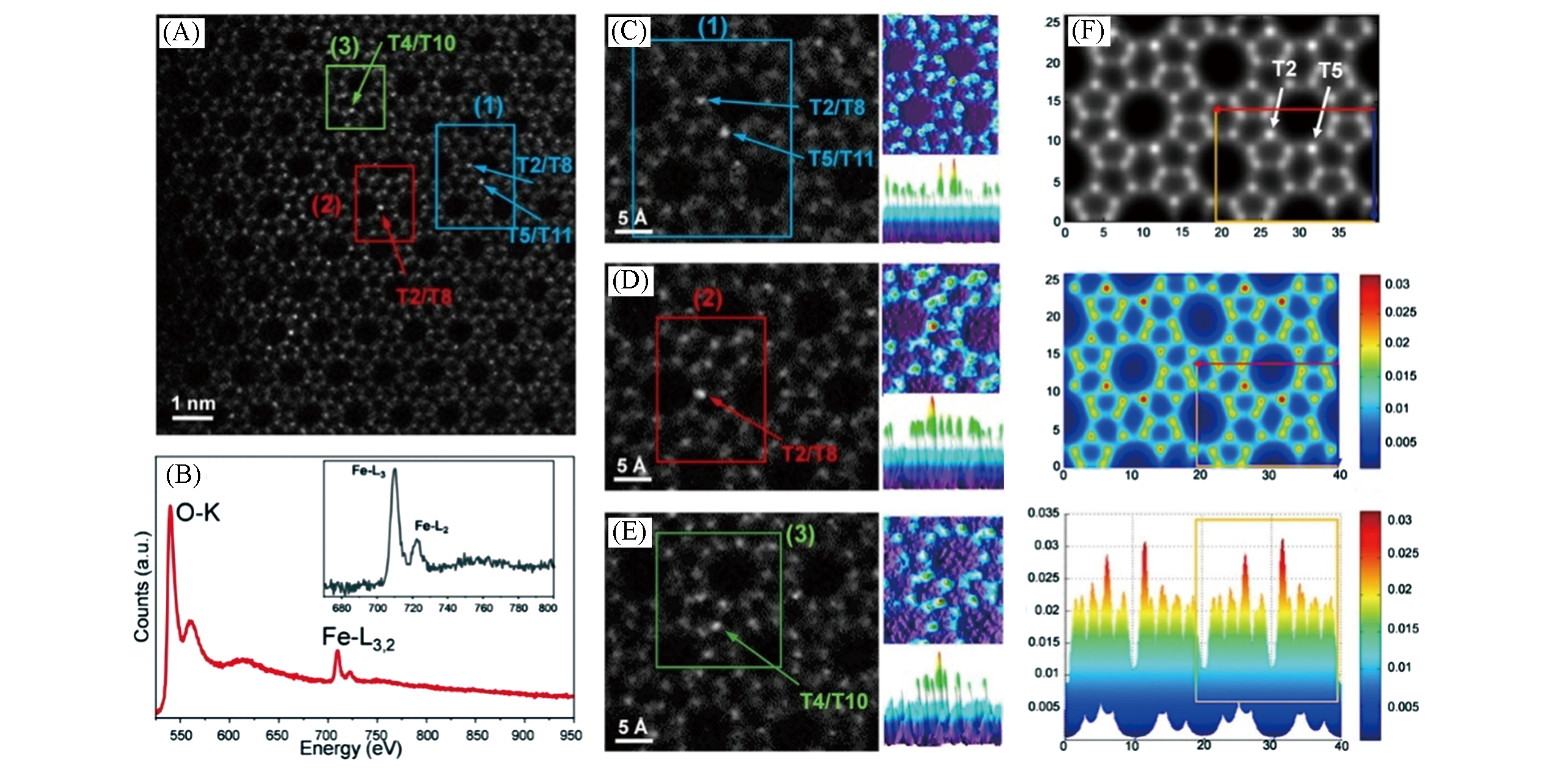
Fig.7 Cs?corrected STEM ADF images and EEL spectrum of Fe?MFI[46](A) High resolution ADF image.(B) EEL spectrum.(C―E) enlarged images corresponding to three regions marked by rectangles in (A) together with surface plots of 2D?intensity distribution map. Bright dots in (A, C, D, E) are marked by arrows with T?site symbols. (F) simulated images of Fe?MFI, where two single Fe atoms are located at T2 and T5 sites corresponding to 2 Fe atoms/unit?cell, at the conditions of probe?size: 1.0 ? and specimen thickness: 105 ?.Copyright 2020, Wiley?VCH.
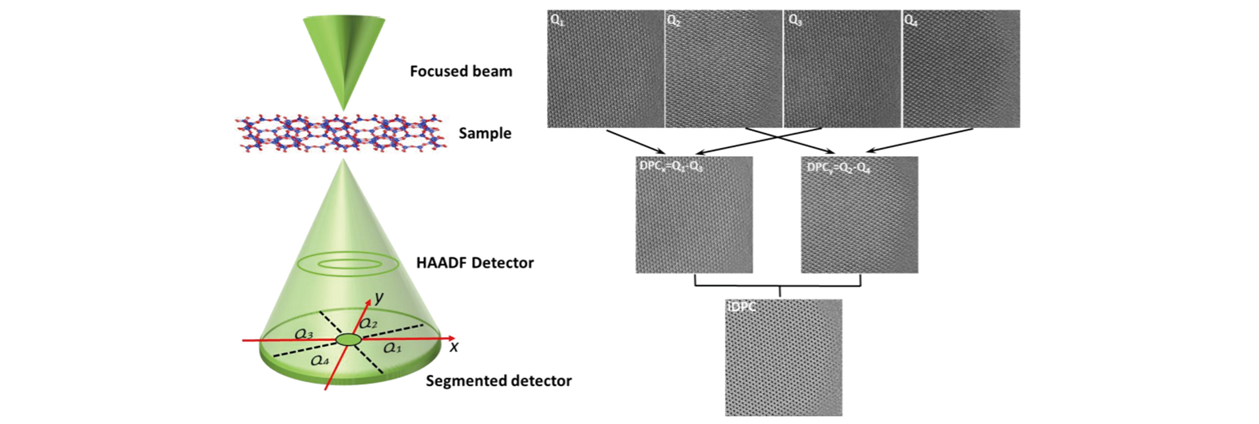
Fig.8 Schematic illustration of iDPC?STEM[17]Copyright 2020, Wiley?VCH.
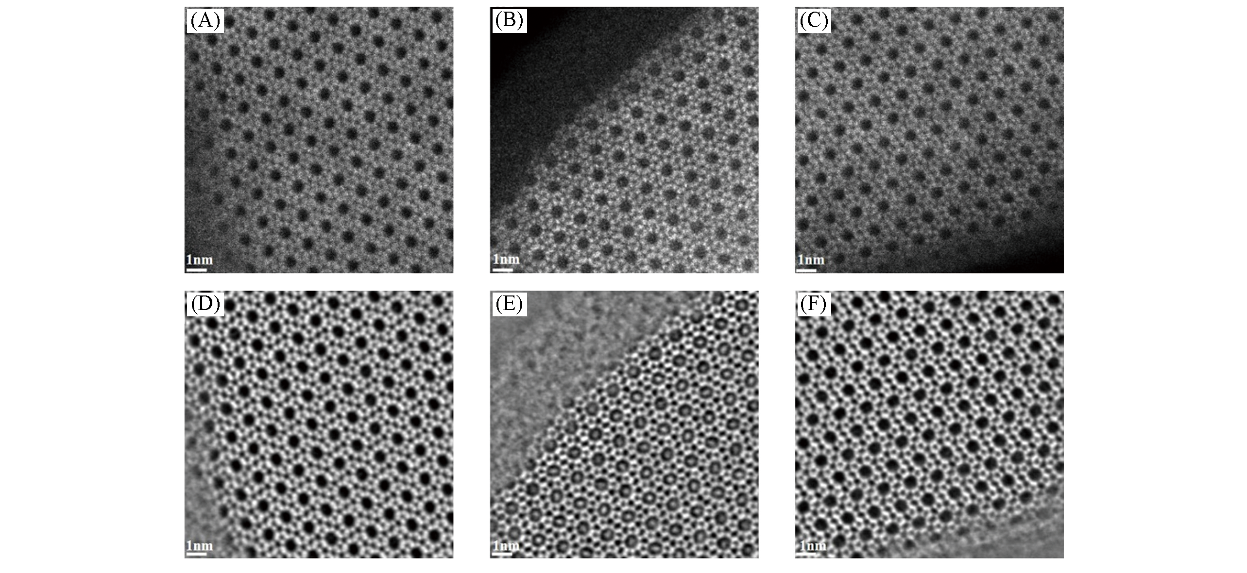
Fig.9 HAADF?STEM(A―C) and iDPC(D―F) images of calcined(A, D), air?exposed(B, E) and re?calcined(C, F) silicalite?1[17]Copyright 2020, Wiley?VCH.
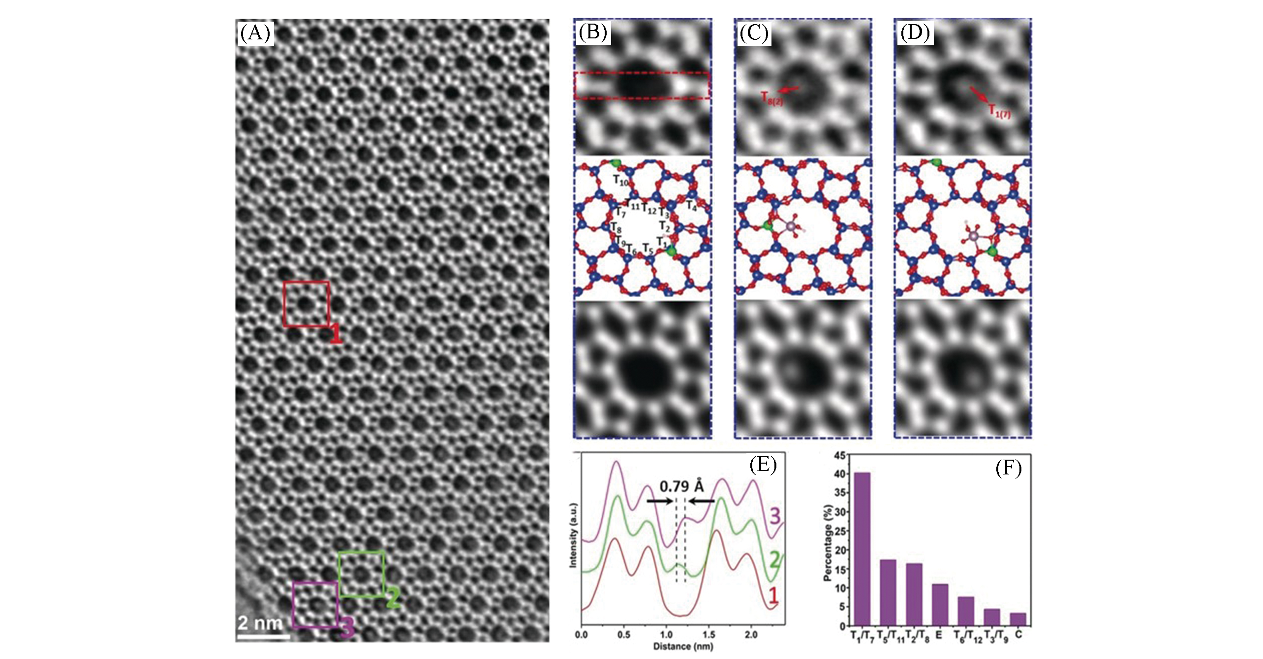
Fig.10 iDPC iamges of Mo?ZSM?5(A) and related analysis(B―F)[17](B―D) Zoomed?in areas 1(B), 2(C), and 3(D) of (A): empty 10 MRs channel(B), a MoO3H cluster bound at the T8 site(C) or at the T1 site(D) in the 10 MRs channel. Each panel includes the raw image(top), the calculated structural model(middle) and the simulated projected electrostatic potential(bottom). Si, blue; O, red; Al, green, Mo, pink.(E) Intensity line profiles of the images in (B―D), across the areas as represented by the red dashed rectangle. (F) Statistics of Al occupancy at different T sites, based on the results of one hundred channels.Copyright 2020, Wiley?VCH.
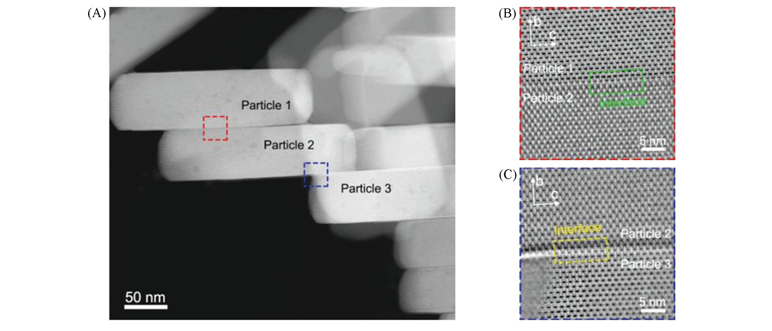
Fig.11 HAADF image of ZSM?5 particles(A) and iDPC images of (010) interfaces taken from red(B)and blue squares(C) in (A)[18]Copyright 2020, Wiley?VCH.
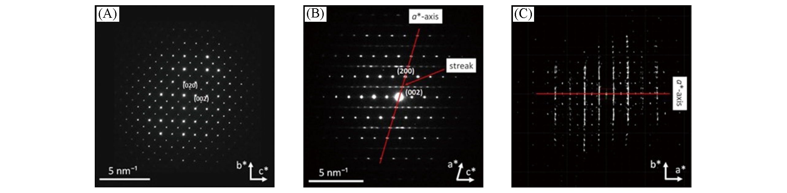
Fig.12 SAED patterns along [100](A), [010](B) and hk0 slice cut from the reconstructed 3D reciprocal lattice showing the diffuse scattering(C) of IM?18 zeolite[10]Copyright 2018, American Chemical Society.
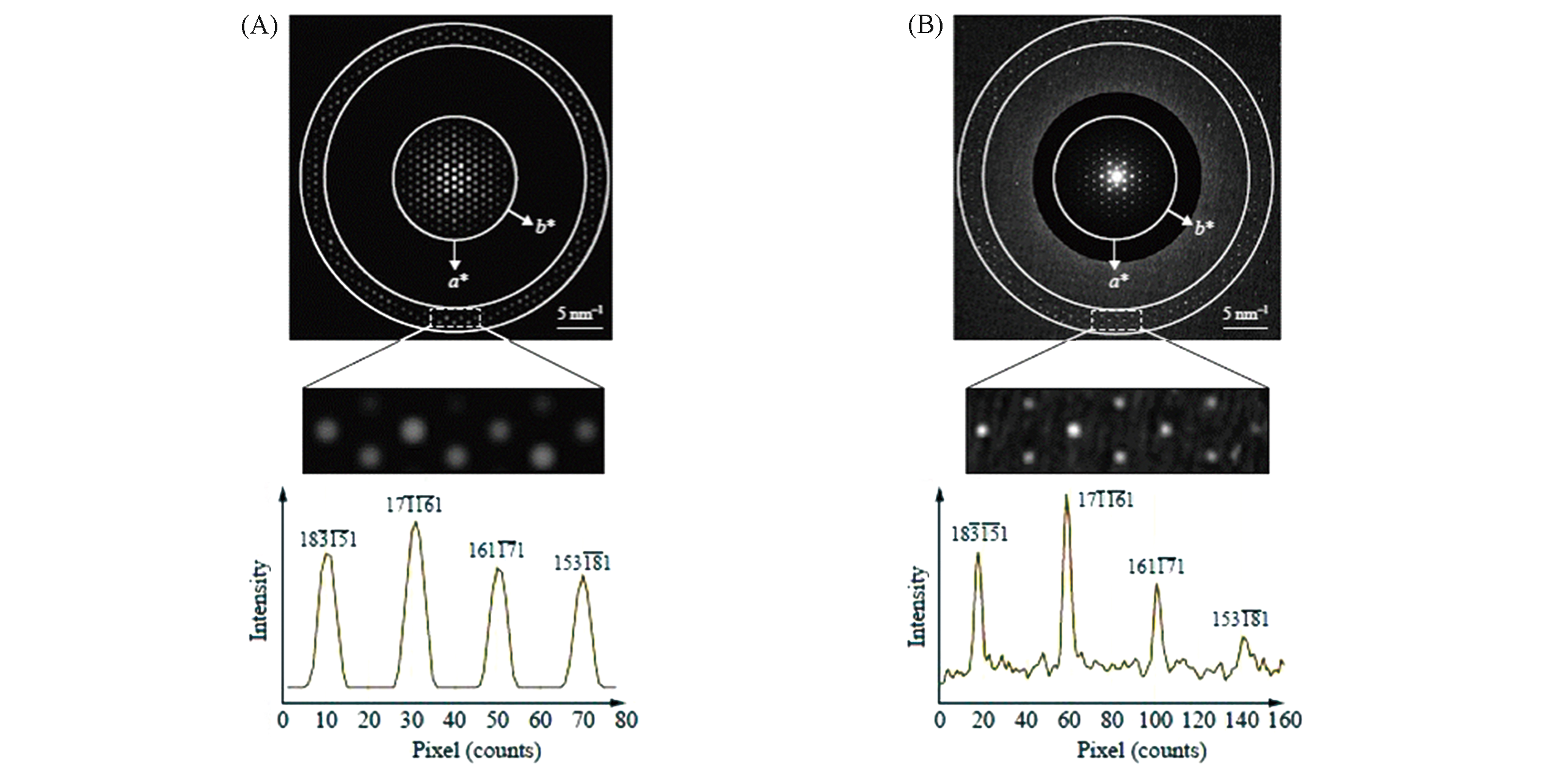
Fig.13 Handedness determination of a chiral zeolite by PED[8]Simulated(A) and experimental(B) PED patterns of STW zeolite along [0001] zone axis, respectively. The area in the white rectangle represents intensity difference of two reflection pairs: (1) 183ˉ15ˉ1 and (1′) 15318ˉ1; (2) 16117ˉ1?and (2′) 171ˉ16ˉ1. Copyright 2019, Springer Nature.
| 3D ED method | Electron beam tilt | Goniometer rotation | Electron beam precession | Data collection |
|---|---|---|---|---|
| ADT(PEDT) | No | Yes | Yes | Stepwise |
| RED | Yes | Yes | No | Stepwise |
| cRED | No | Yes | No | Continuous |
Table 1 Features of different 3D ED methods
| 3D ED method | Electron beam tilt | Goniometer rotation | Electron beam precession | Data collection |
|---|---|---|---|---|
| ADT(PEDT) | No | Yes | Yes | Stepwise |
| RED | Yes | Yes | No | Stepwise |
| cRED | No | Yes | No | Continuous |
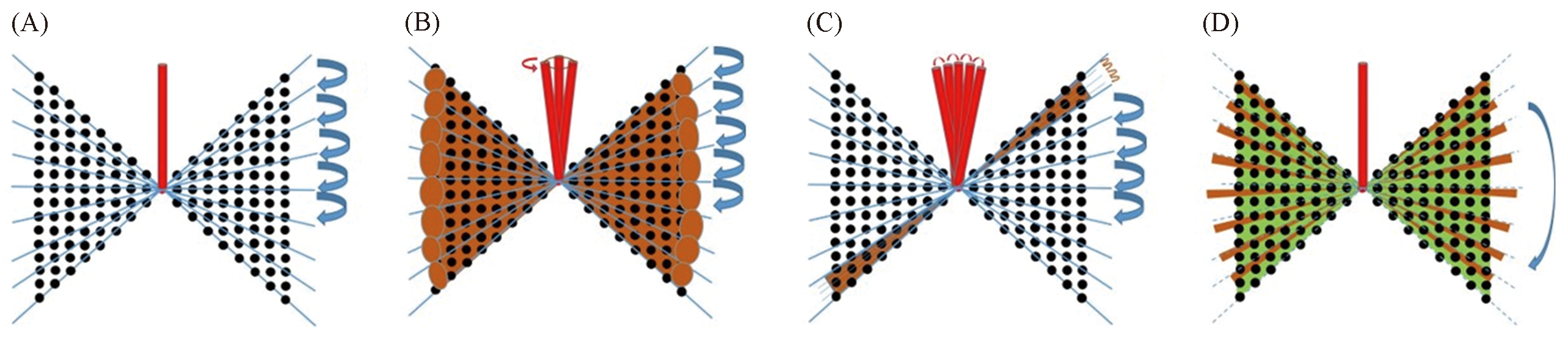
Fig.14 Illustration of simple mechanical tilt steps(A), PEDT(B), RED(C) and cRED(D)

Fig.15 SEM image(A), projection of 3D reconstructed reciprocal spaces along [001] direction(B), 2D 0kl(C), hk0(D) and h0l(E) slices extracted from the reconstructed reciprocal space of UTL?DBU[75]Copyright 2020, American Chemical Society.
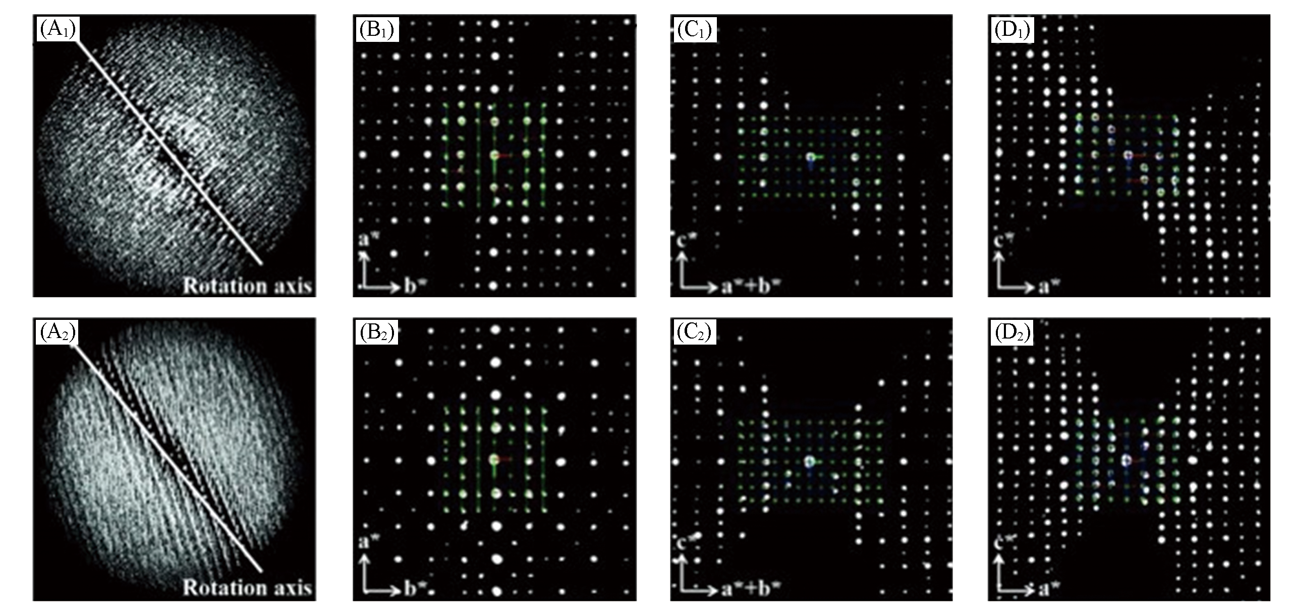
Fig.16 3D reconstructed reciprocal spaces(A1, A2) and 2D slices cut from 3D reconstructed lattice of hk0(B1, B2), hhl(C1, C2) and h0l(D1, D2) collected from PST?13(A1—D1) and PST?14(A2—D2), respectively[24]Copyright 2018, Wiley?VCH.
| 1 | Chester A. W., Derouane E. G., Zeolite Characterization and Catalysis, Springer, Dordrecht, 2009, 169—196 |
| 2 | Qiao Y., Yang M., Gao B., Wang L., Tian P., Xu S., Liu Z., Chem. Commun.,2016, 52(33), 5718—5721 |
| 3 | Sun J., Bonneau C., Cantín Á., Corma A., Díaz-Cabañas M. J., Moliner M., Zhang D., Li M., Zou X., Nature, 2009, 458(7242), 1154—1157 |
| 4 | Weckhuysen B. M., Yu J., Chem. Soc. Rev., 2015, 44(20), 7022—7024 |
| 5 | Wan W., Su J., Zou X. D., Willhammar T., Inorg. Chem. Front., 2018, 5(11), 2836—2855 |
| 6 | Li C., Zhang Q., Mayoral A., ChemCatChem, 2020, 12(5), 1248—1269 |
| 7 | Liu L., Corma A., Chem. Rev., 2018, 118(10), 4981—5079 |
| 8 | Ma Y., Han L., Liu Z., Mayoral A., Díaz I., Oleynikov P., Ohsuna T., Han Y., Pan M., Zhu Y., Sakamoto Y., Che S., Terasaki O., Springer Handbook of Microscopy, Springer, Cham, 2019, 1391—1450 |
| 9 | Smeets S., Xie D., Baerlocher C., McCusker L. B., Wan W., Zou X., Zones S. I., Angew. Chem. Int. Ed., 2014, 53(39), 10398—10402 |
| 10 | Cichocka M. O., Lorgouilloux Y., Smeets S., Su J., Wan W., Caullet P., Bats N., McCusker L. B., Paillaud J. L., Zou X., Cryst. Growth & Des., 2018, 18(4), 2441—2451 |
| 11 | Baerlocher C., Gramm F., Massüger L., McCusker L. B., He Z., Hovmöller S., Zou X., Science, 2007, 315(5815), 1113—1116 |
| 12 | Yu Z. B., Han Y., Zhao L., Huang S., Zheng Q. Y., Lin S., Córdova A., Zou X., Sun J., Chem. Mater., 2012, 24(19), 3701—3706 |
| 13 | Ma Y., Oleynikov P., Terasaki O., Nat. Mater., 2017, 16(7), 755—759 |
| 14 | Mayoral A., Carey T., Anderson P. A., Lubk A., Diaz I., Angew. Chem. Int. Ed., 2011, 50(47), 11230—11233 |
| 15 | Mayoral A., Readman J. E., Anderson P. A., J. Phys. Chem. C, 2013, 117(46), 24485—24489 |
| 16 | Wang N., Sun Q., Bai R., Li X., Guo G., Yu J., J. Am. Chem. Soc., 2016, 138(24), 7484—7487 |
| 17 | Liu L., Wang N., Zhu C., Liu X., Zhu Y., Guo P., Alfilfil L., Dong X., Zhang D., Han Y., Angew. Chem. Int. Ed., 2020, 59(2), 819—825 |
| 18 | Shen B., Chen X., Cai D., Xiong H., Liu X., Meng C., Han Y., Wei F., Adv. Mater., 2020, 32(4), 1906103 |
| 19 | Newsam J., Treacy M. M., Koetsier W., Gruyter C. D., Proc. R. Soc. Lond. A, 1988, 420(1859), 375—405 |
| 20 | Burton A. W., Elomari S., Chan I., Pradhan A., Kibby C., J. Phys. Chem. B, 2005, 109(43), 20266—20275 |
| 21 | Terasaki O., Ohsuna T., Inagaki S., Catal. Surv. Japan, 2001, 4(2), 99—106 |
| 22 | Gemmi M., Lanza A . E., Acta Cryst. B, 2019, 75(4), 495—504 |
| 23 | Luo Y., Smeets S., Peng F., Etman A. S., Wang Z., Sun J., Yang W., Chem. Eur. J., 2017, 23(66), 16829—16834 |
| 24 | Seo S., Yang T., Shin J., Jo D., Zou X., Hong S. B., Angew. Chem. Int. Ed., 2018, 57(14), 3727—3732 |
| 25 | Willhammar T., Su J., Yun Y., Zou X., Afeworki M., Weston S. C., Vroman H. B., Lonergan W. W., Strohmaier K. G., Inorg. Chem., 2017, 56(15), 8856—8864 |
| 26 | Luo Y., Smeets S., Wang Z., Sun J., Yang W., Chem. Eur. J., 2019, 25(9), 2184—2188 |
| 27 | Yuhas B. D., Mowat J. P. S., Miller M. A., Sinkler W., Chem. Mater., 2018, 30(3), 582—586 |
| 28 | Xu L., Ji X., Jiang J., Han L., Che S., Wu P., Chem. Mater., 2015, 27(23), 7852—7860 |
| 29 | Smeets S., Berkson Z. J., Xie D., Zones S. I., Wan W., Zou X. D., Hsieh M. F., Chmelka B. F., McCusker L. B., Baerlocher C., J. Am. Chem. Soc., 2017, 139(46), 16803—16812 |
| 30 | Wan W., Hovmöller S., Zou X., Ultramicroscopy, 2012, 115, 50—60 |
| 31 | Hovmöller S., Ultramicroscopy, 1992, 41(1—3), 121—135 |
| 32 | Momma K., Izumi F., J. Appl. Crystal., 2011, 44(6), 1272—1276 |
| 33 | Choi M., Na K., Kim J., Sakamoto Y., Terasaki O., Ryoo R., Nature, 2009, 461(7265), 246—249 |
| 34 | Jia X., Zhang Y., Gong Z., Wang B., Zhu Z., Jiang J., Xu H., Sun H., Han L., Wu P., Che S., J. Phys. Chem. C, 2018, 122(16), 9117—9126 |
| 35 | Ritsch S., Ohnishi N., Ohsuna T., Hiraga K., Terasaki O., Kubota Y., Sugi Y., Chem. Mater., 1998, 10(12), 3958—3965 |
| 36 | Davis M. E., ACS Catal., 2018, 8(11), 10082—10088 |
| 37 | Che S., Garcia⁃Bennett A. E., Yokoi T., Sakamoto K., Kunieda H., Terasaki O., Tatsumi T., Nat. Mater., 2003, 2(12), 801—805 |
| 38 | Brand S. K., Schmidt J. E., Deem M. W., Daeyaert F., Ma Y., Terasaki O., Orazov M., Davis M. E., Proc. Natl. Acad. Sci. U. S. A., 2017, 114(20), 5101—5106 |
| 39 | Fultz B., Howe J., Transmission Electron Microscopy and Diffractometry of Materials, Springer, Berlin, 2013, 587—615 |
| 40 | Firth D. S., Morris S. A., Wheatley P. S., Russell S. E., Slawin A. M., Dawson D. M., Mayoral A., Opanasenko M., Položij M., Čejka J. I., Chem. Mater., 2017, 29(13), 5605—5611 |
| 41 | Aydin C., Lu J., Liang A. J., Chen C. Y., Browning N. D., Gates B. C., Nano lett., 2011, 11(12), 5537—5541 |
| 42 | Kistler J. D., Chotigkrai N., Xu P., Enderle B., Praserthdam P., Chen C. Y., Browning N. D., Gates B. C., Angew. Chem. Int. Ed., 2014, 53(34), 8904—8907 |
| 43 | Lu J., Aydin C., Browning N. D., Gates B. C., Angew. Chem. Int. Ed., 2012, 51(24), 5842—5846 |
| 44 | Altantzis T., Coutino⁃Gonzalez E., Baekelant W., Martinez G. T., Abakumov A. M., Tendeloo G. V., Roeffaers M. B., Bals S., Hofkens J., ACS Nano, 2016, 10(8), 7604—7611 |
| 45 | Sun Q., Wang N., Zhang T., Bai R., Mayoral A., Zhang P., Zhang Q., Terasaki O., Yu J., Angew. Chem. Int. Ed., 2019, 58(51), 18570—18576 |
| 46 | Mayoral A., Zhang Q., Zhou Y., Chen P., Ma Y., Monji T., Losch P., Schmidt W., Schüth F., Hirao H.,Yu J., Terasaki O., Angew. Chem. Int. Ed., 2020, 59(44), 19510—19517 |
| 47 | Bosch E. G., Lazić I., Ultramicroscopy, 2015, 156, 59—72 |
| 48 | Lazić I., Bosch E. G., Lazar S., Ultramicroscopy, 2016, 160, 265—280 |
| 49 | Ma Y., Wang N., Qian W., Wang Y., Zhang J., Wei F., RSC Adv., 2016, 6(84), 81198—81202 |
| 50 | Song D., Zhang X., Lian C., Liu H., Alexandrou I., Lazić I., Bosch E. G., Zhang D., Wang L., Yu R., Cheng Z., Song C., Ma X., Duan W., Xue Q., Zhu J., Adv. Func. Mater., 2019, 29(45), 1903843 |
| 51 | Shen B., Chen X., Shen K., Xiong H., Wei F., Nat. Commun., 2020. doi:10.1038/s41467⁃020⁃16531⁃y |
| 52 | Han L., Miyasaka K., Terasaki O., Structure from Diffraction Methods: Inorganic Materials Series, John Wiley & Sons,Chichester,2014, 201—258 |
| 53 | Wagner P., Terasaki O., Ritsch S., Nery J. G., Zones S. I., Davis M. E., Hiraga K., J. Phy. Chem. B, 1999, 103(39), 8245—8250 |
| 54 | Guo P., Shin J., Greenaway A. G., Min J. G., Su J., Choi H. J., Liu L., Cox P. A., Hong S. B., Wright P. A., Nature, 2015, 524(7563), 74—78 |
| 55 | Lee H., Shin J., Choi W., Choi H. J., Yang T., Zou X., Hong S. B., Chem. Mater., 2018, 30(19), 6619—6623 |
| 56 | Gemmi M., Mugnaioli E., Gorelik T. E., Kolb U., Palatinus L., Boullay P., Hovmoller S., Abrahams J. P., ACS Cent. Sci., 2019, 5(8), 1315—1329 |
| 57 | Dorset D. L., Weston S. C., Dhingra S. S., J. Phys. Chem. B, 2006, 110(5), 2045—2050 |
| 58 | Dorset D. L., Z. Kristallogr., 2006, 221(4), 260—265 |
| 59 | Kolb U., Gorelik T., Kübel C., Otten M., Hubert D., Ultramicroscopy, 2007, 107(6/7), 507—513 |
| 60 | Kolb U., Gorelik T., Otten M., Ultramicroscopy, 2008, 108(8), 763—772 |
| 61 | Mugnaioli E., Gorelik T., Kolb U., Ultramicroscopy, 2009, 109(6), 758—765 |
| 62 | Zhang D. L., Oleynikov P., Hovmoller S., Zou X. D., Z. Kristallogr., 2010, 225(2/3), 94—102 |
| 63 | Nannenga B. L., Shi D., Leslie A. G., Gonen T., Nat. Methods, 2014, 11(9), 927—930 |
| 64 | Gemmi M., La Placa M. G. I., Galanis A. S., Rauch E. F., Nicolopoulos S., J. Appl. Cryst., 2015, 48(3), 718—727 |
| 65 | Wang Y. C., Takki S., Cheung O., Xu H. Y., Wan W., Ohrstrom L., Inge A. K., Chem. Commun., 2017, 53(52), 7018—7021 |
| 66 | Sun T., Wei L., Chen Y. C., Ma Y., Zhang Y. B., J. Am. Chem. Soc., 2019, 141(28), 10962—10966 |
| 67 | Burla M. C., Caliandro R., Carrozzini B., Cascarano G. L., Cuocci C., Giacovazzo C., Mallamo M., Mazzone A., Polidori G., J. Appl. Cryst., 2015, 48(1), 306—309 |
| 68 | Kolb U., Mugnaioli E., Gorelik T., Cryst. Res. Tech., 2011, 46(6), 542—554 |
| 69 | Wan W., Sun J., Su J., Hovmöller S., Zou X., J. Appl. Cryst., 2013, 46(6), 1863—1873 |
| 70 | Willhammar T., Sun J., Wan W., Oleynikov P., Zhang D., Zou X., Moliner M., Gonzalez J., Martínez C., Rey F., Nat. Chem., 2012, 4(3), 188—194 |
| 71 | Kang J. H., Xie D., Zones S. I., Smeets S., McCusker L. B., Davis M. E., Chem. Mater., 2016, 28(17), 6250—6259 |
| 72 | Bieseki L., Simancas R., Jordá J. L., Bereciartua P. J., Cantín Á., Simancas J., Pergher S. B., Valencia S., Rey F., Corma A., Chem. Commun., 2018, 54(17), 2122—2125 |
| 73 | Liu X., Luo Y., Mao W., Jiang J., Xu H., Han L., Sun J., Wu P., Angew. Chem. Int. Ed., 2020, 59(3), 1166—1170 |
| 74 | Jiang J., Jorda J. L., Yu J., Baumes L. A., Mugnaioli E., Diaz⁃Cabanas M. J., Kolb U., Corma A., Science, 2011, 333(6046), 1131—1134 |
| 75 | Li J., Zhang C., Jiang J., Yu J., Terasaki O., Mayoral A., J. Phys. Chem. Lett., 2020, 11(9), 3350—3356 |
| 76 | Burla M. C., Caliandro R., Carrozzini B., Cascarano G. L., Cuocci C., Giacovazzo C., Mallamo M., Mazzone A., Polidori G., J. Appl. Cryst., 2015, 48(1), 306—309 |
| [1] | 陈晓, 申博渊, 熊昊, 魏飞. 电子束敏感材料的原子尺度结构研究[J]. 高等学校化学学报, 2021, 42(1): 133. |
| [2] | 黄永烽, 黄文婷, 刘文宝, 刘岳峰, 刘伟, 徐成俊. 锌离子电池正极材料V2O5的储能机理和容量衰减原因[J]. 高等学校化学学报, 2020, 41(8): 1859. |
| [3] | 石磊, 于婷婷, 林森, 袁玉斌, 王济凯, 张娜. B掺杂STW结构硅酸盐分子筛的合成与表征[J]. 高等学校化学学报, 2015, 36(8): 1467. |
| [4] | 郭凯. 低分子量聚亚苯基苯并二噻唑在稀溶液中的结晶[J]. 高等学校化学学报, 2011, 32(1): 4. |
| [5] | 姚晰, 董凤霞, 吴杰, 宁杨, 张皓. 用于透射电子显微镜表征的单层纳米晶/聚合物复合薄膜的制备[J]. 高等学校化学学报, 2010, 31(3): 616. |
| [6] | 刘淑芳, 尹俊发, 宋茂勇, 汪海林. 水溶性羟基化单壁碳纳米管与人血清白蛋白之间的相互作用研究[J]. 高等学校化学学报, 2009, 30(9): 1733. |
| [7] | 陈晔, 杨德才, 胡雁鸣, 张学全. 结晶性间同立构1,2-聚丁二烯的单晶结构[J]. 高等学校化学学报, 2003, 24(12): 2321. |
| [8] | 陈晔, 杨德才, 胡雁鸣, 张学全. 间同立构1,2-聚丁二烯的合成和形态结构研究[J]. 高等学校化学学报, 2003, 24(11): 2119. |
| [9] | 杨智, 林建华, 苏勉曾, 尤力平, 王渭, 陶冶. LnBaB9O16:Eu3+(Ln=La,Y)的结构与荧光性质[J]. 高等学校化学学报, 1999, 20(12): 1832. |
| [10] | 黄金满, 杨毅, 杨柏, 刘式墉, 沈家骢. 硫化镉钠米微粒在聚合物网络中的组装[J]. 高等学校化学学报, 1995, 16(12): 1976. |
| 阅读次数 | ||||||
|
全文 |
|
|||||
|
摘要 |
|
|||||