

高等学校化学学报 ›› 2024, Vol. 45 ›› Issue (5): 20240027.doi: 10.7503/cjcu20240027
• 综合评述 • 上一篇
刘康, 潘荣容, 江德臣( )
)
收稿日期:2024-01-17
出版日期:2024-05-10
发布日期:2024-03-12
通讯作者:
江德臣
E-mail:dechenjiang@nju.edu.cn
基金资助:
LIU Kang, PAN Rongrong, JIANG Dechen()
Received:2024-01-17
Online:2024-05-10
Published:2024-03-12
Contact:
JIANG Dechen
E-mail:dechenjiang@nju.edu.cn
Supported by:摘要:
单细胞分析能够更加精准地获取生物学信息, 避免因平均化分析而丢失单细胞异质性特征, 这对于研究阐明细胞代谢和信号通路至关重要. 基于纳米电极的电化学分析技术因其高选择性、 高灵敏度和高时空分辨率的优点而被广泛用于单细胞分析. 本文综合评述了利用纳米电极对单细胞内部生物分子进行定量分析的最新研究进展, 介绍了其在生物学研究中的应用, 并对该领域面临的问题和未来发展进行了总结与展望.
中图分类号:
TrendMD:
刘康, 潘荣容, 江德臣. 基于纳米电极的单细胞内生物分子电化学分析. 高等学校化学学报, 2024, 45(5): 20240027.
LIU Kang, PAN Rongrong, JIANG Dechen. Electrochemical Analysis of Intracellular Molecules at Single Cells Based on Nanoelectrodes. Chem. J. Chinese Universities, 2024, 45(5): 20240027.
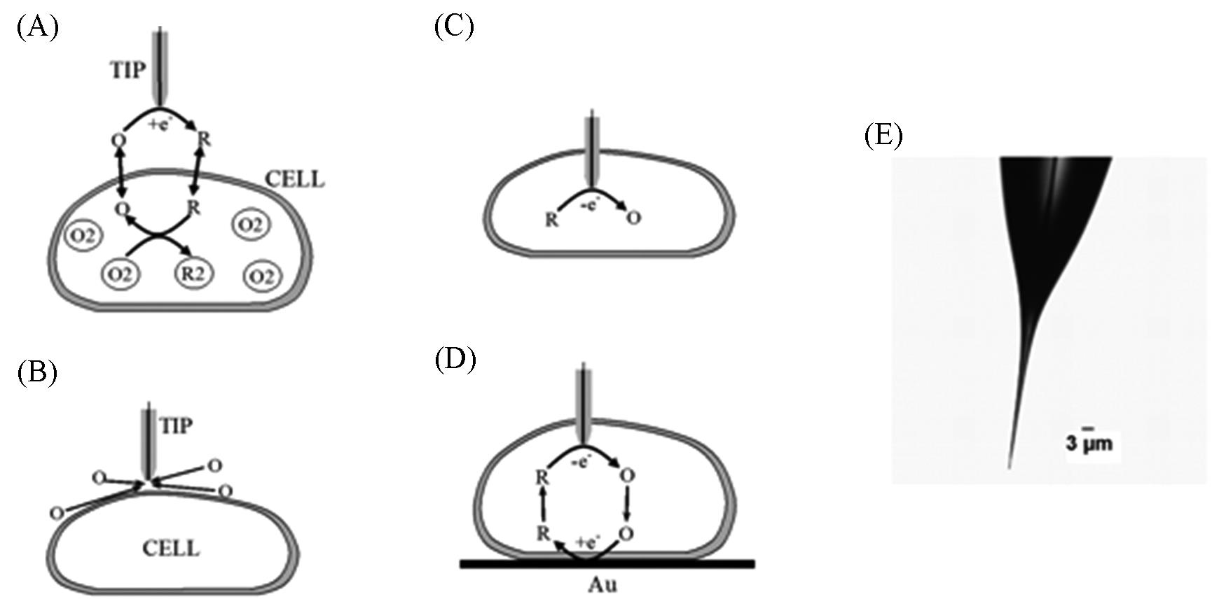
Fig.1 Schematic diagrams of the SECM experiments with single cells(A—D) and an optical micrograph of a typical nanotip used in such experiments(E)[22](A) The tip is positioned in the solution close to the cell surface. Positive feedback is due to bimolecular electron transfer between hydrophobic redox mediator(O/R) and cell-bound redox moieties(O2/R2); (B) the lipid cell membrane is impermeable for a hydrophilic redox mediator. Negative feedback is due to the hindered diffusion of redox species to the tip electrode; (C) nanoelectrode voltammetry inside the cell; (D) positive feedback is produced by mediator regeneration by way of electron transfer at the underlying Au surface.Copyright 2008, National Academy of Science.
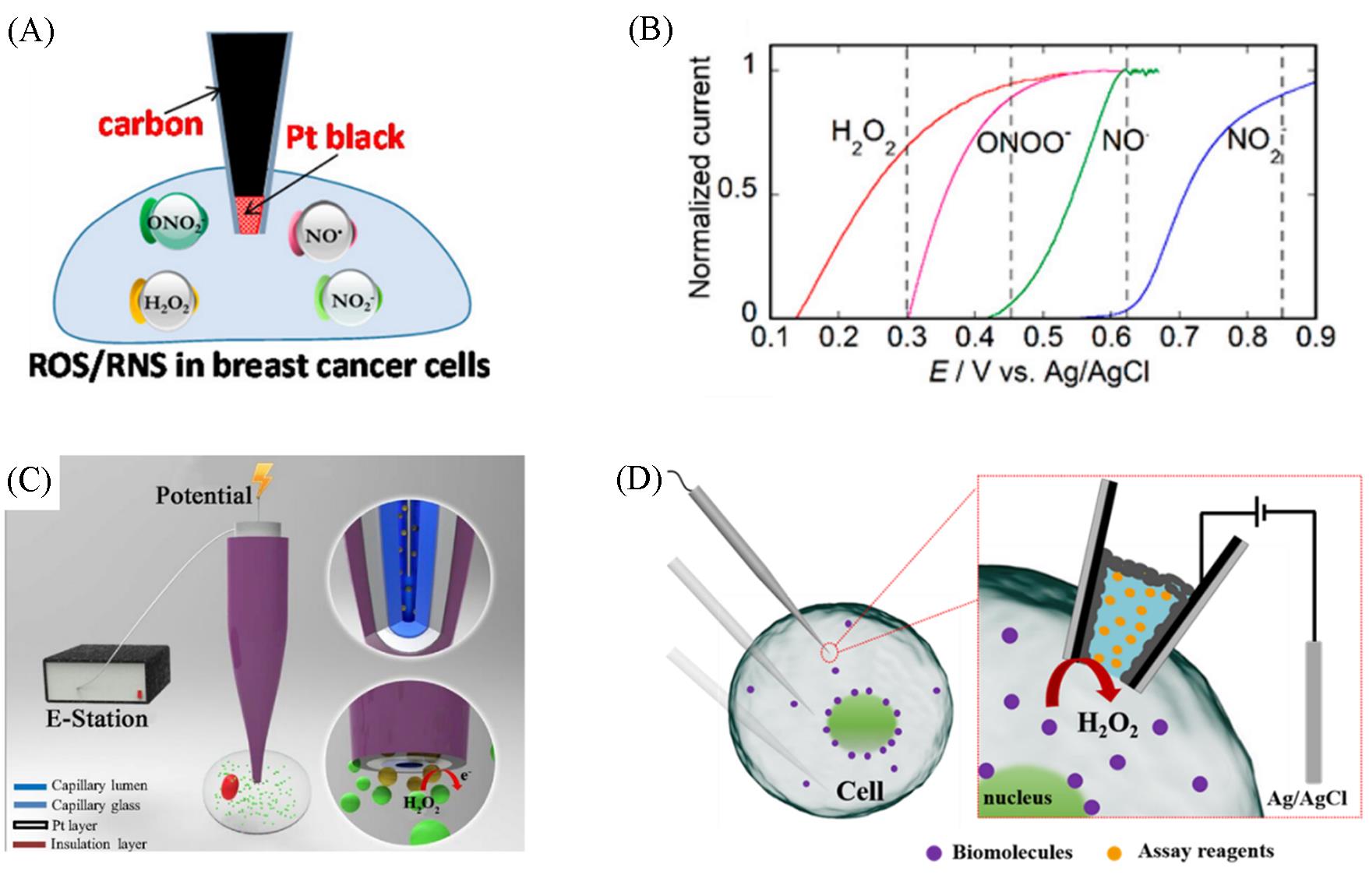
Fig.2 Detection of electroactive(A, B) and non⁃electroactive(C, D) molecules in single cell(A) Intracellular detection of ROS/RNS; (B) normalized oxidation voltammograms of H2O2 (red curve, 1 mmol/L, pH≈7.4), ONOO-(purple curve, 1 mmol/L, pH≈10), NO·(green curve, 1 mmol/L of NO· DEA⁃NONoate donor, pH≈7.4), and NO2- (blue curve, 1 mmol/L, pH≈7.4). Voltammograms were recorded at different platinized tips with a≈100 nm and normalized by their plateau currents. Vertical dashed lines indicate optimal detection potentials for each ROS/RNS species(B)[23]; (C) schematic of the nanokit used for the single⁃cell electrochemical analysis[30]; (D) the schematic liquid⁃phase modified nanopipette for the detection of intracellular molecules in one living cell. The black and silver regions at the inner surface of nanopipette are carbon and Pt layers, respectively[31].(A, B) Copyright 2017, American Chemical Society; (C) Copyright 2016, National Academy of Science; (D) Copyright 2023, Chinese Chemical Society.
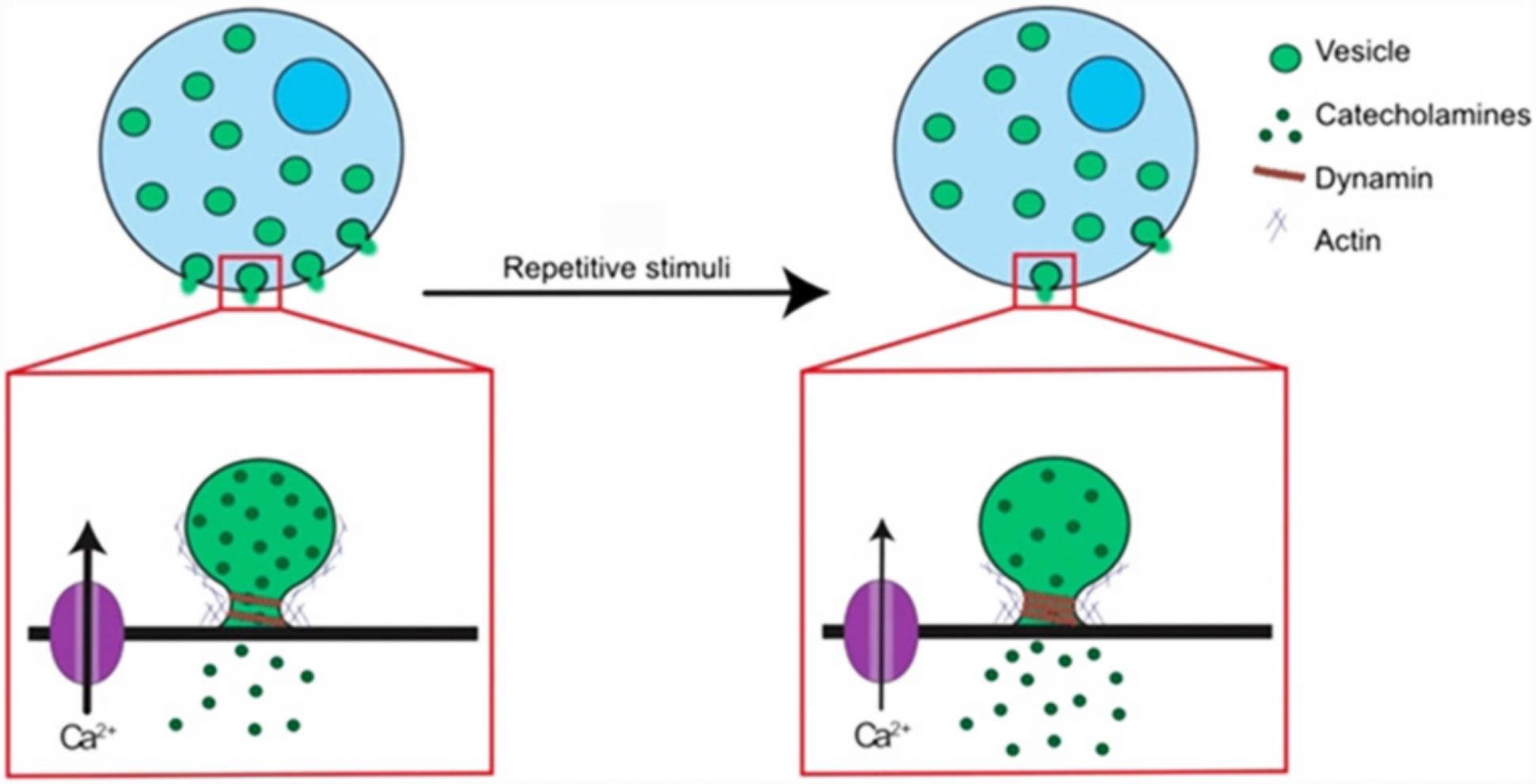
Fig.3 Scheme proposed for the effect of repetitive stimuli on cells and vesicles[42]Copyright 2019, National Academy of Science.
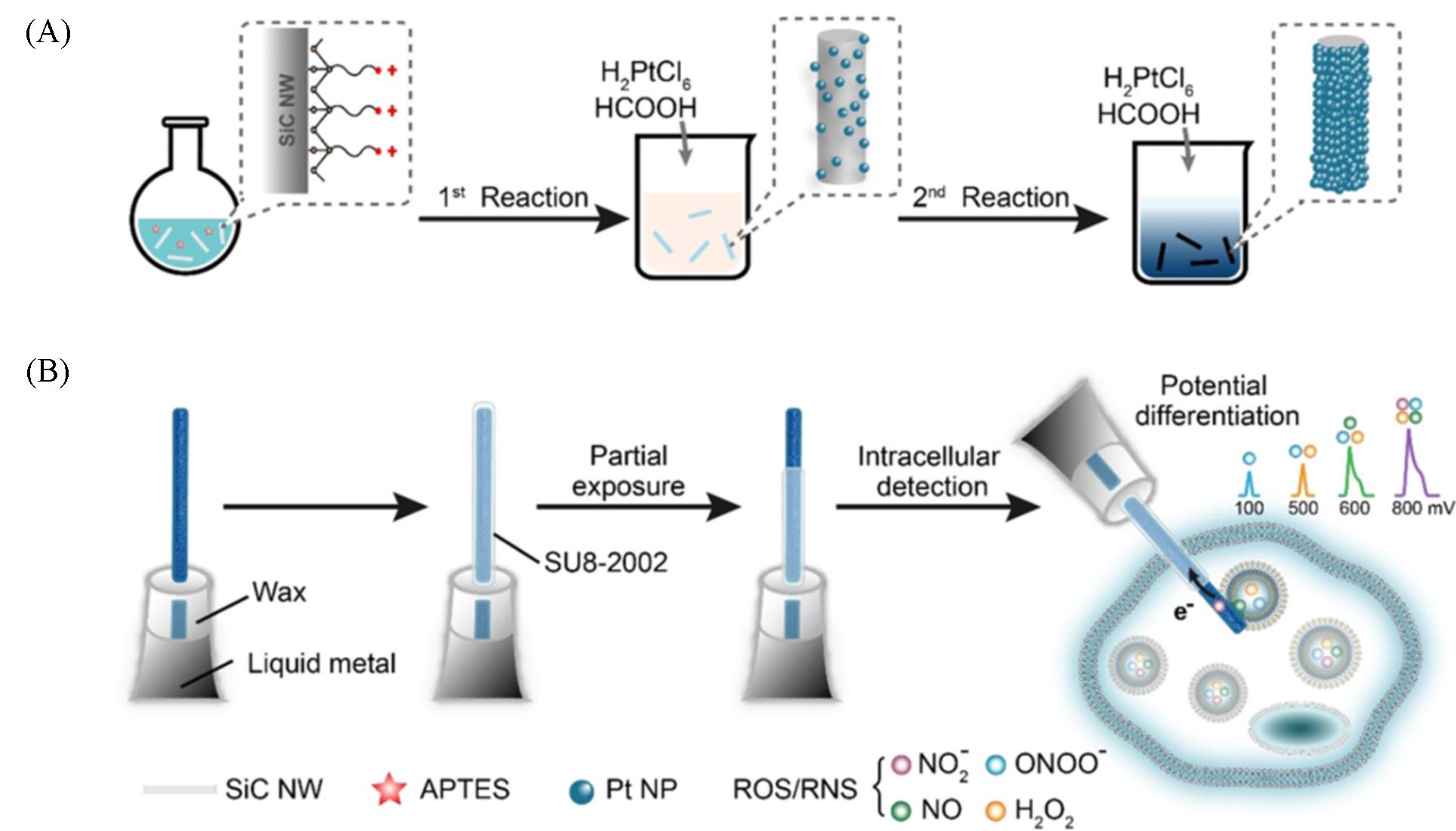
Fig.4 Two⁃step electroless plating of SiC@Pt NWs(A) and mounting a SiC@Pt NW at the tip of a nanopipette for intracellular differential measurement of four primary ROS/RNS (ONOO-, H2O2, NO and NO2- ) at four different electrochemical potentials(B)[59]Copyright 2022, American Chemical Society.
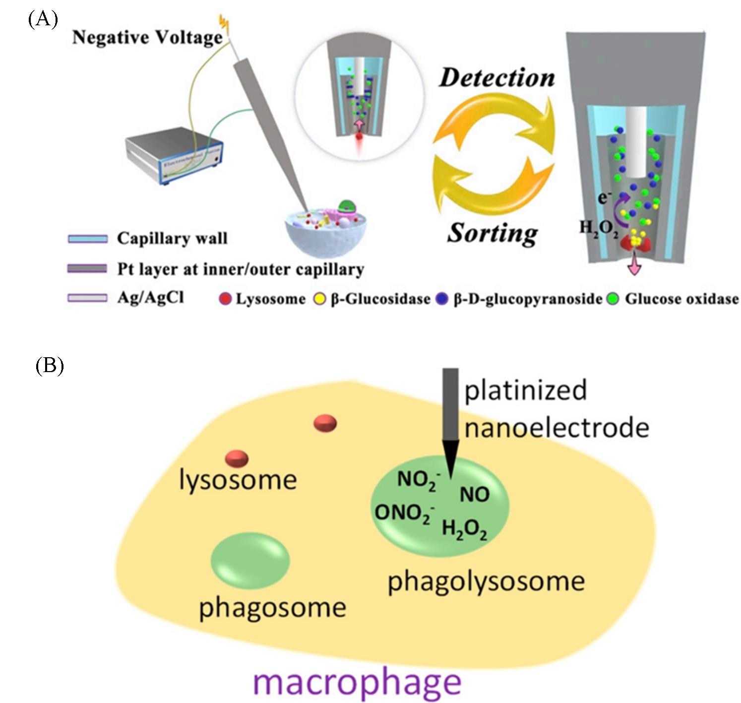
Fig.5 Detection of single organelle based on nanoelectrodes(A) Scheme of electrochemical setup for the detection of glucosidase activity in isolated single lysosomes from a single cell. The capillary coated with a Pt layer(dark shading) and an Ag/AgCl wire(light shading) inserted in the capillary is connected with an electrochemical station. Circle: amplified view of capillary tip with a Pt layer at the edge of the inner surface and the outer surface of the capillary to sort one lysosome(labeled in red). The arrow exhibits the flow direction of buffer with the lysosome. Crosssection view is used to illustrate Pt layer at the inner capillary and kit reaction. (Right) Displaying the release of glucosidase after the lysis of lysosome, the generation of hydrogen peroxide from kit reactions and the following electrochemical detection of hydrogen peroxide at the tip(dark shading). The arrow exhibits the flow direction of previously loaded glucosidase and generated reaction debris outside the capillary[62]; (B) phagolysosome penetration with a 65 nm radius platinized nanotip[63].(A) Copyright 2018, National Academy of Science; (B) Copyright 2019, American Chemical Society.
| 1 | Yang Q., Huang X., Gao B., Gao L., Yu F., Wang F., Analyst, 2022, 148(1), 9—25 |
| 2 | Dittrich P. S., Tachikawa K., Manz A., Anal. Chem., 2006, 78(12), 3887—3908 |
| 3 | El⁃Ali J., Sorger P. K., Jensen K. F., Nature, 2006, 442(7101), 403—411 |
| 4 | Altschuler S. J., Wu L. F., Cell, 2010, 141(4), 559—563 |
| 5 | Shoemaker G. K., Lorieau J., Lau L. H., Gillmor C. S., Palcic M. M., Anal. Chem., 2005, 77(10), 3132—31327 |
| 6 | Oh⁃hora M., Immunol. Rev., 2009, 231(1), 210—224 |
| 7 | Colman⁃Lerner A., Gordon A., Serra E., Chin T., Resnekov O., Endy D., Pesce C. G., Brent R., Nature, 2005, 437(7059), 699—706 |
| 8 | Spiller D. G., Wood C. D., Rand D. A., White M. R., Nature, 2010, 465(7299), 736—745 |
| 9 | Ferrell J. E., Machleder E. M. Jr., Science, 1998, 280(5365), 895—898 |
| 10 | Coralli C., Cemazar M., Kanthou C., Tozer G. M., Dachs G. U., Cancer Res., 2001, 61(12), 4784—4790 |
| 11 | Cohen D., Dickerson J. A., Whitmore C. D., Turner E. H., Palcic M. M., Hindsgaul O., Dovichi N. J., Annu. Rev. Anal. Chem.(Palo Alto Calif), 2008, 1, 165—190 |
| 12 | Thompson M. A., Lew M. D., Moerner W. E., Annu. Rev. Biophys., 2012, 41, 321—342 |
| 13 | Prabhakar A., Puglisi E. V., Puglisi J. D., Cold Spring Harb Perspect Biol., 2019, 11(1), a032714 |
| 14 | Sengupta B., Chaudhuri A., Das N., Sen P., Protein Pept. Lett., 2017, 24(11), 1073—1081 |
| 15 | Xu K., Babcock H. P., Zhuang X., Nat. Methods, 2012, 9(2), 185—188 |
| 16 | Huang B., Bates M., Zhuang X., Annu. Rev. Biochem., 2009, 78, 993—1016 |
| 17 | Betzig E., Patterson G. H., Sougrat R., Lindwasser O. W., Olenych S., Bonifacino J. S., Davidson M. W., Lippincott⁃Schwartz J., Hess H. F., Science, 2006, 313(5793), 1642—1645 |
| 18 | Adams K. L., Puchades M., Ewing A. G., Annu. Rev. Anal. Chem.(Palo Alto Calif), 2008, 1, 329 |
| 19 | Forster R. J., Chemical Society Reviews, 1994, 23(4), 289—297 |
| 20 | Sulzer D., Pothos E. N., Rev. Neurosci., 2000, 11(2/3), 159—212 |
| 21 | Amatore C., Arbault S., Guille M., Lemaître F., Chem. Rev., 2008, 108(7), 2585—2621 |
| 22 | Sun P., Laforge F. O., Abeyweera T. P., Rotenberg S. A., Carpino J., Mirkin M. V., Proc. Natl. Acad. Sci., 2008, 105(2), 443—448 |
| 23 | Li Y., Hu K., Yu Y., Rotenberg S. A., Amatore C., Mirkin M. V., J. Am. Chem. Soc., 2017, 139(37), 13055—13062 |
| 24 | Liu K., Liu R., Wang D., Pan R., Chen H. Y., Jiang D., Anal. Chem., 2022, 94(38), 13287—13292 |
| 25 | Wang Y., Noël J. M., Velmurugan J., Nogala W., Mirkin M. V., Lu C., Guille Collignon M., Lemaître F., Amatore C., Proc. Natl. Acad. Sci., 2012, 109(29), 11534—11539 |
| 26 | Zhang S., Qin H., Cheng S., Zhang Y., Gao N., Zhang M., Angew. Chem. Int. Ed., 2023, 62(16), e202300083 |
| 27 | Wu W. T., Jiang H., Qi Y. T., Fan W. T., Yan J., Liu Y. L., Huang W. H., Angew. Chem. Int. Ed., 2021, 60(35), 19337—19343 |
| 28 | Wu W. T., Chen X., Jiao Y. T., Fan W. T., Liu Y. L., Huang W. H., Angew. Chem. Int. Ed., 2022, 61(15), e202115820 |
| 29 | Jiao Y. T., Kang Y. R., Wen M. Y., Wu H. Q., Zhang X. W., Huang W. H., Angew. Chem. Int. Ed., 2023, 62(51), e202313612 |
| 30 | Pan R., Xu M., Jiang D., Burgess J. D., Chen H. Y., Proc. Natl. Acad. Sci., 2016, 113(41), 11436—11440 |
| 31 | Liu K., Liu R., Wang D., Pan R., Chen H. Y., Jiang D., CCS Chem., 2023, 5(6), 1285—1292 |
| 32 | Wang H. Y., Ruan Y. F., Zhu L. B., Shi X. M., Zhao W. W., Chen H. Y., Xu J. J., Angew. Chem. Int. Ed., 2021, 60(24), 13244—13250 |
| 33 | Shi X. M., Xu Y. T., Wang B., Li Z., Yu S. Y., Dong H., Zhao W. W., Jiang D., Chen H. Y., Xu J. J., Angew. Chem. Int. Ed., 2023, 62(29), e202302930 |
| 34 | Wang H. Y., Xu Y. T., Wang B., Yu S. Y., Shi X. M., Zhao W. W., Jiang D., Chen H. Y., Xu J. J., Angew. Chem. Int. Ed., 2022, 61(47), e202212752 |
| 35 | Zheng J., Li X., Wang K., Song J., Qi H., Anal. Chem., 2020, 92(16), 10940—10945 |
| 36 | Zhang H., Zhao T., Huang P., Wang Q., Tang H., Chu X., Jiang J., ACS Nano, 2022, 16(4), 5752—5763 |
| 37 | Marquitan M., Ruff A., Bramini M., Herlitze S., Mark M. D., Schuhmann W., Bioelectrochemistry, 2020, 133, 107487 |
| 38 | Huang F., Lin M., Duan R., Lou X., Xia F., Willner I., Nano Lett., 2018, 18(8), 5116—5123 |
| 39 | Duan Z., Ouyang Y., Fu Y., Huang F., Xia F., Willner I., Angew. Chem. Int. Ed., 2023, 62(18), e202301476 |
| 40 | Li X., Majdi S., Dunevall J., Fathali H., Ewing A. G., Angew. Chem. Int. Ed., 2015, 54(41), 11978—11982 |
| 41 | Ren L., Pour M. D., Majdi S., Li X., Malmberg P., Ewing A. G., Angew. Chem. Int. Ed., 2017, 56(18), 4970—4975 |
| 42 | Zhu W., Gu C., Dunevall J., Ren L., Zhou X., Ewing A. G., Angew. Chem. Int. Ed., 2019, 58(13), 4238—4242 |
| 43 | Wang Y., Gu C., Patel B. A., Ewing A. G., Angew. Chem. Int. Ed., 2021, 60(44), 23552—23556 |
| 44 | Wang Y., Gu C., Ewing A. G., Angew. Chem. Int. Ed., 2022, 61(20), e202200716 |
| 45 | Larsson A., Majdi S., Oleinick A., Svir I., Dunevall J., Amatore C., Ewing A. G., Angew. Chem. Int. Ed., 2020, 59(17), 6711—6714 |
| 46 | Hu K., Relton E., Locker N., Phan N. T. N., Ewing A. G., Angew. Chem. Int. Ed., 2021, 60(28), 15302—15306 |
| 47 | Hu K., Le Vo K. L., Hatamie A., Ewing A. G., Angew. Chem. Int. Ed., 2022, 61(1), e202113406 |
| 48 | He X., Ewing A. G., J. Am. Chem. Soc., 2022, 144(10), 4310—4314 |
| 49 | Gu C., Larsson A., Ewing A. G., Proc. Natl. Acad. Sci., 2019, 116(43), 21409—21415 |
| 50 | Barut I., He X., Sener E., Sämfors S., Ewing A. G., Fletcher J. S., Angew. Chem. Int. Ed., 2023, 62(15), e202217993 |
| 51 | Yue Q., Wang K., Guan M., Zhao Z., Li X., Yu P., Mao L., Angew. Chem. Int. Ed., 2022, 61(14), e202117596 |
| 52 | Yue Q., Li X., Wu F., Ji W., Zhang Y., Yu P., Zhang M., Ma W., Wang M., Mao L., Angew. Chem. Int. Ed., 2020, 59(27), 11061—11065 |
| 53 | Wu F., Yu P., Mao L., Angew. Chem. Int. Ed., 2023, 62(1), e202208872 |
| 54 | Wei S., Wu F., Liu J., Ji W., He X., Liu R., Yu P., Mao L., Angew. Chem. Int. Ed., 2023, 62(52), e202315681 |
| 55 | Zhang X. W., Qiu Q. F., Jiang H., Zhang F. L., Liu Y. L., Amatore C., Huang W. H., Angew. Chem. Int. Ed., 2017, 56(42), 12997—13000 |
| 56 | Zhang X. W., Oleinick A., Jiang H., Liao Q. L., Qiu Q. F., Svir I., Liu Y. L., Amatore C., Huang W. H., Angew. Chem. Int. Ed., 2019, 58(23), 7753—7756 |
| 57 | Yang X. K., Zhang F. L., Wu W. T., Tang Y., Yan J., Liu Y. L., Amatore C., Huang W. H., Angew. Chem. Int. Ed., 2021, 60(29), 15803—15808 |
| 58 | Yang X. K., Zhang F. L., Jin X. K., Jiao Y. T., Zhang X. W., Liu Y. L., Amatore C., Huang W. H., Proc. Natl. Acad. Sci., 2023, 120(19), e2219994120 |
| 59 | Qi Y. T., Jiang H., Wu W. T., Zhang F. L., Tian S. Y., Fan W. T., Liu Y. L., Amatore C., Huang W. H., J. Am. Chem. Soc., 2022, 144(22), 9723—9733 |
| 60 | Pan R., Hu K., Jiang D., Samuni U., Mirkin M. V., J. Am. Chem. Soc., 2019, 141(50), 19555—19559 |
| 61 | Pan R., Hu K., Jia R., Rotenberg S. A., Jiang D., Mirkin M. V., J. Am. Chem. Soc., 2020, 142(12), 5778—5784 |
| 62 | Pan R., Xu M., Burgess J. D., Jiang D., Chen H. Y., Proc. Natl. Acad. Sci., 2018, 115(16), 4087—4092 |
| 63 | Hu K., Li Y., Rotenberg S. A., Amatore C., Mirkin M. V., J. Am. Chem. Soc., 2019, 141(11), 4564—4568 |
| 64 | Liu K., Zhang Z., Liu R., Li J. P., Jiang D., Pan R., Angew. Chem. Int. Ed., 2023, 62(34), e202303053 |
| [1] | 赵雪琪, 赵越, 薛静, 白敏, 陈锋, 孙颖, 宋大千, 赵永席. 单细胞核酸编码扩增成像分析[J]. 高等学校化学学报, 2022, 43(12): 20220572. |
| [2] | 高成耀, 佟建华, 边超, 孙楫舟, 李洋, 王晋芬, 龚顺, 惠允, 夏善红. 痕量镉离子在原位铋修饰掺硼金刚石电极上的传感分析[J]. 高等学校化学学报, 2018, 39(3): 447. |
| [3] | 何艳艳, 葛军营, 赵常志. 氧化锌纳米棒光电极对核黄素的光致电化学响应及应用[J]. 高等学校化学学报, 2016, 37(12): 2144. |
| [4] | 张江华, 吕英, 贾红亮, 宋银银, 孙晓霞, 柴敦宵, 王兰英. 吲哚二甲川菁的合成、 晶体结构、光谱性质及生物应用[J]. 高等学校化学学报, 2015, 36(10): 1924. |
| [5] | 钟家松, 向卫东, 刘丽君, 杨昕宇, 蔡文, 张景峰, 梁晓娟. 生物分子辅助溶剂热合成硫化锑纳米棒[J]. 高等学校化学学报, 2010, 31(7): 1303. |
| [6] | 程介克, 王玮, 吴文展, 黄卫华, 王宗礼. 探索脑化学纳米电化学监测单细胞、单囊泡、突触间隙释放化学信号分子及形貌分析[J]. 高等学校化学学报, 2008, 29(12): 2609. |
| [7] | 杜富滢, 黄卫华, 吴文展, 王宗礼, 程介克 . 新型微盘传感器的研制及用于单细胞释放NO监测[J]. 高等学校化学学报, 2007, 28(6): 1069. |
| [8] | 凌笑梅,刘一,赖先银,张媛,刘晓明,屠鹏飞,赵玉英,,崔景荣 . 采用毛细管电泳方法以凝血酶为靶筛选天然药物提取化合物[J]. 高等学校化学学报, 2007, 28(2): 234. |
| [9] | 孙悦 卢敏 殷学锋. 纳米脂质体包裹荧光试剂进入单细胞的研究[J]. 高等学校化学学报, 2006, 27(4): 632. |
| [10] | 赵书林, 沈江珊. 单细胞中D-天冬氨酸和D-谷氨酸的毛细管电泳分析[J]. 高等学校化学学报, 2005, 26(9): 1612. |
| [11] | 吴文展, 黄卫华, 王玮, 王宗礼, 程介克, 张蓉颖, 陈宇, 刘杰, 郑从义, 沈超. 纳米电极-膜片钳监测单个活囊泡连续释放[J]. 高等学校化学学报, 2005, 26(5): 829. |
| [12] | 凌云扬, 殷学锋, 方肇伦. 微流控芯片NDA在线衍生测定单细胞中谷胱甘肽[J]. 高等学校化学学报, 2005, 26(2): 247. |
| [13] | 唐宏武, 李涛, 罗美娜, 吴琼水, 陈观铨. 高分辨阿达玛变换显微荧光图像分析(Ⅲ)——系统分辨率和图像恢复过程对单细胞分析结果的影响[J]. 高等学校化学学报, 2005, 26(1): 31. |
| [14] | 姚波, 冯雪, 罗国安, 王义明. 微流控芯片系统流式细胞术及单细胞荧光检测[J]. 高等学校化学学报, 2005, 26(1): 43. |
| [15] | 姚波, 刘科辉, 冯雪, 罗国安, 王义明. 微流控芯片细胞操纵及单细胞荧光检测[J]. 高等学校化学学报, 2004, 25(S1): 173. |
| 阅读次数 | ||||||
|
全文 |
|
|||||
|
摘要 |
|
|||||