

Chem. J. Chinese Universities ›› 2024, Vol. 45 ›› Issue (11): 20240305.doi: 10.7503/cjcu20240305
• Review • Previous Articles Next Articles
JIN Ying1, ZHANG Junjie1, ZHANG Yixin1, YUAN Yue2, HAN Zhenzhen1( )
)
Received:2024-06-24
Online:2024-11-10
Published:2024-08-15
Contact:
HAN Zhenzhen
E-mail:hanzhenzhen@ahmu.edu.cn
Supported by:CLC Number:
TrendMD:
JIN Ying, ZHANG Junjie, ZHANG Yixin, YUAN Yue, HAN Zhenzhen. Research Progress in Exosome Isolation and Proteomics Analysis[J]. Chem. J. Chinese Universities, 2024, 45(11): 20240305.
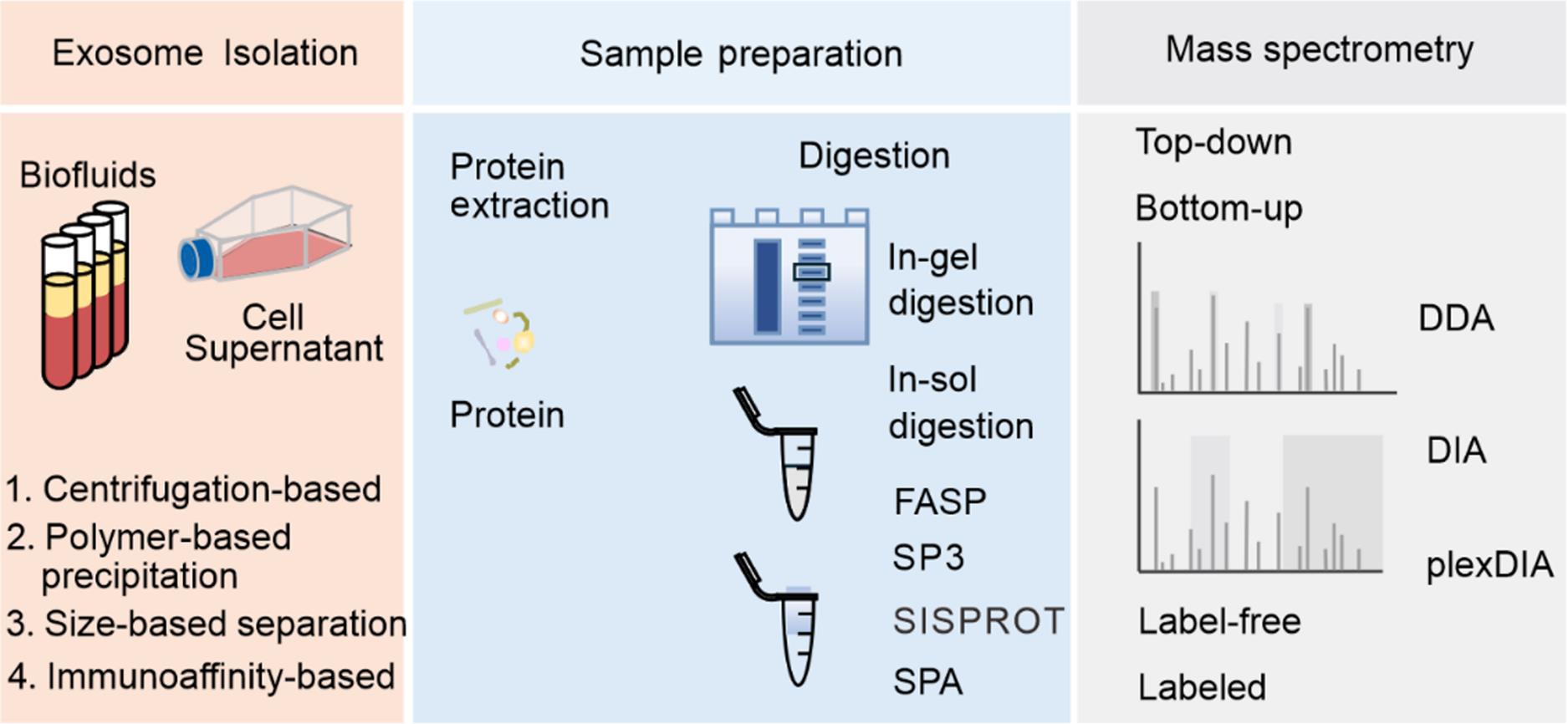
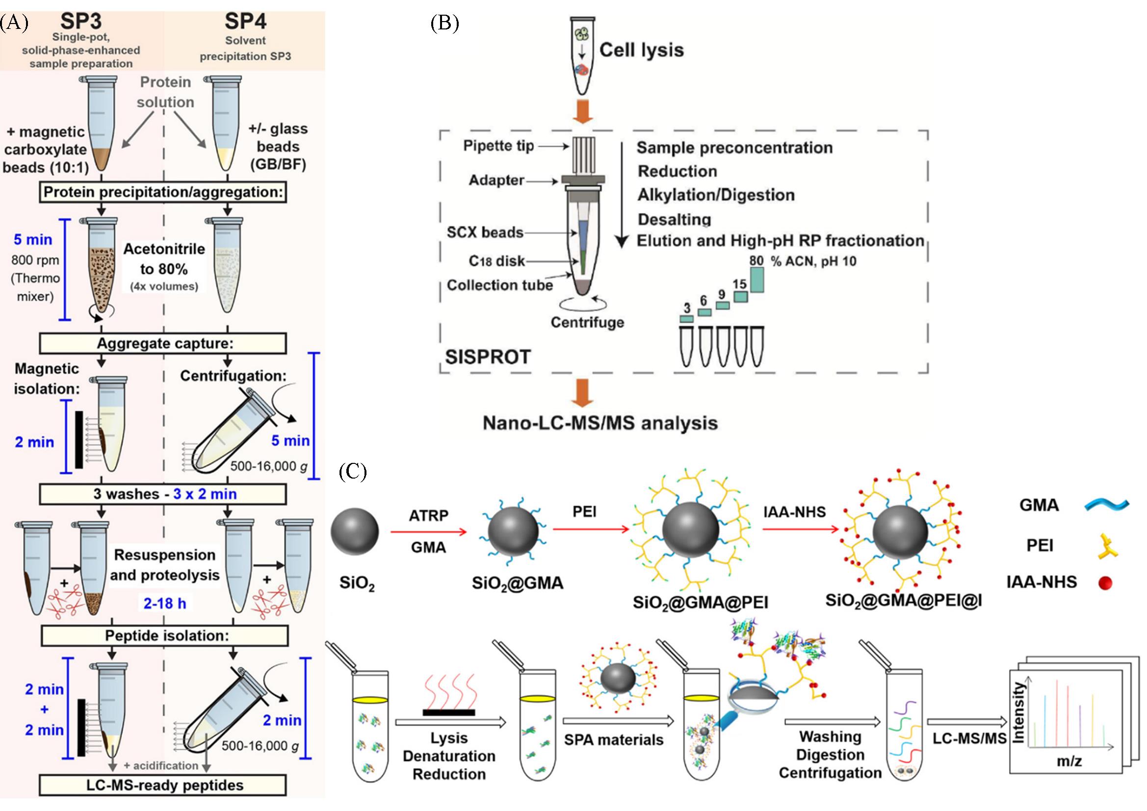
| Sample⁃handling approach | Beneficial feature | Limitation |
|---|---|---|
| In⁃gel digestion | Enzymatic digestion of proteins within the gel environment, enhancing protein recovery rates Simplify operational steps Robustness and efficient impurity removal capability | Time⁃consuming Introduce contamination from the gel, enzymes, or other reagents |
In⁃solution processing methods, such as standard in⁃solution digestion | Applicable to virtually any sample Simple protocols that require minimal handling Generally, afford high recovery of protein input, making them adaptable to a wide range of sample quantities Easily adaptable to high⁃throughput regimes | Limited selection of reagents that can be used for protein extraction and solubilization In specific scenarios, processing volumes required for dilution can challenge downstream handling Diluted chaotropes in solution can hinder proteolysis Downstream removal of acid⁃labile detergents can result in material losses |
| FASP | Compatible with a wide range of lysis and protein solubilization components Effective processing of high input quantities of material Most protocols are simple and flexible for adaptation to different sample types | Processing can be time consuming and laborious When working with small input quantities, material losses can be substantial Reagent compatibility can be limited by consumables(e. g., filter compatibility) |
| SP3 | Applicable to virtually any sample Compatible with a wide range of lysis and protein solubilization components Simple protocol that requires minimal handling Provides high recovery of protein input, making it adaptable to a wide range of sample quantities Rapid processing of large numbers of samples in parallel | Recovery of intact proteins from the paramagnetic beads can be challenging High concentrations of intact chromatin can reduce performance Bead clumping and aggregation can hinder adaptation to high⁃throughput automation |
| SISPROT | The preparation steps of proteomics samples are fully integrated Simple protocol that requires minimal handling Easily multiplexed on standard centrifuges with good reproducibility | It is not compatible with the rare cell proteomic reactor cell lysis buffer which contains 1% Triton Xs100 Costly consumables, high expenses |
| SPA | Simple protocol that requires minimal handling Provides improved efficiency, anti⁃interference ability, and recovery of low⁃input samples | Covalent binding of proteins hinders release of cysteine⁃containing peptides Low sequence coverage of proteins |
Table 1 Comparison between the properties of different proteome sample-handling approaches
| Sample⁃handling approach | Beneficial feature | Limitation |
|---|---|---|
| In⁃gel digestion | Enzymatic digestion of proteins within the gel environment, enhancing protein recovery rates Simplify operational steps Robustness and efficient impurity removal capability | Time⁃consuming Introduce contamination from the gel, enzymes, or other reagents |
In⁃solution processing methods, such as standard in⁃solution digestion | Applicable to virtually any sample Simple protocols that require minimal handling Generally, afford high recovery of protein input, making them adaptable to a wide range of sample quantities Easily adaptable to high⁃throughput regimes | Limited selection of reagents that can be used for protein extraction and solubilization In specific scenarios, processing volumes required for dilution can challenge downstream handling Diluted chaotropes in solution can hinder proteolysis Downstream removal of acid⁃labile detergents can result in material losses |
| FASP | Compatible with a wide range of lysis and protein solubilization components Effective processing of high input quantities of material Most protocols are simple and flexible for adaptation to different sample types | Processing can be time consuming and laborious When working with small input quantities, material losses can be substantial Reagent compatibility can be limited by consumables(e. g., filter compatibility) |
| SP3 | Applicable to virtually any sample Compatible with a wide range of lysis and protein solubilization components Simple protocol that requires minimal handling Provides high recovery of protein input, making it adaptable to a wide range of sample quantities Rapid processing of large numbers of samples in parallel | Recovery of intact proteins from the paramagnetic beads can be challenging High concentrations of intact chromatin can reduce performance Bead clumping and aggregation can hinder adaptation to high⁃throughput automation |
| SISPROT | The preparation steps of proteomics samples are fully integrated Simple protocol that requires minimal handling Easily multiplexed on standard centrifuges with good reproducibility | It is not compatible with the rare cell proteomic reactor cell lysis buffer which contains 1% Triton Xs100 Costly consumables, high expenses |
| SPA | Simple protocol that requires minimal handling Provides improved efficiency, anti⁃interference ability, and recovery of low⁃input samples | Covalent binding of proteins hinders release of cysteine⁃containing peptides Low sequence coverage of proteins |
| Type | Name | Labeling level | Beneficial feature | Limitation |
|---|---|---|---|---|
| Metabolic labeling | SILAC | Cells, organisms | Efficient labeling, one label for(tryptic) peptide, semiautomatic data analysis Applicable to cells but can be expanded to tissues or model organisms using internal standards (e.g., super⁃SILAC) | High costs, especially when applied to whole organisms Super⁃SILAC experiments have reduced quantitative proteome coverage Requires metabolically active cells to introduce labels |
| 15N labeling | Cells, organisms | Efficient labeling Applicable to cells and model organisms | Expensive Complex data analysis Limited multiplexing capability(up to 2⁃plex) Not suitable for clinical samples | |
Chemical labeling (in vitro) ⁃isobaric labeling | iTRAQ | Peptide | Efficient labeling enhanced signal intensity in MS and MS/MS, high multiplexing capability, simple data analysis Applicable to any sample(cells, animal or human tissue) Commercially available | Expensive Does not allow in vivo labeling Quantitative precision dependent on the reproducibility of sample preparation |
| TMT | Peptide | Efficient labeling enhanced signal intensity in MS and MS/MS, high multiplexing capability, simple data analysis Applicable to any sample(cells, animal or human tissue) | Expensive Requires fragmentation with HCD or ETD Does not allow in vivo labeling Quantitative precision dependent on the reproducibility of sample preparation | |
| Type | Name | Labeling level | Beneficial feature | Limitation |
| Enzymatic labeling (in vitro) | 18O labeling | Peptide | Low costs, simple in handling Applicable to any sample(cells, animal or human tissue) | Incomplete labeling complicates data analysis Limited multiplexing capability(up to 2⁃plex) Not suitable for in vivo labeling Overlapping isotopic peaks Varied labeling efficiencies |
| Label⁃free | Spectral counting | NA | Low costs, simple in handling Broad applicability | Less accurate than the labeling methods More time needed for MS analysis |
| Chromatographic peak area | NA | Low costs, simple in handling Broad applicability | Less accurate than the labeling methods More time needed for MS analysis |
Table 2 Comparison between the label-free and labeled quantitative proteomics techniques
| Type | Name | Labeling level | Beneficial feature | Limitation |
|---|---|---|---|---|
| Metabolic labeling | SILAC | Cells, organisms | Efficient labeling, one label for(tryptic) peptide, semiautomatic data analysis Applicable to cells but can be expanded to tissues or model organisms using internal standards (e.g., super⁃SILAC) | High costs, especially when applied to whole organisms Super⁃SILAC experiments have reduced quantitative proteome coverage Requires metabolically active cells to introduce labels |
| 15N labeling | Cells, organisms | Efficient labeling Applicable to cells and model organisms | Expensive Complex data analysis Limited multiplexing capability(up to 2⁃plex) Not suitable for clinical samples | |
Chemical labeling (in vitro) ⁃isobaric labeling | iTRAQ | Peptide | Efficient labeling enhanced signal intensity in MS and MS/MS, high multiplexing capability, simple data analysis Applicable to any sample(cells, animal or human tissue) Commercially available | Expensive Does not allow in vivo labeling Quantitative precision dependent on the reproducibility of sample preparation |
| TMT | Peptide | Efficient labeling enhanced signal intensity in MS and MS/MS, high multiplexing capability, simple data analysis Applicable to any sample(cells, animal or human tissue) | Expensive Requires fragmentation with HCD or ETD Does not allow in vivo labeling Quantitative precision dependent on the reproducibility of sample preparation | |
| Type | Name | Labeling level | Beneficial feature | Limitation |
| Enzymatic labeling (in vitro) | 18O labeling | Peptide | Low costs, simple in handling Applicable to any sample(cells, animal or human tissue) | Incomplete labeling complicates data analysis Limited multiplexing capability(up to 2⁃plex) Not suitable for in vivo labeling Overlapping isotopic peaks Varied labeling efficiencies |
| Label⁃free | Spectral counting | NA | Low costs, simple in handling Broad applicability | Less accurate than the labeling methods More time needed for MS analysis |
| Chromatographic peak area | NA | Low costs, simple in handling Broad applicability | Less accurate than the labeling methods More time needed for MS analysis |
| Disease | Sample | Type of MS | Potential finding | Ref. |
|---|---|---|---|---|
| Pancreatic ductal adenocarcinoma | Plasma | LC⁃MS/MS | CLDN4, EPCAM, CD151, LGALS3BP, HIST2H2BE and HIST2H2BF | [ |
| Multiple cancers | Tissue explants, plasma, and other bodily fluids | LC⁃MS/MS, DDA | ACTB, MSN and RAP1B | [ |
| Osteosarcoma | Plasma | LC⁃MS/MS, MALDI⁃TOF MS | IGLV2⁃23, IGLV4⁃3, IGLV1⁃51, IGKV3⁃15, IGHV4⁃4, IGLV4⁃60, HBA1 | [ |
| Prostate cancer | Serum | LC⁃MS/MS | Vinculin, ECM, Rac, VASP | [ |
| Alzheimer | Cerebrospinal fluid, plasma | Orbitrap, LC⁃MS | Cathepsin B | [ |
| Breast cancer | Plasma | LC⁃MS/MS, label⁃free | 144 Phosphoproteins | [ |
| Colorectal cancer | Serum | DIA, TMT | Fibrinogen α chain, phosphorylated fibronectin 1, haptoglobin | [ |
Hepatocellular carcinoma | Serum | DDA, DIA | Von Willebrand factor, LGALS3BP, TGFB1, SERPINC1, HPX, HP, HBA1, FGA, FGG, FGB | [ |
| Lung cancer | Serum | UPLC⁃MS/MS, Q⁃exactive | Lipopolysaccharide⁃binding proteins | [ |
| Liver cancer | Serum | LC‐MS, LTQ⁃orbitrap XL | Thrombospondin⁃1, fibulin⁃1, fibrinogen gamma chain | [ |
Oral squamous cell carcinoma | Serum | LC⁃MS, Q‐exactive, orbitrap | PF4V1, CXCL7, F13A1, ApoA1 | [ |
| Epithelial ovarian cancer | Plasma | LC‐MS/MS, TMT | Fibrinogen alpha chain, fibrinogen alpha chain | [ |
| Prostate cancer | Serum | LC‐MS/MS | Filamin A | [ |
Table 3 Proteomic analysis of tumor-derived exosomes for clinical disease diagnosis
| Disease | Sample | Type of MS | Potential finding | Ref. |
|---|---|---|---|---|
| Pancreatic ductal adenocarcinoma | Plasma | LC⁃MS/MS | CLDN4, EPCAM, CD151, LGALS3BP, HIST2H2BE and HIST2H2BF | [ |
| Multiple cancers | Tissue explants, plasma, and other bodily fluids | LC⁃MS/MS, DDA | ACTB, MSN and RAP1B | [ |
| Osteosarcoma | Plasma | LC⁃MS/MS, MALDI⁃TOF MS | IGLV2⁃23, IGLV4⁃3, IGLV1⁃51, IGKV3⁃15, IGHV4⁃4, IGLV4⁃60, HBA1 | [ |
| Prostate cancer | Serum | LC⁃MS/MS | Vinculin, ECM, Rac, VASP | [ |
| Alzheimer | Cerebrospinal fluid, plasma | Orbitrap, LC⁃MS | Cathepsin B | [ |
| Breast cancer | Plasma | LC⁃MS/MS, label⁃free | 144 Phosphoproteins | [ |
| Colorectal cancer | Serum | DIA, TMT | Fibrinogen α chain, phosphorylated fibronectin 1, haptoglobin | [ |
Hepatocellular carcinoma | Serum | DDA, DIA | Von Willebrand factor, LGALS3BP, TGFB1, SERPINC1, HPX, HP, HBA1, FGA, FGG, FGB | [ |
| Lung cancer | Serum | UPLC⁃MS/MS, Q⁃exactive | Lipopolysaccharide⁃binding proteins | [ |
| Liver cancer | Serum | LC‐MS, LTQ⁃orbitrap XL | Thrombospondin⁃1, fibulin⁃1, fibrinogen gamma chain | [ |
Oral squamous cell carcinoma | Serum | LC⁃MS, Q‐exactive, orbitrap | PF4V1, CXCL7, F13A1, ApoA1 | [ |
| Epithelial ovarian cancer | Plasma | LC‐MS/MS, TMT | Fibrinogen alpha chain, fibrinogen alpha chain | [ |
| Prostate cancer | Serum | LC‐MS/MS | Filamin A | [ |
| 1 | Li Q., Zhang Z., Wang F., Wang X., Zhan S., Yang X., Xu C., Liu D., Sci. Adv., 2023, 9(15), eadf4568 |
| 2 | Kalluri R., McAndrews K. M., Cell, 2023, 186(8), 1610—1626 |
| 3 | Li Z., Liu C., Cheng Y., Li Y., Deng J., Bai L., Qin L., Mei H., Zeng M., Tian F., Zhang S., Sun J., Sci. Adv., 2023, 9(16), eade2819 |
| 4 | Ding L., Liu X., Zhang Z., Liu L. E., He S., Wu Y., Effah C. Y., Yang R., Zhang A., Chen W., Yarmamat M., Qu L., Yang X., Wu Y., Lab Chip, 2023, 23(6), 1694—1702 |
| 5 | Zhang L., Yin W., Tong Y., Zhang Y., Xu Y., Liu S. Y., Dai Z., Zou X., Anal. Chem., 2022, 94(31), 10991—10999 |
| 6 | Wang X., Xiang Z., Liu Y., Huang C., Pei Y., Wang X., Zhi H., Wong W. H. S., Wei H., Ng I. O. L., Lee P. P. W., Chan G. C. F., Lau Y. L., Tu W., Sci. Transl. Med., 2020, 12(563), eaaz3426 |
| 7 | Zhang K., Cheng K., Nat. Rev. Bioeng., 2023, 12, 1—2 |
| 8 | Zhang M., Hu S., Liu L., Dang P., Liu Y., Sun Z., Qiao B., Wang C., Signal Transduct. Target. Ther., 2023, 8(1), 124 |
| 9 | Das S., Lyon C. J., Hu T., ACS Nano, 2024, 18(14), 9784—9797 |
| 10 | Novikova S. E., Soloveva N. A., Farafonova T. E., Tikhonova O. V., Liao P. C., Zgoda V. G., Molecules, 2021, 26(20), 6145 |
| 11 | Lane R. E., Korbie D., Hill M. M., Trau M., Clin. Transl. Med., 2018, 7(1), 14 |
| 12 | Keerthikumar S., Chisanga D., Ariyaratne D., Al Saffar H., Anand S., Zhao K., Samuel M., Pathan M., Jois M., Chilamkurti N., Gangoda L., Mathivanan S., J. Mol. Biol., 2016, 428(4), 688—692 |
| 13 | Pathan M., Fonseka P., Chitti S. V., Kang T., Sanwlani R., Van Deun J., Hendrix A., Mathivanan S., Nucleic Acids Res., 2019, 47(D1), D516—D519 |
| 14 | Abramowicz A., Widlak P., Pietrowska M., Mol. Biosyst., 2016, 12(5), 1407—1419 |
| 15 | Johnstone R. M., Bianchini A., Teng K., Blood, 1989, 74(5), 1844—1851 |
| 16 | Théry C., Clayton A., Amigorena S., Raposo G., Curr. Protoc. Cell Biol., 2006, Chapter 3, Unit 3.22 |
| 17 | Yan H., Li Y., Cheng S., Zeng Y., Anal. Chem., 2021, 93(11), 4739—4774 |
| 18 | Principe S., Jones E. E., Kim Y., Sinha A., Nyalwidhe J. O., Brooks J., Semmes O. J., Troyer D. A., Lance R. S., Kislinger T., Drake R. R., Proteomics, 2013, 13(10/11), 1667—1671 |
| 19 | Principe S., Kim Y., Fontana S., Ignatchenko V., Nyalwidhe J. O., Lance R. S., Troyer D. A., Alessandro R., Semmes O. J., Kislinger T., Drake R. R., Medin J. A., J. Proteome Res., 2012, 11(4), 2386—2396 |
| 20 | Chia B. S., Low Y. P., Wang Q., Li P., Gao Z., TrAC⁃Trend. Anal. Chem., 2017, 86, 93—106 |
| 21 | Li P., Kaslan M., Lee S. H., Yao J., Gao Z., Theranostics, 2017, 7(3), 789—804 |
| 22 | Crescitelli R., Lasser C., Jang S. C., Cvjetkovic A., Malmhall C., Karimi N., Hoog J. L., Johansson I., Fuchs J., Thorsell A., Gho Y. S., Olofsson Bagge R., Lotvall J., J. Extracell. Vesicles, 2020, 9(1), 1722433 |
| 23 | Sódar B. W., Kittel Á., Pálóczi K., Vukman K. V., Osteikoetxea X., Szabó⁃Taylor K., Németh A., Sperlágh B., Baranyai T., Giricz Z., Wiener Z., Turiák L., Drahos L., Pállinger É., Vékey K., Ferdinandy P., Falus A., Buzás E. I., Sci. Rep., 2016, 6(1), 24316 |
| 24 | Bard M. P., Hegmans J. P., Hemmes A., Luider T. M., Willemsen R., Severijnen L. A., van Meerbeeck J. P., Burgers S. A., Hoogsteden H. C., Lambrecht B. N., Am. J. Respir. Cell Mol. Biol., 2004, 31(1), 114—121 |
| 25 | Keller S., Ridinger J., Rupp A. K., Janssen J. W., Altevogt P., J. Transl. Med., 2011, 9, 86 |
| 26 | Tauro B. J., Greening D. W., Mathias R. A., Ji H., Mathivanan S., Scott A. M., Simpson R. J., Methods, 2012, 56(2), 293—304 |
| 27 | Patel G. K., Khan M. A., Zubair H., Srivastava S. K., Khushman M., Singh S., Singh A. P., Sci. Rep., 2019, 9(1), 5335 |
| 28 | Welton J. L., Webber J. P., Botos L. A., Jones M., Clayton A., J. Extracell. Vesicles, 2015, 4, 27269 |
| 29 | Taylor D. D., Shah S., Methods, 2015, 87, 3—10 |
| 30 | Stranska R., Gysbrechts L., Wouters J., Vermeersch P., Bloch K., Dierickx D., Andrei G., Snoeck R., J. Transl. Med., 2018, 16(1), 1 |
| 31 | Karimi N., Cvjetkovic A., Jang S. C., Crescitelli R., Hosseinpour Feizi M. A., Nieuwland R., Lotvall J., Lasser C., Cell. Mol. Life Sci., 2018, 75(15), 2873—2886 |
| 32 | Yang D., Zhang W., Zhang H., Zhang F., Chen L., Ma L., Larcher L. M., Chen S., Liu N., Zhao Q., Tran P. H. L., Chen C., Veedu R. N., Wang T., Theranostics, 2020, 10(8), 3684—3707 |
| 33 | Lane R. E., Korbie D., Trau M., Hill M. M., Proteomics, 2019, 19(8), e1800156 |
| 34 | Takov K., Yellon D. M., Davidson S. M., J. Extracell. Vesicles, 2019, 8(1), 1560809 |
| 35 | Nordin J. Z., Lee Y., Vader P., Mager I., Johansson H. J., Heusermann W., Wiklander O. P., Hallbrink M., Seow Y., Bultema J. J., Gilthorpe J., Davies T., Fairchild P. J., Gabrielsson S., Meisner⁃Kober N. C., Lehtio J., Smith C. I., Wood M. J., El Andaloussi S., Nanomedicine, 2015, 11(4), 879—883 |
| 36 | Lobb R. J., Becker M., Wen S. W., Wong C. S., Wiegmans A. P., Leimgruber A., Moller A., J. Extracell. Vesicles, 2015, 4, 27031 |
| 37 | Thompson A. G., Gray E., Mager I., Fischer R., Thezenas M. L., Charles P. D., Talbot K., El Andaloussi S., Kessler B. M., Wood M., Turner M. R., Proteomics, 2018, 18(24), e1800257 |
| 38 | Busatto S., Vilanilam G., Ticer T., Lin W. L., Dickson D. W., Shapiro S., Bergese P., Wolfram J., Cells, 2018, 7(12), 273 |
| 39 | Han Z., Peng C., Yi J., Zhang D., Xiang X., Peng X., Su B., Liu B., Shen Y., Qiao L., Sens. Actuators B: Chem., 2021, 333, 129563 |
| 40 | Hua X., Zhu Q., Liu Y., Zhou S., Huang P., Li Q., Liu S., Anal. Chim. Acta, 2023, 1258, 341160 |
| 41 | Bu J., Nair A., Iida M., Jeong W. J., Poellmann M. J., Mudd K., Kubiatowicz L. J., Liu E. W., Wheeler D. L., Hong S., Nano Lett., 2020, 20(7), 4901—4909 |
| 42 | Contreras⁃Naranjo J. C., Wu H. J., Ugaz V. M., Lab Chip, 2017, 17(21), 3558—3577 |
| 43 | Zhang K., Yue Y., Wu S., Liu W., Shi J., Zhang Z., ACS Sens., 2019, 4(5), 1245—1251 |
| 44 | Zhang P., Zhou X., Zeng Y., Chem. Sci., 2019, 10(21), 5495—5504 |
| 45 | Liu C., Guo J., Tian F., Yang N., Yan F., Ding Y., Wei J., Hu G., Nie G., Sun J., ACS Nano, 2017, 11(7), 6968—6976 |
| 46 | Kang K., Lee S. S., Hyun K., Lee S. J., Kim J. M., Nat. Commun., 2013, 4, 2567 |
| 47 | Liu C., Ding B., Xue C., Tian Y., Hu G., Sun J., Anal. Chem., 88(24), 12547—12553 |
| 48 | Liu C., Zhao J., Tian F., Chang J., Zhang W., Sun J., J. Am. Chem. Soc., 2019, 141(9), 3817—3821 |
| 49 | Lee K., Shao H., Weissleder R., Lee H., ACS Nano, 2015, 9(3), 2321—2327 |
| 50 | Wu M., Ouyang Y., Wang Z., Zhang R., Huang P. H., Chen C., Li H., Li P., Quinn D., Dao M., Suresh S., Sadovsky Y., Huang T. J., Proc. Natl. Acad. Sci. USA, 2017, 114(40), 10584—10589 |
| 51 | Wang Z., Wang H., Becker R., Rufo J., Yang S., Mace B. E., Wu M., Zou J., Laskowitz D. T., Huang T. J., Microsyst. Nanoeng., 2021, 7, 20 |
| 52 | Liu C., Zhao J., Tian F., Cai L., Zhang W., Feng Q., Chang J., Wan F., Yang Y., Dai B., Cong Y., Ding B., Sun J., Tan W., Nat. Biomed. Eng., 2019, 3(3), 183—193 |
| 53 | Zhao J., Liu C., Li Y., Ma Y., Deng J., Li L., Sun J., J. Am. Chem. Soc., 2020, 142(11), 4996—5001 |
| 54 | Mallia A., Gianazza E., Zoanni B., Brioschi M., Barbieri S. S., Banfi C., Diagnostics, 2020, 10(10), 843 |
| 55 | Kim H., Kim D. W., Cho J. Y., Proteome Sci., 2019, 17, 5 |
| 56 | Fontana S., Saieva L., Taverna S., Alessandro R., Proteomics, 2013, 13(10/11), 1581—1594 |
| 57 | Ludwig K. R., Schroll M. M., Hummon A. B., J. Proteome Res., 2018, 17(7), 2480—2490 |
| 58 | Manza L. L., Stamer S. L., Ham A. J., Codreanu S. G., Liebler D. C., Proteomics, 2005, 5(7), 1742—1745 |
| 59 | Wisniewski J. R., Zougman A., Nagaraj N., Mann M., Nat. Methods, 2009, 6(5), 359—362 |
| 60 | Hughes C. S., Moggridge S., Muller T., Sorensen P. H., Morin G. B., Krijgsveld J., Nat. Protoc., 2019, 14(1), 68—85 |
| 61 | Johnston H. E., Yadav K., Kirkpatrick J. M., Biggs G. S., Oxley D., Kramer H. B., Samant R. S., Anal. Chem., 2022, 94(29), 10320—10328 |
| 62 | Chen W., Wang S., Adhikari S., Deng Z., Wang L., Chen L., Ke M., Yang P., Tian R., Anal. Chem., 2016, 88(9), 4864—4871 |
| 63 | Xu R., Tang J., Deng Q., He W., Sun X., Xia L., Cheng Z., He L., You S., Hu J., Fu Y., Zhu J., Chen Y., Gao W., He A., Guo Z., Lin L., Li H., Hu C., Tian R., Anal. Chem., 2018, 90(9), 5879—5886 |
| 64 | Huang P., Kong Q., Gao W., Chu B., Li H., Mao Y., Cai Z., Xu R., Tian R., Anal. Chim. Acta, 2020, 1127, 140—148 |
| 65 | Shah P., Zhang B., Choi C., Yang S., Zhou J., Harlan R., Tian Y., Zhang Z., Chan D. W., Zhang H., Anal. Biochem., 2015, 469, 27—33 |
| 66 | Li Y., Yuan H., Cheng M., Zhu X., Yang K., Zhang W., Sui Z., Zhang C., Zhang L., Zhang Y., Sci. Bull., 2022, 67(16), 1628—1631 |
| 67 | Mathew B., Mansuri M. S., Williams K. R., Nairn A. C., Brain Sci., 2021, 11(2), 258 |
| 68 | Wallen H., Nat. Rev. Methods Primers, 2024, 4(1), 39 |
| 69 | Doyle L., Wang M., Cells, 2019, 8(7), 727 |
| 70 | Jalaludin I., Lubman D. M., Kim J., Mass Spectrom. Rev., 2023, 42(2), 844—872 |
| 71 | Toby T. K., Fornelli L., Kelleher N. L., Annu. Rev. Anal. Chem., 2016, 9(1), 499—519 |
| 72 | Aebersold R., Agar J. N., Amster I. J., Baker M. S., Bertozzi C. R., Boja E. S., Costello C. E., Cravatt B. F., Fenselau C., Garcia B. A., Ge Y., Gunawardena J., Hendrickson R. C., Hergenrother P. J., Huber C. G., Ivanov A. R., Jensen O. N., Jewett M. C., Kelleher N. L., Kiessling L. L., Krogan N. J., Larsen M. R., Loo J. A., Ogorzalek Loo R. R., Lundberg E., MacCoss M. J., Mallick P., Mootha V. K., Mrksich M., Muir T. W., Patrie S. M., Pesavento J. J., Pitteri S. J., Rodriguez H., Saghatelian A., Sandoval W., Schlüter H., Sechi S., Slavoff S. A., Smith L. M., Snyder M. P., Thomas P. M., Uhlén M., Van Eyk J. E., Vidal M., Walt D. R., White F. M., Williams E. R., Wohlschlager T., Wysocki V. H., Yates N. A., Young N. L., Zhang B., Nat. Chem. Biol., 2018, 14(3), 206—214 |
| 73 | Zhu Y., Pick H., Gasilova N., Li X., Lin T. E., Laeubli H. P., Zippelius A., Ho P. C., Girault H. H., Chem, 2019, 5(5), 1318—1336 |
| 74 | Buck K. M., Roberts D. S., Aballo T. J., Inman D. R., Jin S., Ponik S., Brown K. A., Ge Y., Anal. Chem., 2022, 94(20), 7164—7168 |
| 75 | Brahmadhi A., Chuang Y. K., Wang S. Y., Kao C. C., Tsai I. L., J. Food Drug Anal., 2022, 30(2), 202—222 |
| 76 | Guan S., Taylor P. P., Han Z., Moran M. F., Ma B., J. Proteome Res., 2020, 19(8), 3230—3237 |
| 77 | Zhang Z., J. Am. Soc. Mass Spectrom., 2012, 23(8), 1400—1407 |
| 78 | Bian Y., Zheng R., Bayer F. P., Wong C., Chang Y. C., Meng C., Zolg D. P., Reinecke M., Zecha J., Wiechmann S., Heinzlmeir S., Scherr J., Hemmer B., Baynham M., Gingras A. C., Boychenko O., Kuster B., Nat. Commun., 2020, 11(1), 157 |
| 79 | Wu A. Y., Ueda K., Lai C. P., Proteomics, 2019, 19(1/2), e1800162 |
| 80 | Zhao L., Shi J., Chang L., Wang Y., Liu S., Li Y., Zhang T., Zuo T., Fu B., Wang G., Ruan Y., Zhang Y., Xu P., ACS Omega, 2021, 6(1), 827—835 |
| 81 | Zheng X., Xu K., Zhou B., Chen T., Huang Y., Li Q., Wen F., Ge W., Wang J., Yu S., Sun L., Zhu L., Liu W., Gao H., Yue L., Cai X., Zhang Q., Ruan G., Zhu T., Wu Z., Zhu Y., Shao Y., Guo T., Zheng S., J. Extracell. Vesicles, 2020, 9(1), 1750202 |
| 82 | Yang Y., Liu X., Shen C., Lin Y., Yang P., Qiao L., Nat. Commun., 2020, 11(1), 146 |
| 83 | Yang Y., Yan G., Kong S., Wu M., Yang P., Cao W., Qiao L., Nat. Commun., 2021, 12(1), 6073 |
| 84 | Derks J., Leduc A., Wallmann G., Huffman R. G., Willetts M., Khan S., Specht H., Ralser M., Demichev V., Slavov N., Nat. Biotechnol., 2022, 41(1), 50—59 |
| 85 | Patel V. J., Thalassinos K., Slade S. E., Connolly J. B., Crombie A., Colin Murrell J., Scrivens J. H., J. Proteome Res., 2009, 8(7), 3752—3759 |
| 86 | Neilson K. A., Ali N. A., Muralidharan S., Mirzaei M., Mariani M., Assadourian G., Lee A., van Sluyter S. C., Haynes P. A., Proteomics, 2011, 11(4), 535—553 |
| 87 | Lobo M. D., Moreno F. B., Souza G. H., Verde S. M., Moreira R. A., Monteiro⁃Moreira A. C., Front. Oncol., 2017, 7, 14 |
| 88 | Lee J., McKinney K. Q., Pavlopoulos A. J., Niu M., Kang J. W., Oh J. W., Kim K. P., Hwang S., Mol. Cells, 2018, 41(3), 179—187 |
| 89 | Xu L., Gimple R. C., Lau W. B., Lau B., Fei F., Shen Q., Liao X., Li Y., Wang W., He Y., Feng M., Bu H., Wang W., Zhou S., Mass Spectrom. Rev., 2020, 39(5/6), 745—762 |
| 90 | Lindemann C., Thomanek N., Hundt F., Lerari T., Meyer H. E., Wolters D., Marcus K., Biol. Chem., 2017, 398(5/6), 687—699 |
| 91 | Wang S., Chen G., Lin X., Xing X., Cai Z., Liu X., Liu J., Oncol. Lett., 2017, 14(6), 8122—8131 |
| 92 | Sun H., Wang C., Hu B., Gao X., Zou T., Luo Q., Chen M., Fu Y., Sheng Y., Zhang K., Zheng Y., Ren X., Yan S., Geng Y., Yang L., Dong Q., Qin L., Signal Transduct. Target. Ther., 2021, 6(1), 187 |
| 93 | Clark D. J., Fondrie W. E., Liao Z., Hanson P. I., Fulton A., Mao L., Yang A. J., Anal. Chem., 2015, 87(20), 10462—10469 |
| 94 | Huang Y., Liu Y., Huang Q., Sun S., Ji Z., Huang L., Li Z., Huang X., Deng W., Li T., Front. Immunol., 2022, 13, 800902 |
| 95 | Dephoure N., Gygi S. P., Sci. Signal., 2012, 5(217), rs2 |
| 96 | Stepath M., Zülch B., Maghnouj A., Schork K., Turewicz M., Eisenacher M., Hahn S., Sitek B., Bracht T., J. Proteome Res., 2019, 19(2), 926—937 |
| 97 | Simpson K. L., Whetton A. D., Dive C., J. Chromatogr. B, 2009, 877(13), 1240—1249 |
| 98 | Li H., Han J., Pan J., Liu T., Parker C. E., Borchers C. H., J. Mass Spectrom., 2017, 52(5), 319—341 |
| 99 | DeSouza L. V., Romaschin A. D., Colgan T. J., Michael Siu K. W., Anal. Chem., 2009, 81(9), 3462—3470 |
| 100 | Rauniyar N., Yates J. R., J. Proteome Res., 2014, 13(12), 5293—5309 |
| 101 | Clark D. J., Fondrie W. E., Yang A., Mao L., J. Proteomics, 2016, 133, 161—169 |
| 102 | Kugeratski F. G., Hodge K., Lilla S., McAndrews K. M., Zhou X., Hwang R. F., Zanivan S., Kalluri R., Nat. Cell Biol., 2021, 23(6), 631—641 |
| 103 | Chen X., Wei S., Ji Y., Guo X., Yang F., Proteomics, 2015, 15(18), 3175—3192 |
| 104 | Schey K. L., Luther J. M., Rose K. L., Methods, 2015, 87, 75—82 |
| 105 | Castillo J., Bernard V., San Lucas F. A., Allenson K., Capello M., Kim D. U., Gascoyne P., Mulu F. C., Stephens B. M., Huang J., Wang H., Momin A. A., Jacamo R. O., Katz M., Wolff R., Javle M., Varadhachary G., Wistuba II, Hanash S., Maitra A., Alvarez H., Ann. Oncol., 2018, 29(1), 223—229 |
| 106 | Melo S. A., Luecke L. B., Kahlert C., Fernandez A. F., Gammon S. T., Kaye J., LeBleu V. S., Mittendorf E. A., Weitz J., Rahbari N., Reissfelder C., Pilarsky C., Fraga M. F., Piwnica⁃Worms D., Kalluri R., Nature, 2015, 523(7559), 177—182 |
| 107 | Hoshino A., Kim H. S., Bojmar L., Gyan K. E., Cioffi M., Hernandez J., Zambirinis C. P., Rodrigues G., Molina H., Heissel S., Mark M. T., Steiner L., Benito⁃Martin A., Lucotti S., di Giannatale A., Offer K., Nakajima M., Williams C., Nogues L., Pelissier Vatter F. A., Hashimoto A., Davies A. E., Freitas D., Kenific C. M., Ararso Y., Buehring W., Lauritzen P., Ogitani Y., Sugiura K., Takahashi N., Aleckovic M., Bailey K. A., Jolissant J. S., Wang H., Harris A., Schaeffer L. M., Garcia⁃Santos G., Posner Z., Balachandran V. P., Khakoo Y., Raju G. P., Scherz A., Sagi I., Scherz⁃Shouval R., Yarden Y., Oren M., Malladi M., Petriccione M., de Braganca K. C., Donzelli M., Fischer C., Vitolano S., Wright G. P., Ganshaw L., Marrano M., Ahmed A., DeStefano J., Danzer E., Roehrl M. H. A., Lacayo N. J., Vincent T. C., Weiser M. R., Brady M. S., Meyers P. A., Wexler L. H., Ambati S. R., Chou A. J., Slotkin E. K., Modak S., Roberts S. S., Basu E. M., Diolaiti D., Krantz B. A., Cardoso F., Simpson A. L., Berger M., Rudin C. M., Simeone D. M., Jain M., Ghajar C. M., Batra S. K., Stanger B. Z., Bui J., Brown K. A., Rajasekhar V. K., Healey J. H., de Sousa M., Kramer K., Sheth S., Baisch J., Pascual V., Heaton T. E., La Quaglia M. P., Pisapia D. J., Schwartz R., Zhang H., Liu Y., Shukla A., Blavier L., DeClerck Y. A., LaBarge M., Bissell M. J., Caffrey T. C., Grandgenett P. M., Hollingsworth M. A., Bromberg J., Costa⁃Silva B., Peinado H., Kang Y., Garcia B. A., O'Reilly E. M., Kelsen D., Trippett T. M., Jones D. R., Matei I. R., Jarnagin W. R., Lyden D., Cell, 2020, 182(4), 1044—1061 |
| 108 | Han Z., Peng C., Yi J., Wang Y., Liu Q., Yang Y., Long S., Qiao L., Shen Y., iScience, 2021, 24(8), 102906 |
| 109 | Wang S., He Y., Lu J., Wang Y., Wu X., Yan G., Fang X., Liu B., ACS Appl. Mater. Interfaces, 2022, 14(32), 36341—36352 |
| 110 | Wang S., He Y., Tian T., Lu J., Lu Y., Huang X., Zou Y., Zhang L., Fang X., Liu B., Anal. Chem., 2023, 95(41), 15276—15285 |
| 111 | Yuyama K., Sun H., Fujii R., Hemmi I., Ueda K., Igeta Y., Brain, 2024, 147(2), 627—636 |
| 112 | Hunter T., Cell, 2000, 100, 113—127 |
| 113 | Chen I. H., Xue L., Hsu C. C., Paez J. S., Pan L., Andaluz H., Wendt M. K., Iliuk A. B., Zhu J. K., Tao W. A., Proc. Natl. Acad. Sci. USA, 2017, 114(12), 3175—3180 |
| 114 | Andaluz Aguilar H., Iliuk A. B., Chen I. H., Tao W. A., Nat. Protoc., 2020, 15(1), 161—180 |
| 115 | Wang N., Song X., Liu L., Niu L., Wang X., Song X., Xie L., Cancer Sci., 2018, 109(5), 1701—1709 |
| 116 | Uzzaman A., Zhang X., Qiao Z., Zhan H., Sohail A., Wahid A., Shang Z., Guan X., Cao C. X., Xiao H., Biochimie, 2020, 177, 132—141 |
| 117 | Li C., Zhou Y., Liu J., Su X., Qin H., Huang S., Huang X., Zhou N., Cancer Epidem. Biomar., 2019, 28(10), 1668—1681 |
| 118 | Zhang W., Ou X., Wu X., Int. J. Oncol., 2019, 54, 1719—1733 |
| 119 | Panigrahi G. K., Praharaj P. P., Kittaka H., Mridha A. R., Black O. M., Singh R., Mercer R., van Bokhoven A., Torkko K. C., Agarwal C., Agarwal R., Abd Elmageed Z. Y., Yadav H., Mishra S. K., Deep G., Cancer Med., 2019, 8(3), 1110—1123 |
| 120 | Wang X., Tian L., Lu J., Ng I. O. L., Oncogenesis, 2022, 11(1), 54 |
| 121 | Chen G., Huang A. C., Zhang W., Zhang G., Wu M., Xu W., Yu Z., Yang J., Wang B., Sun H., Xia H., Man Q., Zhong W., Antelo L. F., Wu B., Xiong X., Liu X., Guan L., Li T., Liu S., Yang R., Lu Y., Dong L., McGettigan S., Somasundaram R., Radhakrishnan R., Mills G., Lu Y., Kim J., Chen Y. H., Dong H., Zhao Y., Karakousis G. C., Mitchell T. C., Schuchter L. M., Herlyn M., Wherry E. J., Xu X., Guo W., Nature, 2018, 560(7718), 382—386 |
| 122 | Garcia⁃Silva S., Benito⁃Martin A., Sanchez⁃Redondo S., Hernandez⁃Barranco A., Ximenez⁃Embun P., Nogues L., Mazariegos M. S., Brinkmann K., Amor Lopez A., Meyer L., Rodriguez C., Garcia⁃Martin C., Boskovic J., Leton R., Montero C., Robledo M., Santambrogio L., Sue Brady M., Szumera⁃Cieckiewicz A., Kalinowska I., Skog J., Noerholm M., Munoz J., Ortiz⁃Romero P. L., Ruano Y., Rodriguez⁃Peralto J. L., Rutkowski P., Peinado H., J. Exp. Med., 2019, 216(5), 1061—1070 |
| 123 | Hirschberg Y., Valle⁃Tamayo N., Dols⁃Icardo O., Engelborghs S., Buelens B., Vandenbroucke R. E., Vermeiren Y., Boonen K., Mertens I., J. Extracell. Vesicles, 2023, 12(12), e12383 |
| 124 | Zhang J., Guan M., Lv M., Liu Y., Zhang H., Zhang Z., Zhang K., ACS Nano, 2023, 17(20), 20120—20134 |
| 125 | Vyhlídalová Kotrbová A., Gömöryová K., Mikulová A., Plešingerová H., Sladeček S., Kravec M., Hrachovinová Š., Potěšil D., Dunsmore G., Blériot C., Bied M., Kotouček J., Bednaříková M., Hausnerová J., Minář L., Crha I., Felsinger M., Zdráhal Z., Ginhoux F., Weinberger V., Bryja V., Pospíchalová V., J. Extracell. Vesicles, 2024, 13(3), e12420 |
| [1] | DONG Peiying, LIU Tong, QIN Weijie. A New Method for Large-scale Enrichment and Stepwise Identification of RNA-protein Complexes [J]. Chem. J. Chinese Universities, 2024, 45(11): 20240091. |
| [2] | HU Yuhong, YU Xiangming, SONG Lili, XING Qinghe, ZHOU Feng. Dynamic Proteome Profiling of Neonatal Hair Shaft Using DEEP SEQ Method [J]. Chem. J. Chinese Universities, 2024, 45(11): 20240326. |
| [3] | FAN Zhirui, FANG Qun, YANG Yi. Single Cell Proteomic Analysis by Mass Spectrometry [J]. Chem. J. Chinese Universities, 2024, 45(11): 20240294. |
| [4] | CAO Yiqing, HOU Jingxin, LIU Jianye, LI Yan. Advances and Challenges of Exosome Metabolomics in Body Fluids [J]. Chem. J. Chinese Universities, 2024, 45(11): 20240324. |
| [5] | XU Xia, QIN Weida, LI Ruomeng, WANG Qianqian, LIU Ning, LI Gongyu. Mass Spectrometry-based Deep Coverage Proteome: Evaluation of Cellular Protein Extraction Methods [J]. Chem. J. Chinese Universities, 2024, 45(11): 20240344. |
| [6] | XU Hongmei, WANG Liangchen, MIN Qianhao. Advances in MALDI MS Matrices for the Detection of Small Molecules [J]. Chem. J. Chinese Universities, 2024, 45(11): 20240285. |
| [7] | HUANG Yuying, YU Chengkun, LIU Siqi, REN Yan. Cell Map of Mouse Peripheral Blood Mononuclear Cells with a Label-free Single-cell Proteomics Method [J]. Chem. J. Chinese Universities, 2024, 45(11): 20240355. |
| [8] | LIU Siying, SU Wen, ZHOU Zhongyan, YANG Zhiyu, PEI Huafu, HE Zhiru, WANG Na, YUE Lei. Protein-Small Molecule Interaction Electrospray Ionization Mass Spectrometry Study of the Ubiquitin/Adenosine Triphoshate Couple over Temperature Variation [J]. Chem. J. Chinese Universities, 2024, 45(11): 20240382. |
| [9] | SHEN Fenglin, FENG Zhaoying, FANG Jing, ZHANG Lei, LIU Xiaohui, ZHOU Xinwen. New Technologies for Mass Spectrometry-based Single-cell Resolved Spatial Proteomics Research [J]. Chem. J. Chinese Universities, 2024, 45(11): 20240299. |
| [10] | HUO Zhiyuan, ZHOU Jinping, MA Xiumin, ZHOU Yan, HUANG Lin. Advances in Single-cell Multi-omics Analysis Based on Mass Spectrometry [J]. Chem. J. Chinese Universities, 2024, 45(11): 20240389. |
| [11] | CAO Ting, SHU Weikang, WAN Jingjing. Plasmonic Composites Aid Semi-quantitative Analysis and Identification of Low-molecular-weight Metabolites by Mass Spectrometry [J]. Chem. J. Chinese Universities, 2024, 45(11): 20240325. |
| [12] | ZHANG Yihan, HUA Tianyu, HOU Shijiao, ZHANG Yangyang, YIN Dan, JI Xiangbo, ZHANG Yanhao, PEI Congcong, ZHANG Shusheng. Tip Fe2O3 Nanorods Driven High-performance Mass Spectrometry Analysis for Constructing Metabolic Fingerprint of PM2.5-exposed Mice [J]. Chem. J. Chinese Universities, 2024, 45(11): 20240376. |
| [13] | SHI Qian, LIU Dongmei, FANG Xiaoni, LIU Baohong. High-throughput Analysis of Tyrosinase Activity and Inhibitors Based on Matrix-assisted Laser Desorption/ionization Mass Spectrometry [J]. Chem. J. Chinese Universities, 2024, 45(11): 20240330. |
| [14] | JIANG Yan, CHEN Yanlin, SONG Gaoyu, CHEN Yanyan, BAI Jing, ZHU Yingdi, LI Juan. Bacterial Protein Profiling [J]. Chem. J. Chinese Universities, 2024, 45(11): 20240345. |
| [15] | YAN Yongjie, GAO Wenbo, LU Chenhui, YANG Cheng, XU Shuting. Detection of Coffee Polyphenols in Nanoliter Cerebrospinal Fluid by Microextraction Coupled to Nanoelectrospray Ionization Mass Spectrometry [J]. Chem. J. Chinese Universities, 2024, 45(11): 20240327. |
| Viewed | ||||||
|
Full text |
|
|||||
|
Abstract |
|
|||||